10551075
Beschreibung
Karteikarten von Marissa Alvarez, aktualisiert more than 1 year ago
|
|
Erstellt von Marissa Alvarez
vor fast 7 Jahre
|
|
| Frage | Antworten |
| The Organization of the Brain: Cerebrum, Diencephalon, Brainstem, and Cerebellum The brain's 6 major divisions include: | 1) Cerebrum (2 hemispheres) 2) Diencephalon 3) Midbrain 4) Pons 5) Medulla oblongata 6) Cerebellum (also has 2 hemispheres) |
| The Organization of the Brain: Cerebrum, Diencephalon, Brainstem, and Cerebellum 4 weeks following fertilization, the forerunner to the brain and spinal cord is formed, the _____ tube. This hollow tube forms all of the tissue found in the CNS. The walls of this neural tube become the neurons and glial cells, while the hollow cavity becomes the ______ canal in the spinal cord and series of interconnected CAVITIES in the brain called ________. The neural tube develops several subdivisions that give rise to the several brain regions. | Neural Central Ventricles |
| The Organization of the Brain: Cerebrum, Diencephalon, Brainstem, and Cerebellum The walls of the tube are made of an inner cluster of _____ matter, consisting of neuronal cell bodies and synapses. More superficially, tracts of _____ matter pass signals to/from different parts of the nervous system. The _____ matter appears lighter in color due to the presence of _____ around its nerve fibers. Gray matter _____ myelin because it is composed of cell bodies, dendrites, and axon terminals; these regions of neurons _____ myelin. | Gray White, white, myelin lack, lack |
| Ventricles of the Brain Cerebrospinal fluid (CSF) is produced as fluid seeps from a specialized capillary bed, the ______ ______, found in the walls of the ________. The ventricles house and circulate the CSF so that the brain is bathed in this fluid. | Choroid Plexus Ventricles |
| Ventricles of the Brain A set of paired ventricles lying within the cerebral hemispheres are known as the ________ ventricles. Anteriorly, these two ventricles come in close contact and are SEPARATED by a thin membrane called the ________ _________, which might be seen in the brains specimens in the lab. | Lateral Septum pellucidum |
| Ventricles of the Brain The lateral ventricles each connect to the _____ ventricle by an ____________ foramen. The third ventricle is surrounded by the __________. Inferiorly, the third ventricle narrows to form the _______ __________, which passes through the _________. | 3rd Inter ventricular foramen diencephalon Cerebral aqueduct Midbrain |
| Ventricles of the Brain The diamond-shaped ______ ventricle is bordered anteriorly by the _____ and posteriorly by the ______. Then, it narrows substantially to become the ______ canal of the medulla and the spinal cord. | 4th Pon Cerebellum central |
| The Meninges The brain is surrounded by the bones of the skull and a series of membranes called _______. The most superficial of the meninges is the _____ ______. This fibrous membrane provides a resilient layer around the brain. In several locations, the dura’s two sub-layers divide to form a route for blood, the dural ______. These ______ function as veins, draining blood _____ from the brain and scalp. | Meninges dura mater sinuses sinuses away |
| The Meninges On the innermost surface of the dura is the thin ________ mater. The fine strands of this membrane extend across the _________ space to the deepest meninx, the ______ mater. The subarachnoid space, filled with ___, provides a watery cushion surrounding the brain. Excess ____ is absorbed by the dural _____ through extensions of the arachnoid mater called _________ __________. | arachnoid subarachnoid pia CSF CSF sinuses arachnoid granulations |
| The Meninges CSF may circulate from the ventricles to the ________ space through small apertures near the _____. Thus, CSF is produced at the _______ _______, moves through the subarachnoid space and the ventricles, and is collected in the dural _______ by the ________ ___________. | subarachnoid space pons choroid plexuses sinuses arachnoid granulations |
| GUTS Mater = -Dura, Arachnoid, Pia Singular form of meninges is: | Mater means mother in latin -Dura =tough -Arachnoid = spider -Pia = delicate Meninx = singular form of meninges |
| The Cerebrum: Overview The cerebral hemispheres are divided into ___ separate lobes, the frontal, parietal, occipital, temporal, and insular. Of all of the lobes, the _____ is the most difficult to visualize. Notice it is tucked away in the _____ sulcus. | 5 insular lobe lateral |
| The Cerebrum: Overview On the surface of the cerebral hemispheres is the _____ cortex, a thin layer of _____ matter, which is composed of neuron cell bodies. The surface of the cerebrum is covered in ridges, called ____ (_____, singular), grooves called ____ (_____, singular), and major grooves called _______. | cerebral gray gyri, gyrus (singular) sulci, sulcus (singular) fissures |
| The Cerebrum: Overview The right and left hemispheres of the cerebrum are separated by the __________ ____________. The _______ ________ separates the frontal from parietal lobes. The _______ ________ divides the temporal from frontal and parietal lobes. | Longitudinal fissure central sulcus lateral sulcus |
| The Cerebrum: Overview The various lobes and the intricate folding pattern created by the ____ and ____ create a very large _____ _____ of the brain, which allows for an expansive network of synapses. | gyri, sulci surface area |
| The Cerebrum: Functional Overview The cortex is organized into functional regions. On one hand, there are regions that produce voluntary movement throughout the body. Such regions are considered “______ areas,” & they are found in the ____ lobe. They send instructions to the _____ division of the ______ nervous system. The _______ _______ on the frontal lobe of each hemisphere is the site of the primary motor cortex, the region that issues motor commands to the body. | motor frontal motor peripheral pre central gyrus |
| The Cerebrum: Functional Overview On the other hand, there are regions concerned with receiving information from the senses, the “______ areas.” The general senses of the body (touch, temperature, pain) arrive at the somatosensory cortex located on the ________ ______ of the _______ lobes. | sensory postcentral gyrus parietal |
| The Cerebrum: Functional Overview The special senses project to other parts of the cortex: Visual cortex: Auditory cortex: Olfactory cortex: Gustatory (taste) cortex: | Visual cortex: occipital lobe Auditory cortex: temporal lobe Olfactory cortex: temporal lobe Gustatory (taste) cortex: insular lobe |
| Additionally, there are domains called “multimodal association areas,” which are found throughout the cortex. These areas integrate inputs from _______ senses. These areas allow us to make _______ of the information we receive. The best understood is the ______ cortex, a region known to be important in social interactions and personality. | multiple meaning prefrontal |
| The Cerebrum : White Matter Beneath the cortex is the _____ matter. These tracts of axons make connections and allow the passage of information to and from the cortex. There are ____ directions that these bundles of axons can take. | white 3 |
| The Cerebrum: White Matter There are three directions that these bundles of axons can take: 1) __________ fibers: connect the cortex to the cortex of the _______ hemisphere. The vast majority of fibers cross at the ______ ________. A smaller commissure is visible inferior to the corpus callosum, is called the ________. 2) _______ fibers: connect the cortex to _____ brain regions or the spinal _____. These fibers control muscles and relay senses to/from most of your body. 3) _________ fibers: connect the cortex to adjacent regions of the ______ hemisphere. | 1) Commissural opposite Corpus callosum fornix 2) Projection lower cord 3) Association same |
| The Cerebrum: Basal Nuclei Lying close to the ventricles in the deepest recesses of the cerebral hemispheres, lie the ______ _____ (sometimes called _____ ganglia). These clusters of _____ matter have many roles, primary among them is the regulation of _______ initiation and the coordinated control of ________ muscle pairs (biceps vs. triceps, for example). | Basal nuclei basal gray movement antagonistic |
| The Cerebrum: Basal Nuclei __________ disease results in the over-activity of these nuclei. Individuals with _______ have uncontrolled muscles contracting on _____ sides of joints. This results in resting _____ and individuals have difficulty ______ movements. The region of the brain directly affected by Parkinson's is actually the _______, a region that regulates activity of the _____ ______. Locate the position of the midbrain relative to the basal nuclei. What classification of white matter tracts would you expect to connect these regions? | Parkinson's Parkinson's both tremors initiating midbrain basal nuclei Projection fibers |
| The Diencephalon: Thalamus, Hypothalamus, and Epithalamus The diencephalon is a brain region that gives rise to what three major structures of the adult brain? | Thalamus Hypothalamus Epithalamus |
| The Diencephalon: Thalamus, Hypothalamus, and Epithalamus Thalamus: the so-called _____ station of the brain, the thalamus is the site of synapse for nearly all sensory pathways. Signals are routed from the spinal cord or lower brain regions to the appropriate region(s) of the cerebral cortex. The ________ _______ is a small spot where the two halves of the thalamus make contact through the ____ ventricle. Interestingly, it is found in just 70% of brains. | relay inter thalamic adhesion 3rd |
| The Diencephalon: Thalamus, Hypothalamus, and Epithalamus Hypothalamus: below the ______, the hypothalamus (hypo = below) contains several nuclei regulating many bodily functions: 1. ___________ control: governs the activity of the autonomic nuclei in the brainstem 2. _______ center: initiates “gut reaction” to various stimuli including hunger, thirst, sex, anger, fear, rage ("drives"). 3. ______ ________ control: initiates sweating or shivering 4. _____ cycle control 5. _______ control: releases hormones that control the release of other hormones from the ______ gland. | Thalamus 1. Autonomic 2. Emotion 3. Body temperature 4. Sleep 5. Pituitary pituitary |
| The Diencephalon: Thalamus, Hypothalamus, and Epithalamus Epithalamus: during development, this region above the _______ (epi = above) develops into the ______ ______ and the ______ ______. The pineal body produces _________, which prepares the body for sleep, and the choroid plexus produces _________ ______ (___). | Thalamus pineal body choroid plexus melatonin cerebrospinal fluid (CSF) |
| The Brainstem: Midbrain The midbrain is a narrow region between the ____ and _______. Internally there are two pairs of pigmented nuclei: 1) The ____ nucleus coordinates gross limb movements 2) The ______ _____ controls the basal nuclei. These dopamine-releasing neurons degenerate in Parkinson's disease leaving the basal nuclei _______. | pons, thalamus 1) Red nucleus 2) substantia nigra unregulated |
| The Brainstem: Midbrain CEREBRAL peduncles: (peduncle = little foot) large bundles of axons containing _______ fibers that permit communication between the cerebral hemispheres and lower parts of the central nervous system. | Projection |
| The Brainstem: Midbrain Corpora quadrigemina: SUPERIOR colliculi: nuclei on the ______ side of the midbrain that initiate visual reflexes, such as the coordination of ____ and head movements when tracking a moving object. INFERIOR colliculi: nuclei on the _____ midbrain that receive ______ information and coordinate reflexive actions in response to sounds. Together the superior and inferior colliculi form the ______ ________. | Posterior eye posterior auditory corpora quadrigemina (corpora = bodies; quadrigemina = four twins). "Your EYES are above your EARS" |
| The Brainstem: The Pons The pons contains mostly tracts of _____ matter. Many tracts passes vertically through the pons and form the ______ _______ of the ______. However, other tracts arriving at the pons enter the cerebellum through large bundles, the _________ ________. | white cerebral peduncles midbrain cerebellar peduncles |
| The Cerebellum The cerebellum receives information from the cerebral hemispheres about its intent to ______ movements, and it receives information from sensory receptors in the muscles and joints (_________) that provide information about the position of the body. With this information from above and from below, the cerebellum _______ coordinated movements of the body. All of this happens before you move a muscle! | initiate proprioceptors calculates |
| The Cerebellum Cerebellum means “_____ _____,” and for good reason. Like the cerebrum, the cerebellum has __ hemispheres that have an outer cortex and an inner white matter. The white matter, when viewed from a midsagittal section exhibits a distinct tree-like branching pattern, named the ____ _____ (___ _ ___). Additionally, the cerebellum has a miniature third “lobe” at the center, the ____ (____). | little brain 2 arbor vitae (tree of life) vermis (worm) |
| The Brainstem: Medulla oblongata The most striking external feature of the medulla is the large ridges on the anterior surface. These are the medullary ______ ______. They contain the very large “________ tracts,” which contain axons directing ________ movements of the body. | pyramidal tracts corticospinal tracts voluntary |
| The Brainstem: Medulla oblongata In addition to the mostly white matter tracts, several important nuclei are present. These centers send and receive information for the _______ nervous system: _________ centers: regulates heart rate and blood vessel diameter __________ centers: control depth and rate of breathing ________ _______ centers: reflex centers for the gastrointestinal tract | autonomic Cardiovascular Respiratory Various autonomic |
| Functional brain systems The Limbic System Two important functional components of the brain span several anatomical boundaries, the _____ system and the ______ _______. They cannot be easily identified in the lab, but you should be aware of their function and constituent parts. | limbic reticular formation |
| Functional brain systems: The Limbic System The limbic system includes structures in the ______ _____, ______ ______, _______, and _________. These structures coordinate to recognize _____ cues (facial expressions, for example) and elicit _____ and _______. Since the hypothalamus is involved in both emotion and autonomic control, emotional _____________ from the limbic system can have affect on autonomic targets, such as nausea, vomiting, and other psychosomatic illnesses. Because many of these structures lie within the brain but outside of the midsagittal plane, they are difficult to see. You won’t be asked to identify the limbic system, but you should know what it does and what parts of the brain it includes. | cerebral cortex basal nuclei thalamus hypothalamus social emotion memory overstimulation |
| Functional brain systems: The Reticular Formation A loosely organized series of nuclei extending throughout the _____, the reticular formation has an important role in ______ sensory information on its way to the cortex. This particularly important part of the reticular formation, the ______ ______ ______ (___) keeps from _______ thought the multitude of ______ coming from all of your tactile, visual, auditory, olfactory, and gustatory receptors. While constantly informing the brain, such information is not always desirable to the conscious mind and can detract from one's focus. | brainstem filtering reticular activating system (RAS) conscious stimuli |
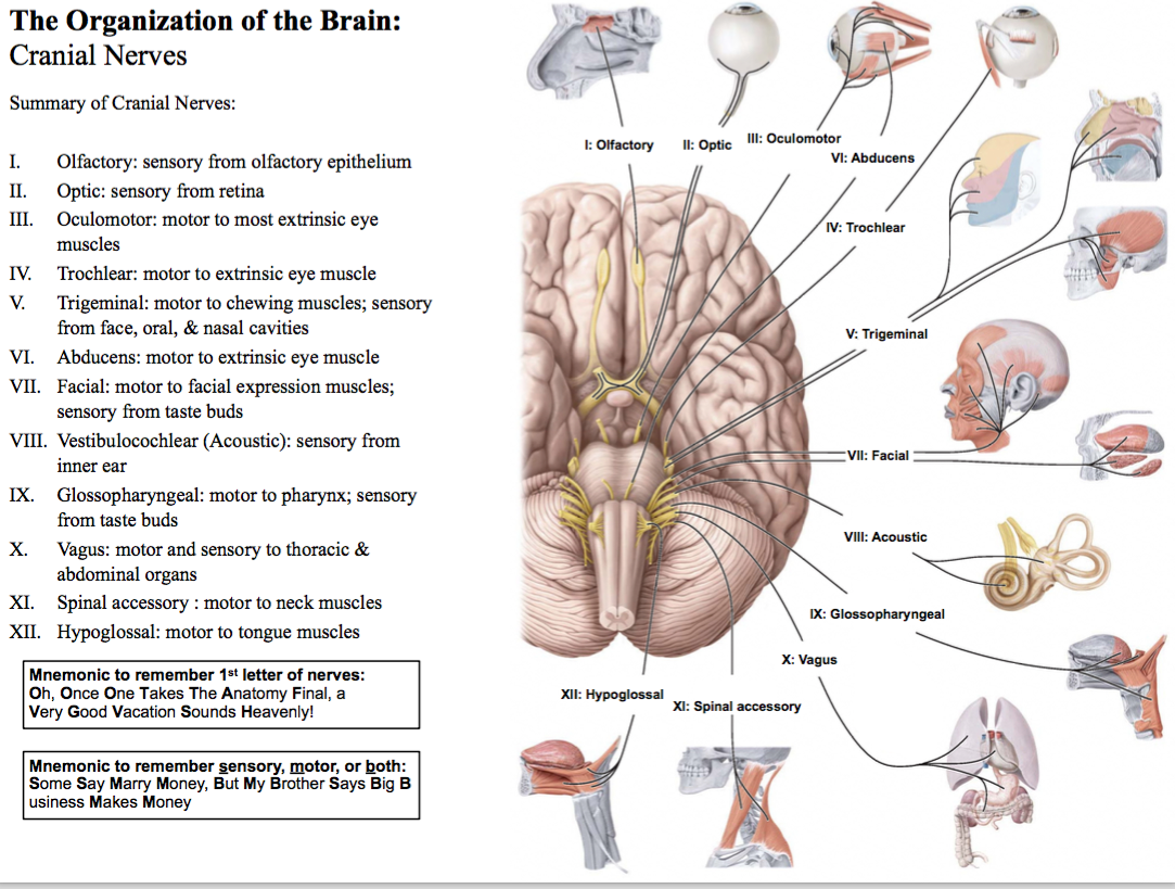
| The Organization of the Brain: Cranial Nerves Cranial nerve I is the ________ nerve. The visible structure on the surface of the brain is actually the olfactory _____. It is an extension of the cerebral hemisphere. The olfactory nerve is very short, running from the _____ epithelium through the _______ plate of the ethmoid bone. The olfactory nerve synapses within the olfactory ____ at the distal end of the olfactory tracts. Thus, the olfactory tracts and bulbs are _____, while the olfactory nerves are not shown here nor on any models in lab. | olfactory tract nasal cribriform bulbs visible |
| The Organization of the Brain: Cranial Nerves Cranial nerve II is the _____ nerve. It carries information from the _____ of the eye to the optic ____, where about half of the fibers cross to the _____ side. From there, the optic _____ carry information to the brain. | optic retina chiasm opposite tract |
| Cerebellar peduncle | Communication to/from cerebellum |
| Cerebral peduncle | Communication to/from cerebrum |
| Hypothalamus | 1. Autonomic control 2. Emotion control 3. Body temperature control 4. Sleep control 5. Pituitary control |
| Inferior colliculus | Auditory reflex center |
| Insular lobe of cerebrum | Gustatory cortex |
| Medulla oblongata | White matter & autonomic reflexes (cardio, resp, etc) |
| Midbrain | Contains corpora quadragemina, coordinates movements, regulates basal ganglia |
| Optic Chiasm | Decussation (hemidecussation, really) of optic nerves |
| Optic nerve | carries info from retina |
| Optic tract | carries info from chiasm to brain |
| Pineal body | regulates sleep, produces melatonin |
| Pituitary | Releases hormones |
| Pons | White matter tracts |
| Pyrimidal tracts | Contains Corticospinal tracts |
| Superior colliculus | Visual reflexes |
| Thalamus | “Relay station” that distributes info to cerebral cortex |
| Cranial nerves? Know 1st and second functions Know all names and whether sensory, motor, or both Use mnemonics! |
Möchten Sie mit GoConqr kostenlos Ihre eigenen Karteikarten erstellen? Mehr erfahren.