12123191
Beschreibung
Karteikarten von Agasana Viengmany, aktualisiert more than 1 year ago
|
|
Erstellt von Agasana Viengmany
vor fast 7 Jahre
|
|
| Frage | Antworten |
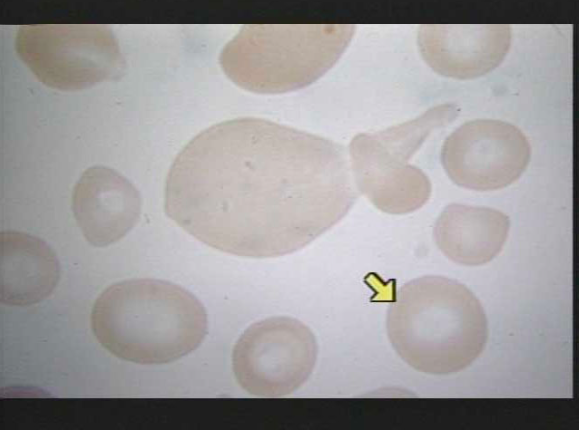
| mature erythrocyte (RBC) *As the red blood cell matures, it destroys most of its own organelles--such as, its nucleus, mitochondria, Golgi, etc.--and fills its cytoplasm with mostly hemoglobin molecules and some important enzymes* | |
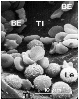
| WBC/Leukocyte "Le" = Leukocytes (they are seen inside of an arteriole) "TI" = tunica intima, the inner surface layer of the arteriole "BE" = biconcave-shaped erythrocytes in the vessel. | |
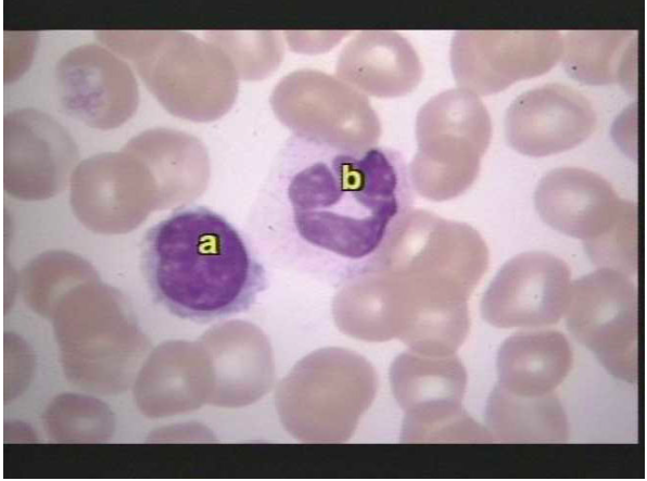
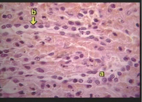
| WBC/Leukocyte Slide 2 shows a light micrograph of two kinds of leukocyte--the lymphocyte "a", seen with a large spherical nucleus, and the monocyte "b", a larger cell with a C-shaped nucleus. | |
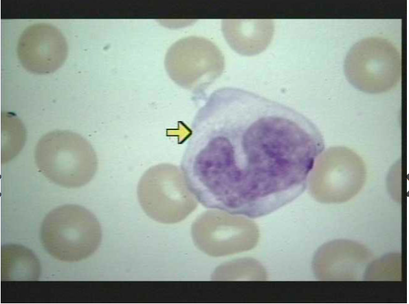
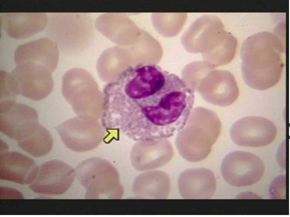
| WBC/Leukocyte Monocytes are the largest of the leukocytes and generally have kidney or horseshoe-shaped nuclei (select slide 3 and note the cell at the tip of the arrow); agranular | |
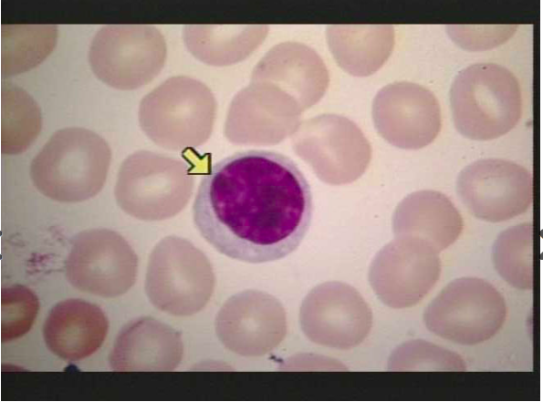
| WBC/Leukocyte Lymphocytes, on the other hand, are smaller cells with large round nuclei surrounded by a halo of cytoplasm (select slide 4 and note the cell at the tip of the arrow). agranular | |
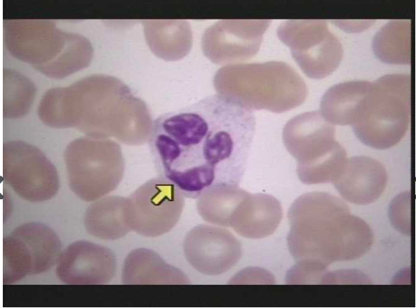
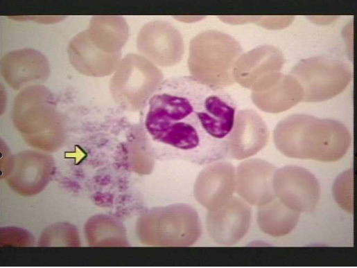
| WBC/Leukocyte Neutrophils are the most abundant type of leukocyte, comprising 50%-70% of the leukocytes in the blood (select slide 5 to see an example of a neutrophil at the tip of the arrow) Note the multi-lobed nucleus and the poorly stained granules. | |
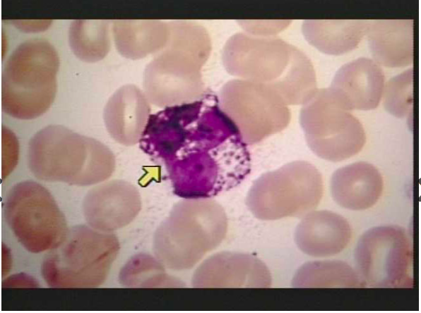
| WBC/Leukocyte Granular leukocytes with pink-staining granules are called eosinophils (select slide 6 to see an eosinophil at the tip of the pointer). Note the striking presence of the pinkish granules in the cytoplasm and the multilobed nucleus. | |
| WBC/Leukocyte leukocytes with blue-staining granules are called basophils (select slide 7 to see a basophil at the tip of the arrow). These cells are believed to be immature forms of mast cells which are known to secrete anticoagulants and inflammatory substances into the blood. | |
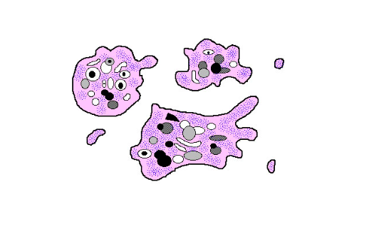
| Thrombocytes/ Platelets Thrombocytes are the smallest of the formed elements of the blood and are actually fragments of large cells called megakaryocytes, found in bone marrow. The fragments that enter the circulation as platelets lack nuclei but, like leukocytes, are capable of amoeboid movement. | |
| Thrombocytes/ Platelets Thrombocytes are the smallest of the formed elements of the blood and are actually fragments of large cells called megakaryocytes, found in bone marrow. The fragments that enter the circulation as platelets lack nuclei but, like leukocytes, are capable of amoeboid movement. | |
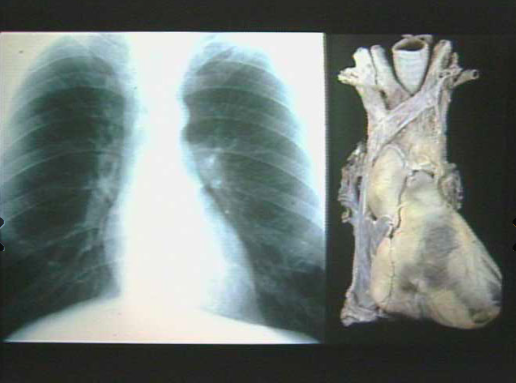
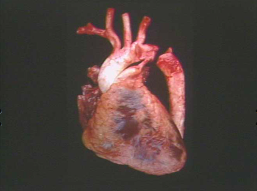
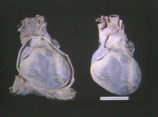
| Heart (Exterior) The heart is a hollow muscular organ located in the thoracic cavity, in an area called the mediastinum note the large blood vessels that enter the superior portion of the heart (called the base of the heart) | |
| view a freshly removed human heart | |
| The heart is surrounded by a tough protective fibrous sac called the pericardium. Select slide 4 to see the pericardium. The pericardium consists of a double-walled serous membrane, with serous fluid between the two layers. | |
| Heart Chambers The heart contains four major cavities, or chambers. Dividing the heart vertically down the middle into a right heart and a left heart is a wall of muscle called the septum. At the top of each half of the heart is a chamber called an atrium. Below it is another chamber, the ventricle. Each atrium has a flap-like appendage called an auricle or "little ear". The atria lead to the ventricles by way of openings called atrioventricular orifices, and in a normal heart the blood always flows from the atria to the ventricles. | |
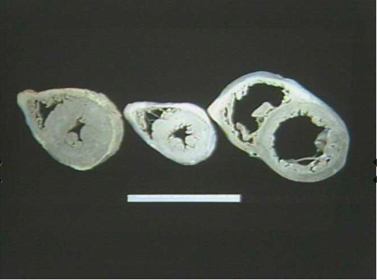
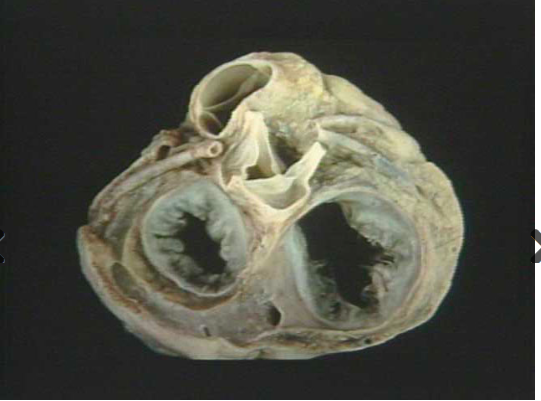
| Heart Chambers Select slide 3 to see cross-sections through the heart at various levels. Note that the wall of the left ventricle (donut-shaped area in the lower right of each cross-section in the photo) is thicker than the wall of the right ventricle (the crescent-shaped area in the upper left of each cross-section in the photo). This is because the left ventricle must be strong enough to supply blood to all parts of the body, whereas the right ventricle supplies only the lungs. As a result, the left ventricular blood pressure is higher (120 mm Hg) during contraction than in the right ventricle (30 mm Hg). The left ventricle is thicker to accommodate the higher pressure required to pump blood a greater distance against a high resistance. The walls of the atria are thinner than the ventricular walls because less pressure is required to pump blood only a short distance (and against less resistance) into the ventricles. | |
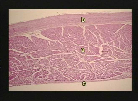
| Heart Walls a = myocardium b = epicardium c = endocardium | |
| Heart Walls a = epicardium b = myocardium | |
| Heart Walls b = endocardium a = myocardium | |
| Heart valves Look at slide 1 and 2. The four heart valves prevent the backflow of blood within the heart. The two atrioventricular valves (tricuspid and mitral valve) prevent blood from flowing back into the atria from the ventricles when the ventricles are in systole. The two semilunar valves (each containing three, half-moon shaped leaflets) prevent blood from returning to the ventricles from the major arteries when the heart enters diastole. The pulmonary semilunar valve (top in both pictures) blocks the return of blood from the pulmonary artery, while the aortic valve (middle of both pictures) blocks the return of blood from the aorta. Also shown on the diagram is the attachment of the two coronary arteries (left and right) to the base of the aorta | |
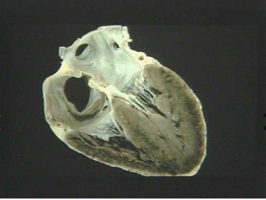
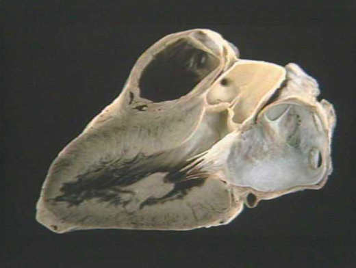
| Heart valves The valve between the left atrium and left ventricle is called the bicuspid, or mitral valve. Select slide 3 to see a frontal section of the heart, showing the position of the mitral valve between the left ventricle (black chamber on the bottom) and the left atrium (white chamber on the top right). Note also the white tendinous cords, called chordae tendineae, that stretch from the valve into the ventricle. These chordae tendineae are seen attaching to a large papillary muscle that is fixed onto the inner wall of the left ventricle. During ventricular systole, the papillary muscles contract, putting tension on the chordae tendineae which supports the valve cusps and prevents the backflow of blood into the atrium from the ventricle. | |
| Coronary vessels Blood is supplied to the heart by the right and left coronary arteries, which are the first branches off the aorta, close to the point where the aorta emerges from the heart. Look at slide 1. The aortic semilunar valve partially covers the openings of these arteries while blood is pumped into the aorta from the heart. Blood can pass through the openings of the coronary arteries only when the left ventricle has relaxed, and the cusps do not cover the openings. Blood enters these arteries as a result of the elastic recoil force of the aorta. | |
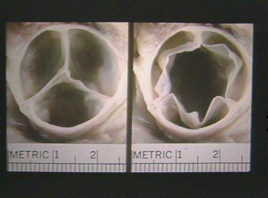
| Heart valves Select slide 6 to see a comparative example of a normal aortic valve, both in the closed and open position. On the left note the complete closure of the valve (all three leaflets tightly apposed to one another) and on the right observe the complete opening (no stenosis) of the valve cusps. | |
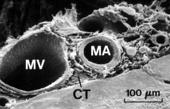
| Blood Vessels the electron micrograph in slide 1 shows a medium sized artery (MA) and vein (MV). In general, arteries have thicker walls than veins. | |
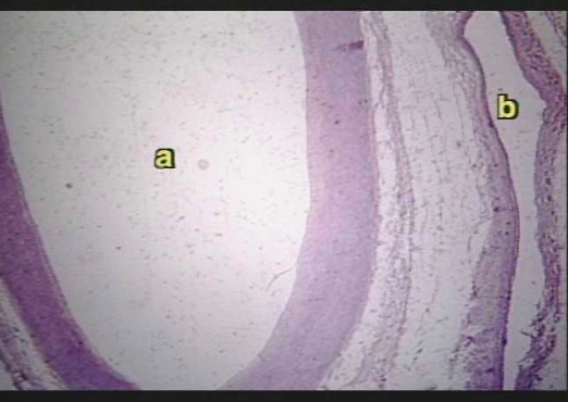
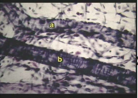
| Blood Vessels slide 2, where "a" is an artery and "b" is a vein. | |
| Blood Vessels slide 3 to see an example of an arteriole "b" and a venule"a". | |
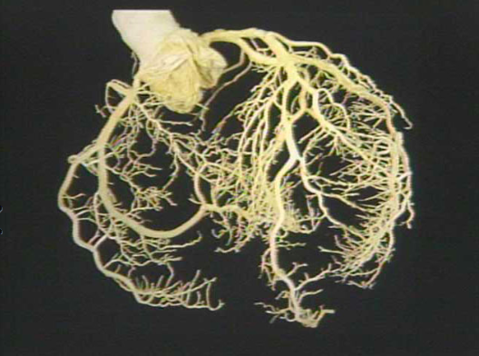
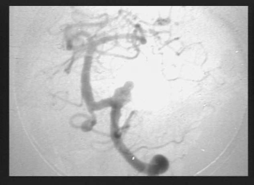
| Blood Vessels slide 4 to see an angiogram of blood vessels in the brain. | |
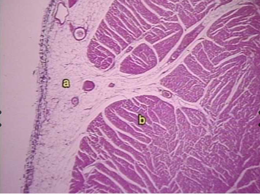
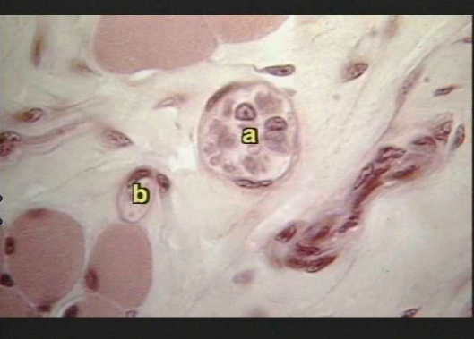
| Blood Vessels select slide 6 to see a light microscope cross-section slide of a small capillary ("b") and a larger arteriole ("a"). Note the flattened endothelial cells which form the walls of these vessels. These vessels are located inside of a loose connective tissue stroma which also contains several adipose cells (solid pink cells on the bottom and top left of the screen). | |
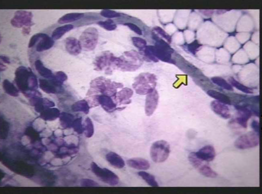
| Blood Vessels Select slide 7 to see a more magnified crossectional view of a capillary. The arrow points to the thin wall of the capillary, a good example of a simple squamous epithelium. Several blood cells can be seen inside the capillary lumen, and adipose cells can be observed outside of the vessel (top right). | |
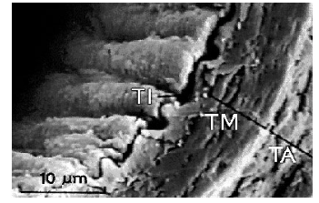
| Arteries TI = tunica intima TM = tunica media TA = tunica adventitia | |
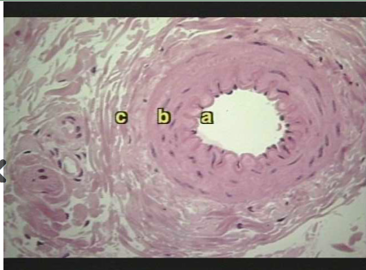
| Arteries a = tunica intima b = tunica media c = tunica adventitia | |
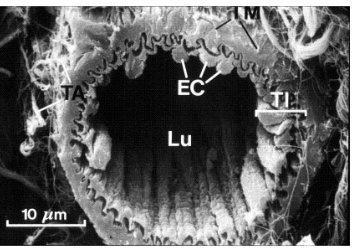
| Arterioles TA= tunica adventitia TI= tunica intima Lu = arteriole lumen | |
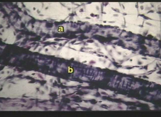
| Arterioles a = arteriole b = capillary | |
| arterioles a= venue b = arterioles | |
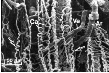
| Capillaries ca = capilliaries ar = arterioles ve= venules arrows= capillaries are coiling | |
| Capillaries arrow= capillary wall | |
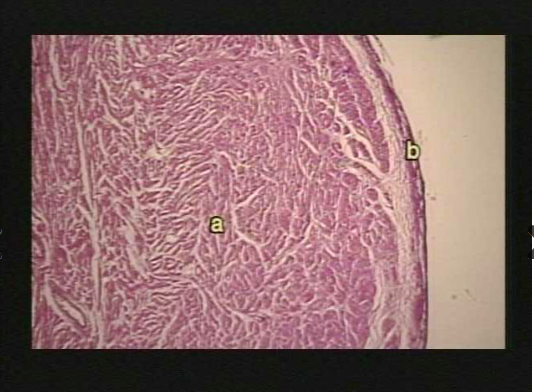
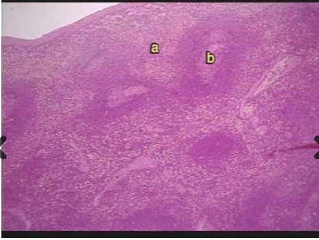
| Spleen a = red pulp b = white pulp | |
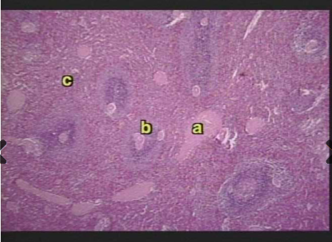
| Spleen Close up view of splenic pulp b = splenic artery c= red pulp a= sinusoids | |
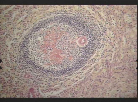
| Spleen germinal center | |
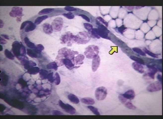
| Spleen a= sinusoid b at tip of arrow = macrophages | |
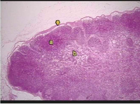
| Lympathic system Lymph nodes are covered by a capsule of fibrous connective tissue (black in the diagram above) and projections of this connective tissue called trabeculae extend inward from the capsule toward the center of the Iymph node, dividing it into compartments. The outer portion of each compartment is the cortex of the node (labeled "a" on slide 2 located directly underneath the subscapular sinus indicated by the arrow). The inner part of a Iymph node is the medulla ("b"). |
Möchten Sie mit GoConqr kostenlos Ihre eigenen Karteikarten erstellen? Mehr erfahren.