RCA Thorax 5: The mediastinum and the root of the neck
Pinned to
1223
1
0
|
|
Created by Ashutosh Kumar
about 8 years ago
|
|
Close
|
|
Created by Ashutosh Kumar
about 8 years ago
|
|

Name the structures in relation to the SVC and right brachiocephalic vein that are
Anterior:
Posterior:
Right:
Left:
The brachiocephalic veins unite to form the SVC behind the right first costal cartilage but what is the surface marking for their origin?
Describe the relation of the phrenic and vagus nerve at the thoracic inlet:
What is the ligamentum arteriosum?
What is the surface marking for the arch of the aorta?
Describe the relations of the trachea
Anterior:
Posterior:
Left:
Right:
What is the surface marking of the tracheal bifurcation?
Name:
Demarcates:
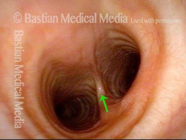
Why might widening of the carina seen at bronchoscopy be a potentially sinister sign in the evaluation of a patient with lung cancer?
Describe the constituents of the trachea:
What clinical relevance does the diameter and orientation of the right main bronchus have to accidental inhalation of a foreign body?
Name:
Constituents:
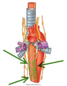
Name:
Source:
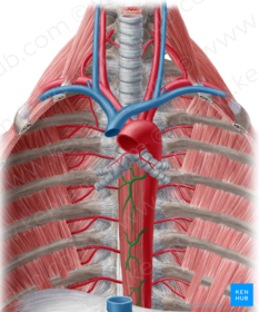
Name G, E and B:
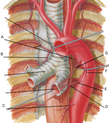
If you were examining the oesophagus with an endoscope, why might it appear indented at three main sites?
Name:
Course:
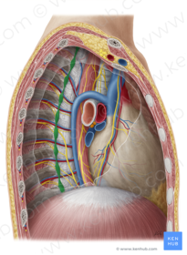
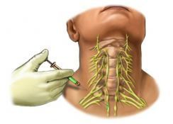
Name the structure being anesthetized:
Location:
Drugs were given to try and encourage the ductus to close but this was unsuccessful and so the baby had the Patent ductus arteriosus tied off surgically via a thoracotomy through the left side of the chest. The baby’s heart failure improved but he remained breathless and a chest X-ray showed a raised left hemidiaphragm due to paralysis of the left side of the diaphragm.
What nerve supplies the diaphragm and why might it have been injured during this operation?
Describe the effect of a patent ductus arteriosus (PDA):

 Hide known cards