12262258
Description
Flashcards by Agasana Viengmany, updated more than 1 year ago
|
|
Created by Agasana Viengmany
over 6 years ago
|
|
| Question | Answer |
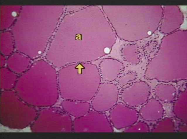
| Thyroid Gland "a" = thyroid follicle filled with a protein substance, the colloid that contains a stored form of thyroid hormones. Arrow = pinpoints the follicular cells that enclose the colloid. | |
| Thyroid Gland In slide 3, the large, solid pink areas are the colloid filled follicles. The purple spots are the nuclei of the follicle cells. | |
| Parathyroid Gland "a" marks a region dominated by principal (chief) cells, one of the two major cell types which secrete parathormone. | |
| Adrenal Gland two regions: the outer adrenal cortex making up the bulk of the gland and the inner adrenal medulla | |
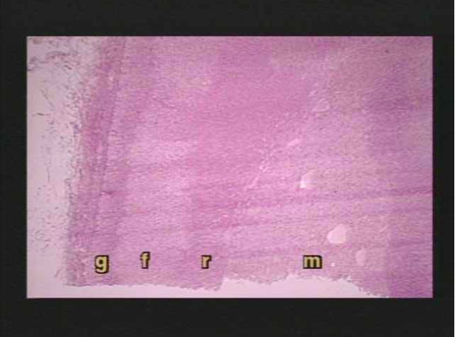
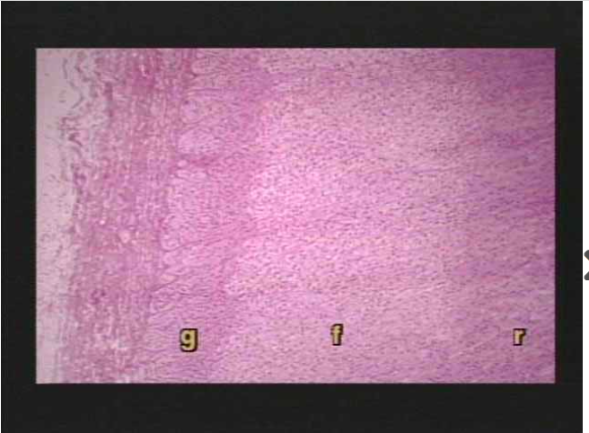
| Adrenal gland "g" is the outermost layer of the cortex, the zona glomerulosa while "f" marks the middle layer of the cortex, the zona fasciculata "r" is the deepest layer of the cortex, the zona reticularis "m" marks the medulla | |
| Adrenal gland "g" is the primary source of mineralcorticoids (zona glomerulosa) "f" mainly secretes glucocorticoids (zona fasciculata) "r" secretes miniscule amounts of gonadocorticoids (zona reticularis) | |
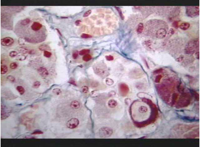
| Adrenal Gland an array of hormone-producing cells called chromaffin cells, which surround bright red sinuses containing blood | |
| Pituitary Gland | |
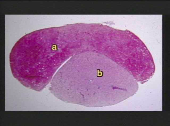
| Pituitary gland "a" marks the anterior lobe which makes up 75% of the pituitary by weight. "b" is the posterior lobe which is actually a neuron fiber extension of the hypothalamus | |
| Pituitary Gland a magnified view of the anterior pituitary. This shows the variety of cell types that compose the pituitary, each of which secretes a specific hormone. | |
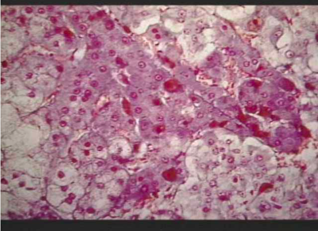
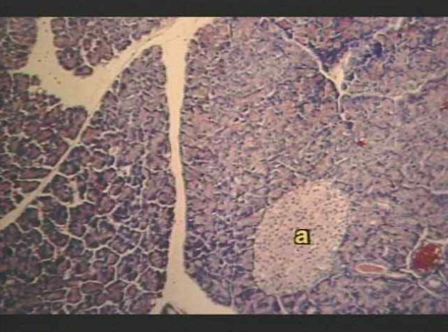
| Pancreas a photomicrograph showing an Islet of Langerhans ("a") (endocrine), among the darker and much more abundant exocrine cells. | |
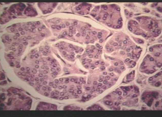
| Pancreas Slide 3 shows an Islet at higher magnification. A lighter, ovoid Islet mass dominates the screen and extends off the left margin of the field. The Islet is surrounded by exocrine cells. | |
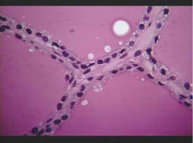
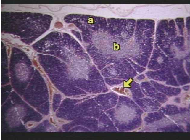
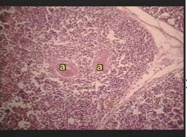
| Thymus gland "a" marks the cortex, and "b" is the lighter stained medulla of a thymic lobule. | |
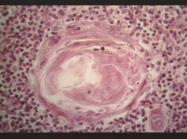
| Thymus gland close-up of a thymic lobule, with "a" marking the characteristic thymic (Hassall's) corpuscles. | |
| Thymus gland a closeup of Hassall's corpuscle which is composed of concentric layers of epithelial cells. There is no known function for these corpuscles, but many think that they are centers of cellular death within the thymus. | |
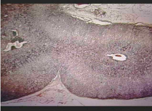
| Pineal gland composed of supportive neuralgia cells and secretory cells. Although many peptides and amines (including serotonin, norepinephrine, and histamine) have been isolated from this minute gland, only melatonin is known with certainty to be a major secretory product. |
Want to create your own Flashcards for free with GoConqr? Learn more.