
38718442
Description
| Question | Answer |
| USED TO DESCRIBE THE DARK AREAS IN RADIOGRAPH ALLOWS PASSAGE OF XRAYS LESS DENSE | RADIOLUCENT |
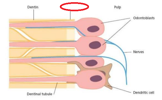
| Innermost soft, connective tissue of the tooth | DENTAL PULP |
| USED TO DESCRIBE THE LIGHT OR WHITE AREAS IN RADIOGRAPH OBSTRUCT PASSAGE OF XRAYS DENSE | RADIOPAQUE |
| IS BONE RADIOPAQUE OR NOT | YES |
| IS SOFT TISSUE A RADIOLUCENT OR NOT? | YES |
| Derived from the dental papilla like the dentin | DENTAL PULP |
| FUNCTIONS OF THE DENTAL PULP | - Formative or developmental -Nutritive -Sensory or Protective - Defensive or reparative |
| Contains nerves, arterioles, venules, capillaries, lymph channels, connective tissue cells, intercellular substance, odontoblasts, fibroblasts, macrophages, collagen and fine fibers | DENTAL PULP |
| ODONTBLAST | |
| *function of pulp* production of primary and secondary dentin by odontoblasts | Formative or developmental |
| *function of pulp* supplies nutrients and moisture to dentin through the blood vascular supply to the odontoblasts and their processes | Nutritive |
| *function of pulp* various stimuli elicit only pain as a response, does not differentiate between heat, touch, pressure and chemicals; control of circulation in the pulp | Sensory and Protective: |
| *function of pulp* ✓ response to irritation by mechanical, thermal, chemical or bacterial stimuli | Defensive or reparative |
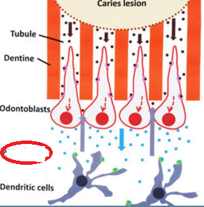
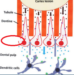
| *function of pulp* deposition of reparative dentin - protective barrier against caries and various other irritating factors | Defensive or reparative |
| *function of pulp* In cases of severe irritation, inflammation may become irreversible; since it is confined in dentin, dentin limits the inflammatory response | Defensive or reparative |
| protective barrier against caries and various other irritating factors | deposition of reparative dentin |
| convenient source of multipotent stem cells | dental pulp |
| Soft connective tissue that supports the dentin | dental pulp |
| Principal cells of dental pulp | Odontoblasts, fibroblasts, undifferentiated ectomesenchymal cells, macrophages, immunocompetent cells |
| NERVES | |
| DENDRITIC CELL | |
| DENDRITIC CELL | |
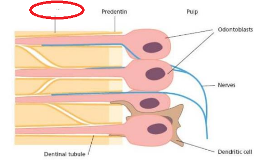
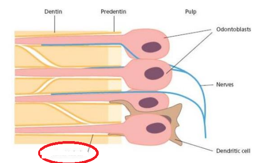
| PREDENTIN | |
| DENTIN | |
| DENTAL PULP | |
| ODONTOBLAST | |
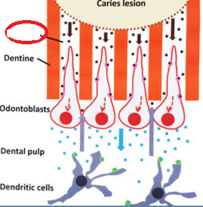
| DENTINE | |
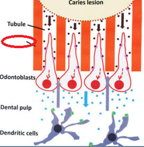
| TUBULE | |
| DENTINAL TUBULE | |
| 4 ZONES OF THE PULP | -Odontoblastic zone -Cell-free zone of Weil -Cell-rich zone -Pulp core |
| ZONE OF THE PULP (pulp periphery) | Odontoblastic zone |
| ZONE high cell density (which again is seen easily in the coronal pulp adjacent to the cell-free zone) | Cell-rich zone |
| zone major vessels and nerves (which is characterized by the major vessels and nerves of the pulp) | Pulp core |
| ZONE OF THE PULP beneath the odontoblasts (which is prominent in the coronal pulp) | Cell-free zone of Weil |
| Form and maintain the dentin | ODONTOBLAST |
| Form a layer lining the periphery of the pulp and have the odontoblastic process extend into dentin | ODONTOBLAST |
| Most distinctive cells of the pulp | ODONTOBLAST |
| odontoblast Midportion of pulp | cuboidal |
| Crown of fully developed tooth: cell bodies are columnar and measure approximately 50 µm in height | ODONTOBLAST |
| odontoblast Apical part | flattened |
| reflects their functional activity and ranges from an active synthetic phase to a quiescent phase | Morphology of odontoblasts |
| begins at the neck of the cells where it begins to narrow as it enters the predentin | Odontoblast process |
| Odontoblasts in the crown is larger than odontoblasts in the root | ODONTOBLAST |
| Soft connective tissue that supports the dentin | dental pulp |
| when differentiated, they cannot undergo further cell division | Odontoblasts are end cells |
| give dentin its viability and ability to respond to various stimuli | Dentinal tubule and its contents |
| 2 DIVISION OF PULP CAVITY | pulp chamber AND root canal |
| convenient source of multi-potent stem cells | dental pulp |
| The space PULP occupies | pulp cavity |
| radicular portion | root canal |
| coronal portion | pulp chamber |
| terminates at the apical foramen, where the pulp and the periodontal ligament meet and the main nerves and vessels enter and leave the tooth | root canaL |
| control of circulation in the pulp | Defensive or reparative |
| LOCATION OF LARGER ODONTOBLASTS | CROWN |
| Most abundant cells in the pulp | FIBROBLASTS |
| When dentin is exposed due to caries, cavity preparation, gingival recession or attrition | DENTIN HYPERSENSITIVITY |
| Represent pool from which pulp connective tissue cells are derived | UNDIFFERENTIATED ECTOMESENCHYMAL CELLS |
| cell bodies are columnar and measure approximately 50 µm in height, | Crown of fully developed tooth |
| Forms and maintains pulp matrix | FIBROBLAST |
| abundant cytoplasm and peripheral cytoplasmic extensions | UNDIFFERENTIATED ECTOMESENCHYMAL CELLS |
| THEORIES OF DENTIN HYPERSENSITIVITY | Nerve theory Odontoblast theory Hydrodynamic theory |
| MECHANISM OF DENTIN HYPERSENSITIVITY The tubular nature of dentin permits fluid movement to occur within the tubule when a stimulus is applied, a movement registered by pulpal free nerve endings close to the odontoblasts * | HYDRODYNAMIC THEORY |
| Numerous in the coronal portion (cell-rich zone) | FIBROBLAST |
| Concentric layers of mineralized tissue formed by surface accretion around blood thombi, dying/ dead cells or collagen fibers | PULP STONES |
| Can be free or unattached to the outer pulpal wall or can be attached to dentin | PULP STONES |
| found throughout the cell-rich area and the pulp core | UNDIFFERENTIATED ECTOMESENCHYMAL CELLS |
| the means by which the pulp and mineralised tissues surrounding the dentine (enamel and cementum) communicate. | DENTINAL TUBULE |
| consists of collagen and ground substance | PULP MATRIX |
| MECHANSIM OF DENTIN The dentin contains nerve endings that respond when it is stimulated | NERVE THEORY |
| Mesenchymal cells that have self-renewal capability | DENTAL PULP STEM CELLS |
| Have the capacity to give rise to osteoblasts and may therefore be a promising tool for bone regeneration | DENTAL PULP STEM CELLS |
| Similar function to the Langerhans’ cells of the epithelium | immunosurveillance through capture and presentation of foreign antigen to the T cells |
| Consists of collagen fibers and ground substance that make up the extracellular matrix of the pulp | MATRIX AND GROUND SUBSTANCE |
| MECHANISM OF DENTIN odontoblasts serves as receptors and are coupled to nerves in the pulp | ODONTOBLASTIC THEORY |
| Reduces the overall number of cells within the pulp | PULP STONES |
| A THEORY WHERE odontoblasts act as a receptor | Odontoblast theory |
| the need for synthesis diminishes and the fibroblasts appear as flattened spindle-shaped cells with dense nuclei | With age |
| PRIMARY FUNCTION OF FIBROBLAST | the maintenance of structural integrity within the connective tissue |
| Macrophages appear as large oval or sometimes elongated cells with dark-stained nucleus microscopically | INFLAMMATORY CELLS |
| FIBROBLAST actively synthesizing matrix and therefore have a plump cytoplasm and extensive amount of organelles | Young pulps |
| Stimulated directly or indirectly by fluid movement | Hydrodynamic theory |
| Endodontic therapy | ROOT CANAL TREATMENT |
| may contain tubules and be surrounded by cells resembling odontoblasts | TRUE PULP STONES |
| ACTS AS Denticles | PULP STONES |
| have the capability of ingesting and degrading collagen when appropriately stimulated | FIBROBLAST |
| Depending on the stimulus, may give rise to odontoblasts or fibroblasts | UNDIFFERENTIATED ECTOMESENCHYMAL CELLS |
| A THEORY WHERE Dentin is innervated directly | Nerve theory |
| Can be a problem during endodontic therapy | PULP STONE |
| promising tool for bone regeneration | DENTAL PULP STEM CELLS |
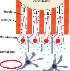
| These cells participate in immunosurveillance and increase in number in carious teeth, where they infiltrate the odontoblast layer and can protect their processes into the tubules. | INFLAMMATORY CELLS |
| T lymphocytes are found HERE | Normal pulps |
| B lymphocytes are scarce HERE | Normal pulps |
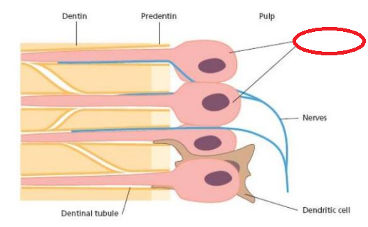
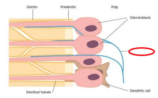
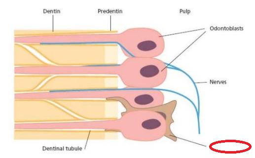
| bone-marrow derived, antigen presentingdendritic cells are found in and around the | Dendritic cells |
| Can differentiate into odontoblasts, chondrocytes, adipocytes and neurons | DENTAL PULP STEM CELLS |
| Discrete calcified masses that have calciumphosphorus ratios comparable to dentin | PULP STONES |
| Overall collagen content increases with age of the pulp | MATRIX AND GROUND SUBSTANCE |
| Decreased volume of the pulp chamber and root canal due to continued dentin deposition | age changes |
| Greatest concentration of collagen generally occurs in the most apical portion of the pulp | Greatest concentration of collagen generally occurs in the most apical portion of the pulp |
| Ground substance resembles other tissues: glycosaminoglycans, glycoprotein and water | MATRIX AND GROUND SUBSTANCE |
| gradual reduction of the tubule diameter; closure of the tubule | Deposition of intratubular dentin |
| Blood vessels enter and exit the dental pulp by way of the apical and accessory foramina | VASCULAR AND LYMPHATIC SUPPLY |
| inceased brittleness and decreased permeability | Sclerotic dentin |
| Fibers are principally type I and type III collagen | MATRIX AND GROUND SUBSTANCE |
| extensive plexus of nerves in the cell-free zone of Weil just below the cell bodies of the odontoblasts in the crown portion of the tooth | Subodontoblastic plexus of Raschkow |
| Nerves enter the pulp through the apical foramen along with the afferent blood vessels and together form the neurovascular bundle | INNERVATION OF THE DENTIN-PULP COMPLEX |
| what age when the cell density has decreased by about half | 70 |
| Circulation establishes the tissue fluid pressure found in the extracellular compartment of the pulp | VASCULAR AND LYMPHATIC SUPPLY |
| Sensory afferent nerves of CN V (trigeminal nerve) and sympathetic branches of the superior cervical ganglion; | myelinate and unmyelinated axons |
| One or sometimes two vessels of arteriolar size enter the apical foramen with the | abt 150 mm) |
| They arise as small, blind, thin-walled vessels in the coronal region of the pulp | Lymphatic vessels |
| reduction in the vascular supply to the pulp | Restriction in pulp volume |
| cells gradually decrease in number | 20 |
| Age changes render the pulp more resistant to environmental injury | tubule occlusion |
| produce more sclerotic dentin, deposit secondary dentin at an increased rate | Response to gradual attrition |
| gradual reduction of the tubule diameter; closure of the tubule | Deposition of intratubular dentin |
| Occurrence of irregular areas of dystrophic calcifications | when the age changes |
| Reparative dentin also contributes to the reducing sensitivity | Increase in dead tracts and sclerotic dentin |
| More severe stimulus | tertiary dentin formation at the ends of the tubules affected by the injury |
| Appearance of fibrous bundles due to change in collagen fibril distribution | WHEN THE AGE CHANGES |
| age lessens the ability of the dentin-pulp complex to repair itself | Age of the pulp determines its ability to repair the damage |
| decreased potential for differentiation of new odontoblasts from the mesenchymal cells of the pulp and the formation of reparative dentin | WITH AGE |
| narrowing of dentinal tubule diameter, deposition of peritubular dentin | AGING |
| have a much more favorable prognosis for surviving pulpal inflammation | Recently erupted teeth with large pulp chambers and short, wide canals with large apical foramina |
| Stimuli are not transmitted as rapidly Complete obliteration of older tubules with mineralization Pulp horns recede Pulp becomes more fibrotic | AGING |
| band of epithelium that gives rise to two subdivisions which ingrow into the underlying mesenchyme colonized by neural crest cells | Primary epithelial band |
| which forms afterwards and is positioned just in front of dental lamina | vestibular lamina |
| Largest portion of the tooth structure, extending almost the full length of the tooth | dentin |
| covered by the enamel in the crown and cementum in the roots | dentin |
| forms the walls of the pulp cavity – pulp chamber and pulp canal | dentin |
| Both dentin and pulp are derived from the | mesoderm |
| Provides elasticity and strength to the tooth; | dentun |
| enables it to withstand loading forces by mastication and trauma | dentin |
| Protects and preserves the integrity of the pulp tissue | dentin |
| • More radiolucent than enamel but more radiopaque than the pulp | dentin |
| Protects and preserves the integrity of the pulp tissue | dentin |
| composition Mature dentin: | 70 inorg , 20 org, 10 water |
| TRUE OR FALSE Dentinal crystallites are smaller than enamel crystallites | T |
| Dentinal crystallites size in bone and cementum | (length: 20-100nm, width: 3nm) |
| Provides greater yield to the pressure of a sharp explorer tine (tends to catch and hold in dentin) | DENTIN |
0 comments
Want to create your own Flashcards for free with GoConqr? Learn more.