8409552
Description
Flashcards by Kayla Price, updated more than 1 year ago
|
|
Created by Kayla Price
over 7 years ago
|
|
| Question | Answer |
| What does the central nervous system consist of? | The brain and spinal chord |
| What does the peripheral nervous system consist of? | All the neurones that connect the CNS to the rest of the body |
| What is the somatic nervous system? | Part of the nervous system that is under conscious control, carries impulses to muscles |
| What is the autonomic nervous system? | Part of the nervous system that is controlled subconsciously controlled, carries impulses to glands, smooth muscle and cardiac muscle |
| What is the difference between the sympathetic and parasympathetic nervous system? | Sympathetic is causing an increase in activity, parasympathetic is causing a decrease |
| What neurotransmitter is present in the neurones of the somatic nervous system? | Acetylcholine |
| What neurotransmitter is present in the neurones of the sympathetic nervous system? | Noradrenaline |
| What neurotransmitter is present in the neurones of the parasympathetic nervous system? | Acetylcholine |
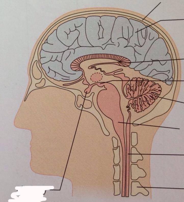
| What are meninges? | Protective membranes surrounding the brain |
| What is the function of the cerebrum? | Controls voluntary actions eg. learning, memory, personality, conscious thought |
| What is the function of the cerebellum? | Controls unconscious functions eg. posture, balance, non-voluntary movement |
| What is the function of the medulla oblongata? | Autonomic control eg. heart rate and breathing rate |
| What is the function of the hypothalamus? | Regulating temperature and water balance |
| What is the function of the pituitary gland? | Stores and releases hormones, divided into two sections (anterior and posterior). Anterior produces six hormones and posterior stores and releases hormones produced by the hypothalamus |
| What is the pathway of the reflex arc? | receptor - sonsory neurone - relay neurone - motor neurone |
| What are the three types of muscle in the body? | Skeletal muscle, cardiac muscle, smooth muscle |
| Describe the structure and function of skeletal muscle? | Striated, responsible for movement, voluntary control, arranged so muscle contracts in one direction, rapid & short contractions |
| Describe the structure and function of cardiac muscle | Specialised striated, involuntary control, cells branch and interconnect for simultaneous contraction, intermediate contraction speed and length |
| Describe the structure and function of smooth muscle | Non-striated, involuntary control, no regular arrangement of cells so they contract in different directions, slow contraction speed and can remain contracted for a long time |
| What is the sarcolemma? | A plasma membrane that encloses bundles of muscle fibres |
| Describe the arrangement of muscle fibres in skeletal muscle | Made up of bundles of muscle fibres enclosed in sarcolemma. The fibres contain a number of nuclei and are very long. Fibres consist of many muscle cells fused together with a shared cytoplasm called the sarcoplasm. |
| What are T-tubules and why are they helpful? | Parts of the sarcolemma that fold inwards to help spread electrical impulses to ensure all the fibres contract at the same time. |
| Why do muscles have many mitochondria? | To provide enough ATP required for contraction |
| What is the sarcoplasmic reticulum? | A modified endoplasmic reticulum in muscle fibres that contains calcium ions required for muscle contraction |
| What are myofibrils? | Long cylindrical organelles made of protein and specialised for contraction. Made up of two types of protein filament: actin and myosin |
| Describe the structure of a sarcomere | - Light bands/I bands: regions where there is only actin present - Dark bands/A bands: regions where myosin and actin overlap - Z lines: found at the centre of each light band, distance between adjacent Z-lines is one sarcomere - H-zone: light region at the centre of a dark band where only myosin is present, part of sarcomere that shortens when muscles contract |
| What happens to a sarcomere during contraction of muscle? | Myosin filament pull actin filaments towards the centre of the sarcomere, making the light band narrower, Z lines move closer together and the H zone narrower. The dark band remains the same width as the myosin filaments don't shorten. |
| Describe the structure of myosin | The filaments have globular heads that are hinged which allows them to move backwards and forwards. On the head is a binding site for each of actin and ATP. The tails of several hundred myosin molecules are aligned together to form the myosin filament. |
| Describe the structure of actin | Actin filaments have binding sites for myosin heads, called actin-myosin binding sites. These are often blocked by a protein called tropomyosin, held in place by troponin. |
| What is striated muscle? | Muscle tissue with repeating units of sarcomeres, giving it a striped appearance |
| How does myosin interact with actin when a muscle is stimulated to contract? | Myosin heads form bonds with actin filaments known as actin-myosin cross-bridges. The myosin heads then flex in unison, pulling the actin filament along the myosin filament. The myosin then detaches and its head returns to its original angle, using ATP. The myosin then reattaches further along the actin filament and the process occurs again. |
| What is a neuromuscular junction? | The point where a motor neurone and a skeletal muscle fibre meet. |
| What is a motor unit? | All the muscle fibres supplied by a single motor neurone. The more motor units stimulated, the stronger the force created when the muscle contracts. |
| How is muscle stimulated? | An action potential reaches the neuromuscular junction, which stimulates calcium ion channels to open. Calcium ions diffuse from the synapse into the synaptic knob, where they cause synaptic vesicles to fuse with the presynaptic membrane. Acetylcholine is released into the synaptic cleft via exocytosis and diffuses across the synapse. It binds to receptors on the postsynaptic membrane (sarcolemma) opening sodium ion channels and resulting in depolarisation. ACh is then broken down by acetylcholinesterase into choline and ethanoic acid, preventing overstimulation. |
| Label the diagram | |
| How is tropomyosin moved away from the actin-myosin binding sites on actin filaments? | When the sarcolemma becomes depolarised, calcium ion channels on the sarcoplasmic reticulum open, causing them to diffuse out into the sarcoplasm. The calcium ions bind to troponin, causing it to change shape. This pulls on the tropomyosin moving it away from the actin-myosin binding site. |
| How does the myosin head detach from the actin filament? | The molecule of ADP bound to the myosin head it released. This allows an ATP molecule to take its place, causing the myosin head to detach. |
| Describe the sequence of events that occurs when myosin and actin interact during muscle contraction | 1) Tropomyosin prevents myosin head from attaching to the binding site on the actin molecule 2) Calcium ions released from the endoplasmic reticulum cause the tropomyosin molecule to pull away from the binding sites of the actin molecule 3) Myosin head now attaches to the binding site on the actin filament 4) Head of myosin changes angle, moving the actin filament along. The ADP molecule bound to the myosin head is released 5) ATP fixes to myosin head, causing it to detah from the actin filament 6) The calcium ions present activate ATPase which hydrolyses the ATP to ADP, providing the energy for the myosin to return to its normal position 7) Head of myosin reattaches to another binding site further along |
| How does creatine phosphate help generate ATP? | It acts as a reserve supply of phosphate to regenerate ATP from ADP. |
| What is an electromyogram? | A record of the electrical activity in a muscle during an activity |
Want to create your own Flashcards for free with GoConqr? Learn more.