4755015
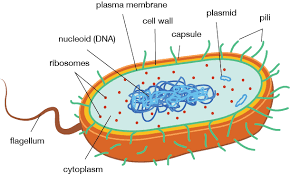
Structure of prokaryotes
- features of a prokaryotic cell
- Flagella
- allow for the movement of the cell
- clockwise rotation
- cell tumbles
- cell tumbles
- anticlockwise rotation
- develops movement for cell
- develops movement for cell
- Allows for directional
movement toward or away
(Chemotaxis)
- clockwise rotation
- allow for the movement of the cell
- Frimbrie
- The hairs of the cell
- Allows the cell to stick to
surfaces/adohernce e.g hard
surfaces
- Allows for Genetic transfer from one bacterium to another
- The hairs of the cell
- Capsule
- slimy layer on the cells surface
- carbohydrate envelope of glycocalx
- carbohydrate envelope of glycocalx
- prevents the cell/organism to becoming dehydrated/dessicatted in non aqueous environments
- resistance to phagocytosis (bacteria being indigested)
- slimy layer on the cells surface
- Ribosomes (Prokaryotic cell)
- 50% smaller than ribosomes found in eurkarotic cells
- allows for the synthesis of proteins
- target site for some antibiotics to inhibit
protein synthesis of the cell and therefore
kill it
- 50% smaller than ribosomes found in eurkarotic cells
- Nucleoid
- Large strands of DNA (loop) not enclosed by a nuclear membrane
- Large strands of DNA (loop) not enclosed by a nuclear membrane
- Plasmid
- Small loops of extrachromosomal DNA
- important in contributing extracelluar DNA in bacterium
- V; Virulence (Can make organism cause diseases)
- R; Can cause bacterium to become resistant to antibiotics
- B; Bacteriocin (natural antibiotic produced by cell)
- F; Fetility (transfer of genes)
- Small loops of extrachromosomal DNA
- Endospores
- cells form spores during unfavourable conditions
- condensation of nuclear material such as DNA
- material is surrounded by two coats of protein
- allows DNA to resits damage caused by Heat, Chemicals and desiccation
- allows DNA to resits damage caused by Heat, Chemicals and desiccation
- cells form spores during unfavourable conditions
- Flagella
- Establishing the difference between Gram Positive (+) and Gram negative (-)
- Gram Postive
- Thick outer layer of polytidglycon
- Thick outer layer of polytidglycon
- Gram Negative
- Thin layer of peptidoglycan
- surrounded by two membranes. Outer Membrane and Plasma Membrane
- Thin layer of peptidoglycan
- peptidoglycan is a protein only found in bacteria therefore is a good
indicator in determining Gram Positive and Negative bacteria
- Gram Staining
- the method used to determine Gram positive and negative bacteria
- Use of two stains
- treated with iodine to fuse the proteins of the peptidoglycan
- colour of stain identifies type bacteria (positive or negative)
- colour of stain identifies type bacteria (positive or negative)
- treated with iodine to fuse the proteins of the peptidoglycan
- Use of two stains
- violet = positive
- pink = negative
- the method used to determine Gram positive and negative bacteria
- Gram Postive
Media attachments
Want to create your own Mind Maps for free with GoConqr? Learn more.