Description
|
|
Created by Ella Broomer
almost 8 years ago
|
|
Page 1
Divisions of the nervous system
Central nervous system includes the brain and spinal chord Peripheral nervous system includes all nerve tissue outside the CNS Brain- maintains life and is involved in higher functions and psychological processes Spinal chord- receives information to and from the brain CNS- concerned with all life functions and psychological processes Peripheral nervous system- transmits information to and from the CNS Somatic nervous system- Transmits information to and from the senses, and to and from the CNS. Voluntary activities under conscious control and some unconscious movements such as reflexes. Autonomic nervous system- Transmits information to and from internal organs to sustain life processes. Involuntary activities not under conscious control or reflexes. Sympathetic nervous system- increases bodily activities. Deals with emergency situations e.g. prepares body for fight or flight. Parasympathetic nervous system- maintains and decreases bodily activities. Controls involuntary activities not emergencies e.g. organs of the digestive system breaking down food.
Page 2
Structure and function of nerves
Interneuron = relay neuronSensory neurone Carry messages PNS snd CNS Have long dendrites and short axons Unipolar (only transmit information) Relay neurone Connects sensory neurones to motor and other relay neurones Short dendrites and short axons Multipolar (send and receive messages from many sources) Motor neurone Connects CNS to effectors in the body Short dendrites and long axons Multipolar Process of synaptic transmission (process for transmitting messages from neurone to neurone) Electrical nerve impulse travels down the neurone and is translated into a chemical signal in the form of a neurotransmitter (chemicals in the brain) at the pre-synaptic terminal Chemical is released into the synaptic fluid where it difffuses across the synase (specialised gap between neurones through which the electrical impulse from a neurone is chemically transmitted) Postsynaptic neurone contains receptors and they quickly take up neurotransmitters from the fluid and convert them into electrical impulses to travel down the next neurone
Excitation and inhibition When the neurotransmitter meets the receptor an ion channel is opened in the membrane This causes ions to flow through the membrane into the neurone and the flooding of neurones cuauses a 'potential' in the dendrites The potential produced due to the ions entering the neurone is either excitatory or inhibitory Excitatory neurones Increases the chance of a neurone firing Neurotransmitters such as noradrenaline Inhibitory neurones Decreases the chance of a neurone firing and the message is likely to be stopped Generally responsible for calming the body, inducing sleep and filtering out unnecessary excitatory signals To find the likelihood of the cell firing the excitatory and inhibitory synaptic input is added together. The sum of this then determines whether the cell fires or not (threshold it needs to reach to fire is -55 mV)
Page 3
Endocrine system
Consists of glands which release chemicals (hormones) throughout the body via blood and other bodily fluids Each hormone is thought to affect behaviour in different ways Hormones travel via the bloodstream to target sites throughout the body A given hormone usually only affects a limited number of cells known as target cells Specific hormones e.g. testosterone must be picked up by specific receptors, cells that do not contain the receptor cannot be directly influenced by the hormone When enough receptor sites are stimulated it results in a physiological reaction in the target cell
Thymus (below the thyroid gland and parathyroid)- helps build resistance to diseaseImportant glands to know about:Pituitary gland- some of the hormones released help regulate the endocrine system. Controls other glands in the body.Adrenal gland- Important for the fight or flight response as it helps trigger the fight or flight responseTestes- triggers the release of testosterone (male hormone)Ovaries- triggers the release of oestrogen and progesteronHormones level and timings of release are critical to normal functioning. Too much or little at one time would cause dysfunction e.g. Cushings syndrome where too much cortisol (often due to taking corticosteroids) which causes weight gain, thinning skin which brusies easily and muscle or bone weakness.
Page 4
Fight or flight
Prepares the body to fight or to flee from perceived attack, harm or threat to our survival Primitive respone Automatic respone Can be useful if you are experiencing threat or danger from humans or animals such as threat from a bear, being mugged or a fireCan be not useful in the modern day as having an adrenaline rush can cause illogical behaviour to occur and the fight or flight response in the modern day is not always a suitable responseSteps which occur in the fight or flight response: Hypothalamus recognises there is a threat Hypothalamus sends a message to the adrenal gland (specifically adrenal medulla) Triggering the release of noradrenaline in the brain and adrenaline to the endocrine system This causes changes to occur within in the body: Increased heart rate- speeds up blood flow to vital organs and improve spread of adrenaline around the body Faster breathing- increases oxygen intake Muscle tension- improve reaction time and speed Pupil dilation- improve vision Production of sweat- facillitate temperature regulation Reduced function of the digestive and immune system- save energy for prioritised functions e.g. running Adrenal glands Adrenal cortex produces corticosteriods (a hormone) The corticosteroids influence or regulate the body's response to stress, metabolism and immune system Inner part of the adrenal gland is the adrenal medulla Adrenal medulla produces catecholamines such as adrenaline (adrenaline increases heart rate and blood prssure) Amygdala and hypothalamus role in fight or flight The amygdala is mobilised when an individual is faced with a threat Amygdala associates sensory signals with fight or flight emotions such as anger Amygdala then sends signals of distress to the hypothalamus (a command centre in the brain) communicating with the rest of the body through the sympathetic nervous system Body responds to the stressers via 2 systems, one for acute stressors (sudden) such as an attack and another for chronic (ongoing) such as stress for exams
Page 5
Localisation of function in the brain
Brain has 2 hemispheres The hemispheres are bridged by the corpus callosum which is a bundle of fibres The corpus callosum enables both sides of the brain to exchange information Phineas Gage Localisation of function originally came from case studies Gage was a railway workers in the USA who worked by using dynamite to blow a clear path so a railway track could be laid Dynamite was placed into position using an iron rode (called a tamping iron) to bed the dynamite into place using sand Rod caused a spark one day and caused the dynamite to blow up This caused the rod to go through the chin of Phineas and out the top of his forehead Phineas was conscious and could speak but lost sight in his left eye Psychologically he had changed too he had gone from being calm and well-mannered to unreliable, hostile and rude Dr Bigelow believed that there was no localisation and Phineas' recovery supported that. However Dr Harlow believed there was localisation of function within the brain as Phineas' personality had changed so it was believed the area damaged must have held the reasoning and planning of an individual. Dr Harlow was proved to be correct in that there was localisation of function within the brain Issues with Gage as evidence Little evidence Reported a while after it happened Case study- only looking at the effects on 1 person so how can you apply it to everyone? Hemispheric (division of function between the 2 hemispheres)lateralisationIn most people the brain is contralateral (left part controls the right side of the body and the right part controls the left side of the body)
Left hemisphere Most people, language processing is here Focuses on detail Right hemisphere Recognising such as recognising emotions of others Spatial relationships - Clarke, Assal and de Tribolet- found that a woman got lost in familiar places however if given verbal instructions she did not get lost. Focuses on patterns Motor cortex Causes movement Sends messages through the brain stem and spinal chord to the muscles Important for complex movemeents such as running but not basic movements such as coughing Motor cortex instructs the body on a specific outcome however the spinal chord and other areas of the body co-ordinate the body's movement in relevant areas Areas of the brain involved in movement- spinal chord and brain (co-ordinate movement), premotor cortex (plans a movement before it is done) and the prefrontal cortex (stores sensory information before the moevement and works out the likelihood of the movement outcome Somatosensory cortex Makes sense of external information from our senses 2 parts to the sensory cortex- primary sensory cortex where the brain first processes information from sensory organs and the second sensory cortex which is involved in higher order processing needed for hand-held objects Amount of neural connections needed to the somatosensory cortex in specific areas is dependent on the amount of the somatosensry cortex needed in that specific area e.g. face would need a lot however trunk of the body would need very little. Visual centresPrimary visual cortex In the occipital lobe Main visual area An area within the main visual area called Area V1 is seen to be very important for visual perception Receives and processes sensory nerve impulses from the eyes Those with damage in V1 are said to not have any vision including conscious vision, dreams or visual imagery while awake (Hurrovitz et al- 1999) Visual information is transmitted along 2 pathways- 1 is components of the visual field and the other is to do with locations within a visual field Individuals with Are V1 damage can have 'blindsight'. This is where someone appears blind as they do not have vision but they can find objects within a visual field by pointing. Shows how some processing in the visual cortex may not be conscious (Bridgeman and Staggs- 1982) Auditory centresPrimary auditory cortex 2 primary auditory cortices, one in each hemisphere Converts sound waves from our ears into understandable data by electric impulses Auditory cortex in both hemispheres receives information from both ears via 2 pathways that transmit information about what a specific sound is and the location of the sound Information from the right ear mainly goes to the left hemisphere but some is transmitted to the left primary auditory complex too (happens in the same way to the left ear) Primary auditory cortex damage does not lead to total deafness as sounds can still be heard but sounds that need complex processing such as music cannot be heard Primary cortex also processes auditory image- when individuals watch silent films the primary auditory cortex in both hemispheres of the brain was activated e.g. if a door was shut with force as the 'bang' that would go with the closing of the door had been imagined Disorders due to damage to this area include classical auditory agnosia (inability to process environmental sounds such as animal noises) Language centresBroca's area Speech production Not all words are equally affected if there is damage to this area of the brain- nouns and verbs seem to be unaffected in some patients with Broca's area damage and other word categories such as prepositions and conjunctions (e.g. and, if, but) are unable to be spoken e.g. cannot read out 'to be or not to be' but can read put 'two bee oar knot two bee' (Gardner and Zurif- 1975) Louis Leborgne suffered from epilepsy and eventually was unable to speak other than to say the word tan. Following his death, Broca (a neurologist treating Leborgne before his death) found a lesion (area which has damage) on the left temporal lobe. It was the only area of the brain with visible damage so Broca concluded this was the area involved with speech production. Since then Leborgne's brain has been scanned using modern techniques and although there is more extensive damage the area of the brain Brocca found for speech production was correct Disorders associated with damage in this area include stuttering (underactivity in the area) Language centresWernicke's area Separate area of language processing Understanding language and assessing words Found by Wernicke who found that those with damage in the area were unable to comprehend language and struggled to find the words they needed but could speak fluently Case study- Phil had a stroke and was unable to speak in understandable language as the Wernick's area of the brain was damaged. He suffered from Wernicke's aphasia Evaluation of research into localisation of function Helps understand people's injuries when brain damage occurs Help to find new treatments and cures for illnesses and disorders caused by damage to a specific area of the brain Can be scientifically studied in a controlled environment Can be studied using scientific equipment There are few cases that can be studied to find the localisation of function Case studies are often outdated Lashley found that on rats' brains there was no specific area involved with memory and it seems memory is stored all over the brain. However caution should be taken when applying this to humans as there are differences physiologically Evidence from case studies suggests there is locationalisation and lateralisation of functions. If functions were all over the brain then specific defects would not occur Case studies only give evidence and cannot provide proof Split brain research To help individuals with epilepsy that cannot be controlled with drugs the area of the brain associated with the epilepsy can be removed If there are several areas of the brain which are causing the epilepsy the corpus callosum can be cut so that the epilepsy is contained within one hemisphere. This reduces the number of epileptic fits as it stops the hemispheres rebounding from each other which can prompt seizure This method of 'splitting the brain' can reduce epileptic fits but can affect behaviour and perception Alien-hand syndrome Where the 2 hemispheres are in competition between parts of the brain The feeling that an individual has that their hand is possessed by a force outside of their control, they can feel sensation in their hand but believe the hand is not a part of their body and have no control over their movement The right side of the brain is usually dominated by the left side of the brain, but in this case the right side refuses to be dominated by the left e.g. left hand may tie the shoelaces and right hand untie the shoe laces without an individual realising Split brain research- Richard Sperry- 1974AimTo investigate the hemispheric functioning of split-brain patientsProcedure Quasi-experiment 11 participants who all had had their corpus callosum severed Performed tasks and the results were compared with those who had their corpus callosum intact The tasks involved presenting information to one hemisphere by sending it to 1 visual field so they were blindfolded so only one eye recieved the information Findings Information shown to 1 hemisphere can only be recalled if shown to that hemisphere again Visual information shown to the left hemisphere (right visual field) can be described in speech Visual material shown to the right hemisphere (left visual field) will deny seeing anything but are able to pick the correct object out with their left hand 2 different figures shown to the hemispheres, the participants could draw what they had seen in the left visual field with their left hand (right hemisphere) but if asked what they had drawn they would say what they saw in their right visual field (left hemisphere) Object placed in the right hand can be described or named (left hemisphere) but an object placed in the left hand cannot be described or named, only picked out from other objects (right hemisphere) Objects can only be selected by the hand which they were originally placed Left hand will ignore objects the right hand is looking for and vice versa Conclusions There is some lateralisation of function between the hemispheresEvaluation of split brain research Small sample size so cannot be generalised accurately Small sample size is representative of the number of individuals who undergo the operation High level of control and variables controlled so is scientific and valid Research evidence Low ecological validity as the research does not involves problems encountered normally so is an artificial setting Cannot compare brains of severely epileptic people to normal people as the brain could be different due to the epilepsy or could be different due to surgery Brain plasticity (ability of the brain to change and adapt synapses, pathways and structures after various experiences e.g. positive (e.g. learning) or negative (e.g. recovering after brain trauma)Rosenzweig, Bennet and Diamond- 1972- role of environmental stimulation on brain plasticityAimInvestigate if environmental factors would affect development of neurones in the cerebal cortexProcedure Rats either in enriched condition (EC) or impoverished condition (IC) EC researchers placed 10-12 rats in a cage with different stimulus objects to explore and play with and received maze training IC researchers placed each rat into individual cages with no maze training or toys Standard condition (SC) where rats were kept in a standard lab cage with food and water Spent 30-60 days in the environment and then were killed to study the brain anatomy differences Results Anatomy of the brain was different between IC and EC Higher thickness and weight in cortex of EC than IC Rats in EC had greater activity in the neurones in the cerebal cortex which were involved with the transmission of acetylcholine (important neurotransmitter for learning and memory) Just 2 hours a day in EC produced the same changes in rats that had been in the EC for a long period of time Evaluation Challenged the belief that brain weight cannot change Controlled environment Animals used so could be hard to generalise to humans Replicated a number of times so is reliable Ethical issues with the use of animals Case study: Jody Miller Aged 3 she started having frequent seizures from her right hemisphere that were nearly fatal Had to have the right hemisphere removed Plasticity of the brain enabled the left hemisphere to take over some of the right hemisphere functions so Jodie was now able to walk (previously she had difficulty doing this) Neurone plasticity Axonal sprouting- when an axon is damaged the connect between the neighbouring neurone is lost. Some cases the other axons connected to the neurone will create extra connections to the neurone to replace connections lost from the damaged axon. Occurs for about 2 weeks after damage and will only work if the damaged and compensatory axons have similar functions Growth of new neurones (neurogenesis)- occurs in the human olfactory bulb and hippocampus. Not yet known how this helps New behavioural strategies- Not direct recovery of function but is about using other means to have the function e.g. memory loss so writing down memories and not relying on your memory. Kaspar- 1997- recovery after brain damage is better in doctors this could be due to them having greater cognitive resources to draw on and leads them to develop better alternative starategies Denervation supersensitivity- axons that do a similar job become more aroused so that they can compensate for axons lost. It can cause consequences such as over-sensitivity to messages such as pain Evaluation of plasticity research Tested on animals sometimes so is not representative Not a large population size Lots of different research has been done How the brain attempts to recover Drugs- chemicals in the brain can help control brain growth and organisation of neurones in the developing brain. Some of the chemicals it is hoped, could be used after damage to prevent the degeneration and death of neurones and increase regeneration and axon sprouting Neuron transplant- new area. Stem cells implanted into a damaged area have the potential to grow into neurones and make functional synaptic connections to help to restore behavioural functions Rehabilitation and brain re-organisation- practising skills can alter the brain's organisation, so practice of a skill affected by brain damage may lead to significant recovery Evaluation Not a large population size Effects are seen under controlled environments not real life situations Different researchers have studied this Could have bias effects with medicine Hard to know how much it has recovered as often functioning before the injury is not known Hard to make generalisations of recovery as each individual is different and recovery varies according to the extent of damage and localisation Factors affecting brain recovery Perseverance- function may be lost but that could be due to the individual not properly trying and believes it is unable to be recovered Physical exhaustion, stress and alcohol consumption- Stress and alcohol consumption can affect the ability an individual has to use a function which has been regained. When function of an individual is recovered it has to be remembered that often the function is used with lots of effort and means the person can do the task but are often fatigued by it Age- deterioration of the brain during old age can affect the extent and speed of recovery. Gender- research suggests women can recover better from brain injuries as their functions are not as lateralised (concentrated in 1 hemisphere). However there are mixed views on women recovering better from brain injuries so no overall conclusions can be made
Page 6
Ways to study the brain
fMRIs (functioning magentic resonance imaging) Operate the same way as MRIs but show activity as it occurs Records the amount of haemoglobin as it haemoglobin reacts differently with oxygen and when an area of the brain is activated it needs more oxygen. Around a 1 second time lapse in the image seen Gives a dynamic (moving) picture Accurate within 1-2 mm in the brain Evaluation Provides a moving picture of activity so can measure activity rather than just physiology Complexity of the brain means analysis of brain scans is hard and complex Dynamic nature of the brain is important and fMRI is useful for this Machines are expensive to buy and operate Often small sample sizes so is difficult to generalise EEGs (electroencephalogram) and ERPs (event-related potentials)EEGs Measure electrical activity of the brain through placing electrodes on the scalp Electrodes measure activity of cells immediately under the scalp Brain cells communicate via electrical impulses and are active all the time so activity shows up as lines on EEG recording ERPs Records information in the same way as EEGs but focuses on brain acitvity in response to stimuli Helps researchers to understand how the brain responds to specific events and can help measure the effectiveness of treatments Costa, Braun and Birbaumer- 2003- found when young people (both sexes, aged 19-29) were asked how they felt about nude pictures men said they were generally aroused by females and females had neutral feelings for both sexes pictures. However when an ERP was done it was found they had a higher response to opposite-nude pictures than reported. More accurate than self-report studies Evaluation Both fairly accurate but activity close to the electrode is only measured and finer detail could be missed Non invasive Cheaper and more readily avaliable than fMRIs Useful to test the reliability of self-report studies Output must be interpreted by someone with expertise Post-mortem examination Examination of a dead body See where damage has occurred in the body and brain and the behaviour shown due to this e.g. case of Leborgne (Tan) Evaluation No discomfort of an individual Looks at actual brain tissue so there is no time lag Brain could be affected by the person's death e.g. disease Issues with function comparison before and after death Brain activity cannot be measured
Page 7
Biological rhythms
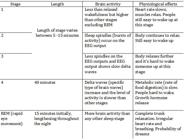
Circadian rhythms (biological rhythms lasting a day)e.g. sleep/wake cycle measured by reading the time and regular events like meals. Our body clock is regulated by internal system including the release of hormones e.g. melatonin, metabolic rate and body temperatureSiffre- 1975AimDiscover how natural rhythms of human life could be affected by living where there was no timeProcedure Lived underground in isolation for 63 days No natural light or cues at to whether it was day or night Had an artificial light to help with navigation through the cave He would check his pulse and do a psychological test every time he called up to the team outside the cave. The tes involved counting from 1-120 at a rate of 1 digit per second. Findings His psychological time had changed His sleep/wake cycle had changed to about 25-30 hour cycle Lost track of the days and thought he had been in the cave for a month less than he had ConclusionsSuggests natural light is important for keeping your 24-hour cycleAschoff and Weber- 1962 Studied participants in a bunker with no lights or windows Participants could turn lights on and off as they wished so the slight source fitted with their body clocks Eventually their body clocks settled to a sleep/wake cycle of 25-27 hours Suggests that natural light is used to adjust our pacemakers with the environment and a 24 hour clock is not inline with our natural body rhythm Effect of endogenous pacemaker and exogenous zeitgebers on the sleep/wake cycleEndogenous pacemaker Rhythms from the internal body system Can be affected by the environment specifically light Do function without environmental clues but will vary without environmental cues Example is of endogenous pacemakers for light:Low light → Optic chiasm in the eye → stimulates SCN → Stimulates pineal gland → Produces melatonin → Enhances production of serotonin → Brain acitivity falls → Sleep Superchiasmatic nucleus (SCN)- is a pacemaker of the biological clock in the sleep-wake cycle, it has it own internal biological rhythm due to protein synthesis. It is connected to the optic chiasm so receives input about the amount of light in the environment. Influences the sleep cycle as light stimulates nerve impulses that travel from the eye along the optic nerve to the brain Pineal gland- structure contains receptors that respond to external light. It is linked to the SCN by neural pathway. Electrical simulation of SCN is transferred to pineal gland and so releases melatonin. Light and melatonin are inversely proportional so when light levels are high, melatonin levels are low. Melatonin induces sleep as it enhances the production of seratonin causing the nervous system to slow down so brain activity falls and sleep begins Exogenous zeitgebers Cues from the environment which play an important role in regulating time External cues They are cues for endogenous pacemakers Help regulate the body clock so it is inline with the environment e.g. light, blue light, noise Research to support Campbell and Murphy- 1998 Monitored the body temperature of 15 volunteers sleeping in a lab Were introduced to light and night with beams of light shone on the backs of their knees at intervals Circadian rhythms were disrupted by up to 3 hours Shows its not necessary for just light to enter the eyes for it to have a psychological effect on biological rhythms Steel et al - 2008 Investigated effects of constant daylight on circadian rhythms by monitoring 6 participants living in isolation in Artic for 6 weeks Constant daylight Participants kept sleep logs Researchers found that 5 of 6 participants developed a sleep/wake cycle longer than 24 hours Also found sleep patterns were individual and no synchronised patterns emerged Social cues may have a stronger effect in absence of zeitgebers Evaluation of research into circadian rhythms Little external validity as conditions are artificial Sleep patterns could be caused by being monitored not the effect of the zeitgeber Animals used in research and cannot generalise findings to animals and humans as there are physiological differences Research can be used to find treatments Infradian rhythms (biological rhythms lasting more than 24 hours)e.g. menstrual cycle dictated by endocrine system and hibernation Not purely dictated by the release of hormones Zeitgebers such as light and odour have an effect also McClintock and Stern- 1998 Women (1) recieved 'odourless compounds' from armpits of women (2) in the latter 1/2 of the menstrual cycle Women (1) found their menstrual cycle shortened possibly due to the effect of the other women's pheromones as they reached the end of their cycle Women's (2) odourless compounds from armpit in beginning of their cycle lengthened their cycle. Menstrual cycle can be altered by pheromones Reinberg- 1967 A woman spent 3 months in a cave with only a lamp for light Her days lengthened to 24.9 hours and her menstrual cycle shortened to 25.7 days Shows light can affect the menstrual cycle After the study it took a year for the body to readjust her menstrual cycle so infradian rhythms can be affected by external zeitgebers Evaluation Helps to explain menstrual synchronicity could be due to pheromones Evolutionary advantage for menstrual synchronicity as there could be synchronised pregnancies and so childcare can be shared once the baby is born Not known what pheromones have an effect and how close and how long women have to live together to experience this Wilson- 1992- challenged the idea of menstrual synchrony saying experimental evidence suggests it's existance is exaggerated Ultradian rhythms (last less than 24 hours)e.g. respiration, rapid eye movement, heart beat, sleep/wake cycleSleep has 5 stages through the night:
Over a course of 90 minutes an individual goes through stages 1-4 then returns to 3 and then 2 and then REM. The sleep cycle then begins again and the number of cycles depends on how long the person sleeps for.Dement and Kleitman- 1957AimsInvestigate brain activity change throughout night-time sleepProcedure 7 adult males and 2 adult females Participants reported to a lab at bedtime to have an EEG EEG measurements taken throughout the time asleep at night Participants told not to drink caffinated drinks before sleep was to be investigated Findings Everyone had REM periods during every night High incidences of dream recall when participants woke during REM periods of sleep. If woken in other stages dreams were not reported as often. Participants were woken up during 5-15 minutes into the start of REM sleep Brain activity of vivid dreams different to less clear dreams Rapid eye movements of participants during REM sleep varied according to dream type and mirrored rapid eye movements when awake and completing a similar task to the task dreamt about So they managed to identify the stage when dreams were most likely to occur Conclusion Sleep stages follow a typical pattern throughout the night Dreams occur mostly during the REM stage Evaluation Individual differences so findings cannot be generalised Useful as it helps to understand medical conditions and find new treatments Small sample sizes used so cannot be generalised Can be applied to finding out the best way to live life e.g. the best time to get to sleep or wake up etc.
Page 8
Key definitions
Action potentialAdrenal glandAdrenal medullaAutonomic nervous systemAxonAxon terminalBrain plasticity (neuroplasticity)Central nervous systemCircadian rhythmsContralateralDendritesElectroencephalogram (EEG)Endocrine systemEndogenous pacemakersEvent-related potentials (ERPs)ExcitationExogenous zeitgebersFunctional magnetic resonance imaging (fMRI)Glia/GialHemispheric lateralisationHormonesHypothalamusInfradian rhythmsInhibitionIonsLimbic systemLocalisationMotor neuronNeuronNeurotransmittersParasympathetic nervous systemPeripheral nervous systemRelay neuronResting potentialReticular formationSensory neuronSomaSomatic nervous systemSubstantia nigraSympathetic nervous systemSynapsesThalamusUltradian rhythms
Want to create your own Notes for free with GoConqr? Learn more.