
Página 1
Carbohydrates
There are polymers and monomers. Polymers invollve polysaccharides, lipids and proteins whilst monomers involve glucose, fructose, glycerol and fatty acids and amino acids.Carbohydrates come as monosaccharide's which are single sugar subunits and disaccharides which are 2 sugar subunits joined together. Polysaccharides have 3 or more sugar subunits joined together. Monosaccharide's are sweet and soluble, contain carbon, hydrogen and oxygen and they are classified according to the number of carbons a molecule has.The general formula of monosaccharides is (CH2O)n where n is the number of carbons. Glucose, galactose and fructose are hexose sugars and examples of monosaccharides.If you join 2 glucose molecules together, you get a disaccharide and you join this by performing a condensation reaction (water is removed) This then forms a glycosidic bond between the 2 monosaccharides. - The inverse of a condensation reaction is a hydrolysis reaction. The image below shows alpha glucose and beta glucose.
When alpha glucose and alpha glucose react together, they form maltose. The bond in maltose is known as a 1,4 glycosidic bond because it forms between carbon 1 on one monosaccharide and carbon 4 on the other monosaccharide.
Other examples of disaccharides include: Sucrose - formed from alpha glucose and fructose Lactose - formed from galactose and glucose In all of these disaccharides, a condensation reaction is performed.Monosaccharides themselves provide a rapid source of energy. They are readily absorbed and require little or no change (for glucose) before being used in cellular respiration. I fmonosaccahrides are eaten (which they commonly are) the quick absorption causes a sharp rise in blood sugar. Polysaccharides and disaccharides have to be digested into monosaccharides before being absorbed which takes time so the monosaccharides are released slowly. This is why eating compelx carbs, do not cause swings in blood sugar.
Polysaccharides
Polymers with sub-units of monosaccharides Repeated condensation reactions Normally 1000's of monomers They are NOT sugars Three examples of polysaccahrides are: Cellulose - most abundant Glycogen Starch More on Starch:Starch is a plant storage polysaccharide and it is s polymer of alpha glucose. It is made up of 2 substances; amylose and amylopectin.Amylose -- Condensation reaction between alpha glucose, forming an alpha 1,4 glycosidic bond. Its shape is a coiled springAmylopectin -- condensation reactions between alpha glucose, forming alpha 1,4 glycosidic bonds but also, alpha 1,6 glycosidic bonds. Its shape Is a coiled spring with branches and these branches are the alpha 1,6 glycosidic bonds.
Glucose can either be made up of alpha glucose or beta glucose. With alpha glucose, the hydroxyl groups on the 1 and 4 carbons are below whereas in beta glucose, one of the hydroxyl groups has switched sides, so one hydroxyl group is at the top and another is on the bottom. Starch is an insoluble store of glucose. A typical starch grain contains 70-80% amylopectin, the rest is amylose (20-30%). If it were soluble then it would interfere with osmosis. Its only found in plant cells and the animal equivalent is glycogen - the storage polymer (glycogen is also found in bacteria and fungi). Glycogen is a much tighter spiral structure and has a lot more 1,6 glycosidic bonds. We store glycogen as its a compact structure with lots of branches making for a higher number of glucose sub units. The numerous side branches means that it can be rapidly hydrolysed to glucose.
Página 2
Surface area to volume ratio
Why do organisms need special exchange surfaces? Absorb substances Remove waste products All living organisms rely on exchanges with the environment to survive. Diffusion only works efficiently if the distance over which the substances have to diffuse is small and the organism has a large surface area compared to its volume. For larger organisms, they have a small surface area : volume ratio and a bigger distance from the surface to the cells and diffusion alone is insufficient to meet the need of the cells.Small organisms such as insects have a large surface area to volume ratio so oxygen can diffuse straight through their bodies. Large organisms need a large surface area to exchange more substances however it cannot diffuse straight through their skin because the surface area to volume ratio is too small and they would die before the oxygen for example, reached the lungs.In order to have an efficient exchange system, animals must have a large surface area, thin membrane and a large diffusion gradient. The greater the surface area, the larger the particle exchange, The more particles there are on one side of the membrane the higher the diffusion rate. Large surface area - the greater the surface area the more particles can be exchanged at the same time. Alveoli in lungs are adapted with an increased surface area so diffusion can take place. Concentration gradient - The more particles there are on one side of the membrane, the faster the diffusion. A reduced concentration slows diffusion so its important that diffused substances are carried away to maintain the gradient, Diffusion distance - If the membrane through which the particles must diffuse is wide, it will take longer (resistance) so the membrane should be thin and permeable.
Examples of specialised exchange surfaces: Alveoli Capillaries root hair cells hyphae of fungi small intestine villi
Diffusion is defined as:The movement of molecules along a concentration gradient from a region of higher concentration to a region of lower concentrationSurface area = (length x width)nVolume = length x width x heightSurface are to volume ratio = surface area / volumeFIcks Law states:Rate of diffusion is proportional to (surface area x diffusion) / length of diffusion path
Página 3
Phospholipids
A phospholipid bilayer is a double layer that surrounds cells and cell organelles. There is a phosphate group on the outside and 2 fatty acid tails on the inside. The fatty acid tails hate water, so they are hydrophobic whilst the phosphate group love water, so they are hydrophilic.Proteins in the cell membranesMany different types of proteins are found within membranes, they can either be extracellular or intracellular. Each type can have a specific function. Some proteins function as enzymes, others as carrier and channel proteins involved in the transport of substances in and out of a cell. Glycoproteins and glycolipids have important roles in cell-to-cell recognition and as receptors. A saturated fatty acid has the maximum possible number of hydrogen atoms attached to every carbon atom. It is therefore said to be saturated with hydrogen atoms and all of the carbons are attached to each other with single bonds. Unsaturated fatty acids have one or more double bonds between carbon atoms. A saturated fatty acid is straight while an unsaturated fatty acid causes a double bond which makes the phospholipids structure have a slight bend. This allows for space between the fatty acids. The molecules can then move around hence the term fluid mosaic model.In some fatty acids, a pair of hydrogen atoms in the middle of a chain is missing. This creates a gap that leaves two carbon atoms connected by a double bond rather than a single bond. Because the chain has fewer hydrogen atoms, it is said to be unsaturated. A fatty acid with one double bond is monounsaturated and with more than one double bond its polyunsaturated.The membrane is more fluid with unsaturated phospholipids than saturated because the kinks in the fatty acid prevent them lying close together. In turn, this creates more space in which the molecules can move.
The proteins in the phospholipid bilayer come in 3 broad forms; Stuck to the inner or outer surface of the membrane Within one layer of the membrane Pass all the way through the membrane
Página 4
The Different Types of Diffusion
How does the cell membrane allow the transport of molecules through diffusion?Simple passive diffusion occurs when small molecules pass through the phospholipid bilayer of a cell membrane. There is no effort involved to transport these molecules.Facilitated diffusion depends on carrier proteins embedded in the membrane, allowing specific substances to pass through that would otherwise not be able to diffuse through the membrane. However once these proteins are saturated, the rate of diffusion decreases. The role of the carrier protein is carried out without any input of energy. An example of facilitated diffusion is when glucose molecules bind to the end of a GLuT transporter and move to the other side of the membrane allowing glucose molecules to pass through easilyHow does the cell membrane allow the transport of molecules through diffusion?Water molecules can (to some extent) pass through the lipid bilayer even though water is a polar molecule. Water is able to enter and exit the cell through pores in the protein membrane called aquaporins. The reason why water molecules cannot otherwise pass through the lipid bilayer is because they are polar and proteins need to be used in order for the transport of these molecules to occur.How does the cell membrane allow the transport of molecules through active transport?Active transport is the net movement of molecules from an area of low concentration to an area of high concentration. Active transport mechanisms draw their energy from the hydrolysis of ATP. ATP is known as adenosine triphosphate and this is a molecule that stores energy and moves the ions through the plasma membrane. Active transport is used to move amino acids and some ions, including potassium and sodium.Specifically, sodium and potassium move through ion channels trying to achieve a balance on both sides of the membrane. In order to do this, allow concentration of sodium and potassium is needed on wither side (potassium - outside, sodium- inside). To get these concentrations, a sodium - potassium pump is used to export sodium ions outside the membrane and potassium ions inside the membrane. How does the cell membrane allow the transport of molecules through exocytosis?Exocytosis is the process by which cells release particles from within the cells into the extracellular space. In exocytosis, waster material is enveloped in a membrane vesicle and fuses with the inside of the plasma membrane. The fusion then opens this membrane envelope and expels the waste material outside of the cell (extracellular space). Exocytosis occurs to get rid of waste and to release chemical transmissions.How does the cell membrane allow the transport of molecules through endocytosis?Endocytosis takes up particles into the cell by invaginating the cell membrane (turn it inside out) to release materials from inside the cell. Endocytosis consists of phagocytosis and pinocytosis. Phagocytosis is the taking in of large food particles and pinocytosis is the ingestion of liquid.Essentially, endocytosis is a type of active transport that moves particles such as large molecules into a cell. There are different variations but they all involve the plasma membrane invaginating (folded inside out forming a pouch) and forming a pocket around the target particle. The pocket is pinched off, resulting in a intracellular vesicle formed from the plasma membrane.
Página 5
Lipids
Lipids contain the elements carbon, hydrogen and oxygen They provide over twice the energy of carbohydrates per gram = 37KJ/g Primarily energy stores in animals and plants Lipids consist of two types of molecules - glycerol and fatty acids. The structure of a lipid is broken down into a glycerol and 3 fatty acid tails (carboxylic acid).
The glycerol molecule forms 3 ester bonds between the 3 fatty acid tails, after a condensation reaction occurs and water is removed. The ester bond is between the oxygen from the glycerol bonded to the carbon on the fatty acid and the other oxygen double bonded to the oxygen. This forms a type of lipid known as a triglyceride and is the most common lipid we eat.Saturated FatsWhen a fatty acid chain contains the maximum number of hydrogen's, they are saturated. In saturated fats, the hydrocarbon chain is long and straight. Animal fats such as meat and dairy are saturated fat sources. There are no carbon to carbon double bonds so the chain is straight and these chains can pack closely together. The strong intermolecular bonds between triglycerides made of saturated fatty acids result in fats that are solid at room temperature.Unsaturated FatsMonounsaturated fats have one double bond between 2 of the carbon atoms in each fatty acid chain. Polyunsaturated fats have a large number of double bonds. This double bond causes a kink in the chain and prevent the unsaturated chains packing close together. This increase in distance between the molecules, weakens the intermolecular forces between unsaturated triglycerides and thus causes oils that are liquid at room temperature. Olive oil is high in monounsaturated fats and vegetable oils, nuts and fish are good sources of polyunsaturated fats.Unsaturated fats can be made solid at room temperature by adding hydrogen to the double bond, making them saturated. These hydrogenated - or trans-fats are produced by the food industry and used in processed food. Trans-fats occur naturally at low levels in meat and dairy products.
Página 6
Water
Polar Molecule Solvent properties Thermal properties Liquid at room temp Necessary for life Medium in which all chemical reactions occur in living organisms Hydrogen bonding holds molecules together Hydrogen has a slightly positive charge (delta +) whilst the oxygen (which is more electronegative) has a slightly negative charge (delta -). The slight negative oxygen atoms attract the slightly positive hydrogen atoms of other water molecules. This is hydrogen bonding in its simplest form.
The biological properties of water are: Acts as a solvent - due to the dipole nature of water, ionic substances can dissolve in it because of the positive and negative attraction High specific heat capacity - It requires a lot of energy to raise the temperature of 1 gram of water by 1 degree. A lot of heat must be lost before the temperature of the same mass of water falls by 1 degree. This means water can maintain relatively constant temperature. This is important in living organisms as it avoids sudden temperature changes (which would otherwise affect metabolic reactions) and this is important for organisms living in water. Normally liquid - for the same reasons above, it takes energy for it to change state Ice is less dense - When water is a liquid, the molecules are constantly moving around and the intermolecular forces are constantly breaking and reforming, however when the temperature starts to drop, the molecules move around with less energy and become a fixed state where the bonds are spread apart evenly. There is a greater space between the molecules now, causing the density to decrease, therefore ice can float on water. Water is very cohesive - Cohesion is the attraction of water molecules to water molecules allowing water to flow. Water molecules are very cohesive due tot he hydrogen bonds and this helps in transport within organisms such as: The xylem of plants where water and ions move up the plant The transport of the soluble products of digestion in the blood plasma of animals. Adhesion is the attraction of water to other polar substances Water has a high surface tension - When air meets water, the cohesive forces between the molecules result in hydrogen bond formation, in an inward direction, giving the water a high surface tension. This force causes the water surface to occupy the least possible area enabling small organisms to land on the water surface and move over it.
Página 7
DNA Base Structure
MONONUCLEOTIDES:These are the monomers that join together to form DNA and RNA. Each monomer carries a single base - so one nucleotide base. The structure of the monomers is similar for both DNA and RNA, the difference is the sugar. DNA is deoxyribose sugar and RNA is ribose sugar.The nucleotide forms from 2 condensation reactions where there is an ester bond and a glycosidic bond.The image below is in its most basic form:
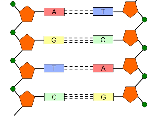
Mononuclotides become polynucleotides through polymerisation and this is catalysed by DNA polymerase or RNA polymerase. The bond that forms between 2 nucleotides is a phosphodiester bond.Between the bases there are hydrogen bonds and the number of bonds depends upon the base: Adenine and thymine = 2 bonds Cytosine and guanine = 3 bonds
Remember ADENINE must bond with THYMINE (DNA) or URACIL (RNA) and GUANINE must bond with CYTOSINE (triple bond). This is the complementary base pairing. In DNA, this complementary base pairing causes two strands to line up parallel to one another, with the bases on the inside - bonded to one another. The weak hydrogen bonds keep the strands together. Bonding also takes place up the strand, causing it to spiral into an alpha helix.
Remem
Página 8
Circulation in the Body
Humans have a double circulated system. A systemic circuit and a pulmocutaneous circuitWithin the circulatory system, there are different vessels which have different features related to their specific function:
ARTERY:Carry blood away from the heart at high pressure Structure: relatively thick wall smooth muscle elastic fibres lined with smooth layer of endothelial cells narrow lumen Functions: withstand high pressure alters diameter of lumen allows walls to stretch and then recoil low friction surface to ease blood flow
CAPILLARYUsed for exchanging substances and form a highly branches networkStructure:very thin wall (one cell thick)Functionallows diffusion to take place of gases and nutrients
VEINCarry blood to the heartStructure: relatively thin wall very little smooth muscle or elastic fibres wide lumen valves Function: blood under low pressure no pulse, stretching and recoiling large volume of blood carried stop backflow
Página 9
The Cardiac Cycle
The cardiac cycle consists of: Atrial systole Ventricular systole Complete cardiac diastole
Atrial Systole (sometimes known as ventricular diastole) The ventricles both relax Blood flows into the atria, via the vena cava and pulmonary veins As the atria fill, the pressure increases against the atrioventricular valves, forcing them to open and blood flows into the ventricles. The atria then contract, decreasing their volume, more blood forces itself into the ventricles. (some blood flows straight from the veins into the atria and into the ventricles, the rest flows now)
Ventricular Systole (also known as atrial diastole) After a very short delay, the atria relax The thick muscular walls of the ventricles then contract from the apex upwards Higher pressure in the ventricles than the atria cause the atrioventricular valves to shut (first heart beat) stopping the backflow. The higher pressure causes the semilunar valves to open and blood is forced into the pulmonary artery and aorta.
Complete Cardiac Diastole The ventricles and the atria both relax Increasing the volume and lowering the pressure in the heart (due to elastic recoil) The higher pressure in the pulmonary artery and the aorta cause the semilunar valves to shut (second heartbeat) The atria then fill with blood again due to the high pressure in the vena cava and pulmonary vein and the cycle starts again
Página 10
Blood Clotting
The main components of blood : Plasma - liquid medium, transports the blood Red blood cells (erythrocytes) - carries oxygen White blood cells (leucocytes) - fight infection Platelets (thrombocytes) - blood clotting
The Blood Clotting Cascade Platelets and damaged tissue release a protein called thromboplastin Thromboplastin activates an enzyme that catalyses the conversion of the protein prothrombin into an enzyme, thrombin. A number of other protein factors, vitamin K and calcium ions must be present in the blood plasma for this conversion to happen. Thrombin then catalyses the conversion of a soluble plasma protein fibrinogen into insoluble protein, fibrin. A mesh of fibrin forms, trapping platelets and red blood cells, also known as a blot. When platelets come into contact with damaged vessel wall, they change shape and stick to the damaged wall and each other, forming a platelet plug. This is where the thromboplastin is released from, which triggers the blood clotting cascade.
Página 11
Protein Synthesis
What are proteins? They are made up of amino acids There are 20 different types of amino acids that occur commonly in proteins Plants can make all the amino acids, animals can only make some, getting the rest from their diet Amino acids that are obtained from diet are called essential amino acids Amino acids in long chains fold into unique shapes which give the function of the protein Proteins have a wide range of functions from antibodies, enzymes and hormones to muscles, ligaments, tendons and hair. They are also found in membranes The bond holding amino acids together is known as a peptide bond
Proteins are made in the cytoplasm but the DNA that codes for them is contained within the nucleus. Protein synthesis is required to make the protein and the two steps involved are transcription and translation. TranscriptionThis is the first stage and it takes place in the nucleus. RNA polymerase (enzyme) attaches to DNA DNA unzips and untwists forming a DNA coding or sense strand and a template or antisense strand The template (antisense) strand is transcribed as RNA polymerase attaches RNA nucleotides to the exposed bases with complementary base pairing (uracil instead of thymine) Phosphodiester bonds form to produce a molecule of mRNA When complete, mRNA leaves through the nucleus through a nuclear pore in the nuclear envelope and the DNA twists and zips back up again. Translation mRNA attaches to the small ribosome subunit The codons are read by the ribosome (three bases) tRNA molecules bind to the codon with an anticodon The first codon is a start codon and the last codon is a stop codon Each tRNA molecule has an amino acid attached at the opposite end of the anticodon. Enzymes form peptide bonds between amino acids forming a dipeptide to a polypeptide via a condensation reaction. Once the amino acids are bonded together, the tRNA molecule is released and can be used again The amino acid chain continues to grow until the stop codon is reached Remember transcription takes place in the nucleus and translation takes place in the cytoplasm.IF there is a single base mutation then it would change the sequence of amino acids and therefore the way the shape folds. IF there is an insertion or deletion then there would be a whole shift in the sequence.The basic structure of an amino acid: (note the R group changes depending on the amino acid)
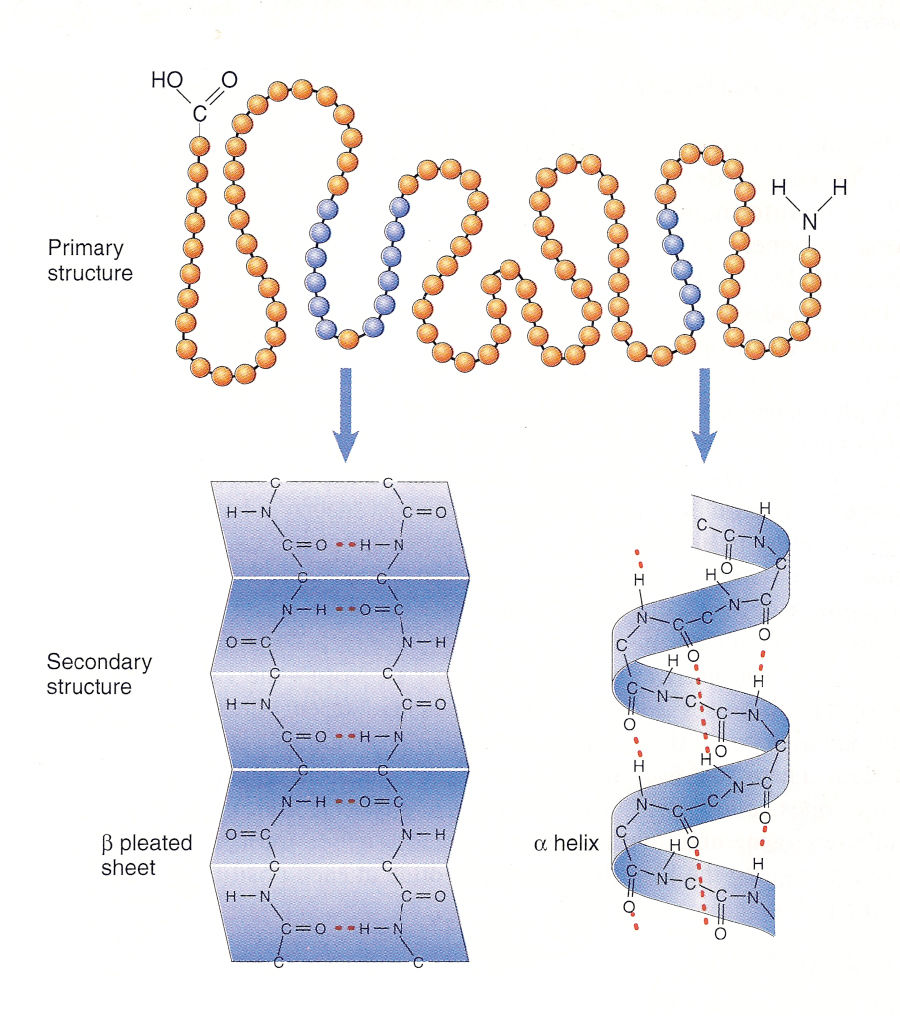
The Different Structure of Amino Acids: Primary Structure - This is like a string of beads and is the amino acid in its most simplest form. It is just the order or sequence of amino acids, the polypeptide chain Secondary Structure; Alpha Helix - This is the primary structure but it has been folded into its shape - alpha helix is held in place by hydrogen bonds between every N-H group and the oxygen of a C=O group in the next turn of the helix, 4 amino acids down the chain
2. Secondary Structure; Beta Pleated Sheets - the other shape the primary structure can be folded into, where hydrogen bonds occur again with each fold3. Tertiary Structure - This is a 3D structure formed by more bonds, depending on the function: Disulphide bonds - strong covalent Ionic bonds - broken by pH changes Hydrogen bonds - easily broken down Polar interactions - hydrophilic groups arrange on outside of protein, hydrophobic groups arrange on inside 4. Quaternary Structure - Arrangement of more than one tertiary structure Proteins come in two types:Globular ProteinsIn globular proteins, the polypeptide chain is folded into a compact, spherical shape. The proteins are soluble due to the hydrophilic side chains which project from the outside of the molecules. This means they're important in metabolic reactions. An example of a globular protein is an enzyme and their shape is important with enzyme - substrate complexes and their ability to catalyse reactionsThe 3D shapes of globular proteins are critical to their role of binding to other substances. An example is transporting proteins within membranes and the oxygen transport pigments haemoglobin.Fibrous ProteinsThey don't fold up into a ball shape but remain as long chains. Several polypeptide chains can be cross linked for additional strength. Insoluble proteins are important structural molecules for example, keratin in your hair.
Errors in transcription and translation leads to incorrect proteins. An error in mRNA effects protein produced from that single mRNA strand on one occasion. If the error is in DNA then the problem is recurrent. The mistake occurs each time it replicates.There are three theories of DNA replication: Semi-conservative - this is the accepted and correct one Conservative Dispersive The Semi Conservative ModelThe two parental strands separate and each makes a copy of itself. After one round of replication, the two daughter molecules each compromises one old and one new strand. After two rounds, two of the DNA molecules consist only of new material while the other two contain one old and one new material: DNA untwists and hydrogen bonds break DNA polymerase links free complementary bases to the exposed bases DNA polymerase catalyses phosphodiester bonds between bases to form two identical strands of DNA, identical to the original molecule. There was an experiment done to determine this and was conducted by Meselson and Stahl.
Página 12
Cystic Fibrosis
Cystic Fibrosis or CF is caused by a gene mutation which makes the CFTR protein. CFTR is coded for by the gene. It can occur in two forms, F and f. F is the normal allele which is dominant f is the mutated CFTR allele which is recessive Cystic fibrosis is what's known as a monohybrid inheritance which means that there is only one gene mutation. Other examples og=f monohybrid inheritance is: Albinism Achondroplasia Huntington's Disease There are some relationships between genetic diseases and other diseases. For example, thalassemia affects the production of haemoglobin and is caused by recessive alleles of a gene and mutations affect this gene. Someone whose homozygous for these recessive alleles either makes no haemoglobin or useless haemoglobin. However people who are carriers (heterozygous for this disease) show no symptoms but they do have protection against malaria. What does the CFTR Protein do?Surrounding the ciliated epithelial cells is a fluid called extracellular fluid. Within this fluid there are chloride ions and sodium ions. The water content regulation in mucus, controlled by osmosis effects the viscosity and the sodium and chloride ions are controlled by the CFTR protein. If the CFTR protein mutates, then this effects the movement of chloride and sodium ions and thus the viscosity of the mucus.
How is the water content of mucus regulated in normal patients? If the mucus layer contains too much water, this is detected by the epithelial cells and causes sodium-potassium pumps (in the basal membrane) to actively pump sodium ions out of the cell. The concentration of sodium ions in the cell falls, causing a concentration gradient to occur across the apical membrane. This causes sodium ions to diffuse down the concentration gradient. The ions then pass into the cell through facilitated diffusion through the sodium channels in the apical membrane There is now a raised concentration of Na+ in the tissue fluid on the basal membrane side and this creates a potential difference between this tissue fluid and the mucus on the apical membrane. The tissue fluid contains more Na+ than the mucus. An electrostatic gradient is created and this causes the chloride ions to diffuse out of the mucus into the tissue fluid via the gaps between neighbouring epithelial cells Now there are elevated Na+ and Cl- concentrations in the tissue fluid and so water is drawn out of the cell by osmosis, across the basal membrane into the tissue fluid. As there is now less water in the cell than in the mucus, water is drawn out from the mucus through osmosis across the apical membrane into the cell.
When there is too little water in the mucus, chloride ions are transported across the basal membrane into the epithelial cell. This creates a concentration gradient across the apical membrane, with the concentration of chloride ions being higher inside the cell than out. The CFTR channel opens and chloride ions diffuse out of the cell, down the concentration gradient, into the mucus. When the CFTR channel opens, the sodium ion channels close in the apical membrane. The build up of Cl- ions in the mucus creates an electrical gradient, into the mucus and the tissue fluid. Sodium ions diffuse out of the tissue fluid. down the electrical gradient into the mucus. The Na+ and Cl- in the mucus draws water out of the cells by osmosis until the solutions on either side of the apical membrane have the same concentration of free water molecules. The movement of water prevents the mucus that lines the airways from becoming too viscous.
Why CF lungs cannot regulate the water in mucusIn a person that has CF, the CFTR protein doesn't function correctly. When there is too little water in the mucus, Cl- cannot be secreted across the apical membrane and there is no blockage of the epithelial sodium ion channels.. This means there is a continuous absorption of Na+ by the epithelial cells. The raised level of Na+ draws chloride ions and thus water out of the mucus, making it more viscous and harder for the cilia to move it. Mucus isn't effectively cleared and sticky mucus builds up in the airways.
Chloride ions are pumped into the cell across the basal membrane Chloride ions diffuse through the open CFTR channels Sodium ions diffuse down the electrical gradient into the mucus Elevated salt concentrations in the mucus, draws water out of the cell by osmosis Water is then drawn into the cell by osmosis
Página 13
Cholesterol
Cholesterol is essential for the body in small amounts. It is needed for maintaining the correct level of fluidity in cell membranes. Cholesterol is also needed in the manufacture of steroid hormones and some of the components of bile. We normally obtain 25% of our blood cholesterol from food and our liver makes the other 75%. High levels of fat in the diet can impact on blood cholesterol levels and the development of cardiovascular diseases.Different types of blood cholesterol are significant in terms of your health because if you have a higher amount of LDL cholesterol, then you are at risk of having coronary heart disease, or you have coronary heart disease. This is the same as HDL cholesterol but this time, having a low value of this in your blood increases the risk. Overall if you have an increased amount of cholesterol in your blood than is necessary then this greatly effects your health.The interaction between HDL cholesterol and LDL cholesterol in the development of cardiovascular disease is opposite. If you have a low amount of LDL and a high amount of HDL then the risk of CHD is very low. Alternatively, if you have a high amount of LDL and a low amount of HDL then the risk of CHD is very high.
CVD
Symptoms of CVD Pain in your chest, tightness spreading chest pain to shoulders/neck dizziness, sweating shortness of breath
How to Avoid CVD? Stop smoking Decrease your BMI Increase activity Decrease alcohol consumption
Página 14
Enzymes
Definitions Enzyme - biological catalysts that speed up reactions in the body Anabolic Reactions - Reactions that build up, the substrate molecules are being added to Catabolic Reactions - reactions that break down the larger substrate molecules Metabolism - Sum of all the enzyme catalysed reactions occurring within it. Combination of anabolic reactions and catabolic reactions Catalyst - speeds up chemical reactions in the body without getting used up in the reaction Metabolic Pathway - The sequence of enzyme controlled reactions Specificity - only able to catalyse specific reactions Substrate - molecules that enzymes work on Product - molecules produced by the enzyme Enzymes can be found inside the cell where they are intracellular or outside the cell where they are extracellular.Examples: Decarboxylase - removal of carboxyl group in respiration with the formation of CO2 - intracellular, catabolic Maltase - breakdown of maltose to glucose in digestion - catabolic, extracellular DNA polymerase - joins nucleotides together in DNA replication - intracellular, anabolic There are two theories about how enzymes fit together: Lock and Key - assumes that the active site of an enzyme is rigid in shape. substrate has a complementary shape and change to active site substrate binds to active site and forms an enzyme/substrate complex substrate converted into product product is no longer complementary shape, so is released. Induced Fit - suggests that the active site is flexible and only assumes its catalytic conformation after the substrate molecules bind to the site. When the product leaves, it reverts back to its original form. substrate binds to active site active site changes shape to accommodate substrate and more bonds form forms enzyme/substrate complex change in active site, weakens bonds in the substrate and activation energy is reduced further shape change of enzyme after products form. An enzyme reduces the activation energy during a reaction. The reaction rate can be measured by:rate of reaction = amount of reactant used or amount of product formed / time takenThe energy required to break bonds and start a reaction is known as the activation energy. Without an enzyme, heating could provide this energy. In cells, enzymes reduce the amount of activation energy without raising the temperature of cells. Scientists calculate the rate at which an enzyme works. By determining the quantity of substrate used or quantity of product forms in a given time, rate can be calculated. The initial rate of reaction is used when comparing rate of enzyme controlled reactions.If you change the concentration of either enzyme or substrate, then they become limiting factors which means the factor is stopping the reaction from preceding at a higher rate. If it is the limiting factor, increasing concentration will increase the reaction rate up to a point, after which any increase will not affect the rate of reaction. This is because it will no longer be the limiting factor and another factor will be limiting the maximum rate of reaction. As a reaction proceeds, the rate of reaction will decrease since the substrate will get used up. The highest reaction rate is the initial reaction rateIncreasing Substrate Concentration:This increases the rate of reaction as there are more substrate molecules colliding with enzymes so more product is formed. However, after a certain concentration, any increase will have no effect since substrate concentration wont be the limiting factor. The enzymes have been saturated.Increasing Enzyme Concentration:Increase rate of reaction as more enzymes are available for substrate collision but this will only effect reaction rate to a certain concentration as the enzyme will no longer be the limiting factor as all the substrates have been used up.
Página 15
Studies
Cohort StudiesThese studies follow a large group of people over time, to see who develops the disease and who doesn't. They are prospective - at the start no one has the disease, scientists are interested in their future. Peoples exposure to potential risk factors and disease development is recorded so correlations can be seen. It takes a long time for the condition to develop so the studies can take years and be expensive. They can come in two types; Population --------> Group who develop condition vs. people who don't develop condition Population exposed to risk factor ----------------->follow over time--------------> Group who develop disease vs. group who don't develop disease .. Population not exposed ----------> follow over times----------->Group who develop disease vs. group who don't develop disease Cohort Studies for CVD are common one of the most well known is the Framingham study. A random sample 5209 were taken to study (age 30-62) and three generations have been involved. Every two years the participants are asked to provide a detailed medical history. The data is then used to look for common features contributing to CVD.
Case-Control StudiesCauses with condition vs. causes without condition ----------> Take histories of exposure to risk factor in past, compare, look for correlationsIn this study, a group of people with a disease are compared with a control of individuals that don't have the disease. Information is collected about the risk factors they've been exposed to, allowing factors to be confirmed if they have contributed.The control group should be representative of the population from which the case group was drawn. Its important not to match any variables which could potentially turn out to be risk factors. There have been many case control studies investigating risk factors of CVD.
Features of a good study Clear aim hypothesis Representative sample, selected from the wider population Differences between people asked to take part and those who respond should be considered The proportion of individuals who drop out must be kept to a minimum Valid and reliable results Data had to be valid - if it measures what it is suppose to measure Method used to collect results must be reliable. A reliable method produces measurements that are repeatable and reproducible. Sample Size - has to be large enough to produce results that could not have occurred by chance Controlling variables - the potential effect of all variables should be considered. This ensures the factor under investigation is influencing the outcome.
Página 16
The Structure of Mononucleotides
Mononucleotides are the monomers that join together to form DNA and RNA. The structure of the monomer is similar for both DNA and RNA, the difference is the sugar. The first image below is the mononucleotide for DNA. This is why it has deoxyribose sugar. Each mononucleotide consists of a phosphate group, a base (either adenine, thymine, cytosine or guanine) and a pentose sugar. The second picture shows the polymerisation of all the mononucleotides. They are joined together through condensation reactions catalysed by the enzymes RNA polymerase or DNA polymerase. This is where water is removed and the bond that forms is known as a phosphodiester bond. This bond occurs between the pentose sugar and the phosphate group. In terms of bases: Adenine must bond with Thymine in DNA or Uracil in RNA Guanine must bond with Cytosine This is complementary base pairing and the bases pair up with hydrogen bonds between them. Cytosine and Guanine have 3 hydrogen bonds while Adenine and Thymine/Uracil have 2 hydrogen bonds
In DNA, the complementary base pairing causes the two strands to line up parallel to one another, with the bases on the inside bonded to one another. The weak hydrogen bonds keep the strands together while bonding also takes place up the strand, causing it to spiral in an alpha helix.
Página 17
CVS and Amniocentesis
CVS carried out during pregnancy - checks your body for disorders such as Down Syndrome In CVS, a small sample of placenta is taken for testing and it is most commonly performed between 11 and 13 weeks How is CVS performed?A tiny amount of the developing placenta is taken. A placenta contains tissue that is genetically identical to the baby. There are two ways to perform it; through the abdomen or through the cervix Through the abdomenAn obstetrician uses local anaesthetic to numb the area, your skin is cleaned where the needle is inserted. An ultrasound is used to guide the direction, a fine needle is pushed through the abdomen and wall of womb to placenta. A tiny amount of placental tissue is sucked into a syringe. The needle is then taken out and the baby is observed with ultrasound.Through the CervixA speculum instrument which separates vaginal walls) is inserted into your vagina enabling doctors to see your cervix. They are cleaned and using ultrasound, fine forceps or tube is passed through cervix to placenta. A small amount of tissue is removed using forceps or suction catheter and the baby is observed via ultrasoundsEither of these methods is chosen depending on the position of the placenta. The mother has to fill out a consent form before procedure is carried out.
AmniocentesisAn ultrasound probe is used as guidance, a fine needle is pushed into you skin, through abdomen and uterus. The person doing the procedure will avoid the placenta. A small sample of fluid is removed using a syringe, the needle is taken and the baby is observed on ultrasoundFor fewer than 7 in every 100 women, not enough fluid can be taken at the first attempt and it has to be reinserted. This is most likely due to the position of the baby.
Página 18
¿Quieres crear tus propios Apuntes gratis con GoConqr? Más información.