20185882
Descripción
Test por Kaysie Gonzalez, actualizado hace más de 1 año
|
|
Creado por Kaysie Gonzalez
hace alrededor de 5 años
|
|
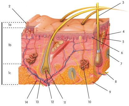
Pregunta 1
Respuesta
-
Sweat Pores
-
Hypodermis
-
Hairs
-
Dermal papilla
Pregunta 2
Respuesta
-
Motor nerve fiber
-
Hair follicle
-
Hairs
-
Sensory nerve fibers
Pregunta 3
Respuesta
-
Sweat pores
-
Dermis
-
Hair follicle
-
Hair Bulb
Pregunta 4
Respuesta
-
Sensory nerve fibers
-
Dermal papilla
-
Motor nerve fibers
-
Hair follicle
Pregunta 5
Respuesta
-
Apocrine Sweat glands
-
Dermal papilla
-
Dermis
-
Pressure receptor
Pregunta 6
Respuesta
-
Dermis
-
Epidermis
-
Sensory nerve fibers
-
Sweat pores
Pregunta 7
Respuesta
-
Dermis
-
Epidermis
-
Hypodermis
Pregunta 8
Respuesta
-
Pressure receptor
-
Apocrine sweat glands
-
Epidermis
-
Hair follicle
Pregunta 9
Respuesta
-
Pressure receptor
-
Sensory nerve fiber
-
Dermal papilla
-
Cutaneous blood vessels
Pregunta 10
Respuesta
-
Sensory nerve fibers
-
Hair follicle
-
Sweat pores
-
Motor nerve fibers
Pregunta 11
Respuesta
-
Hair follicle
-
Pressure receptor
-
Epidermis
-
Motor nerve fibers
Pregunta 12
Respuesta
-
Hypodermis
-
Hairs
-
Dermis
-
Epidermis
Pregunta 13
Respuesta
-
Dermis
-
Pressure receptor
-
Sweat pores
Pregunta 14
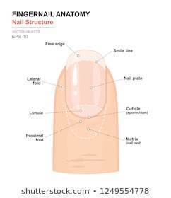
Pregunta
Where is the label at?
Respuesta
-
Cuticle
-
Free edge
-
Nail bed
Pregunta 15
Pregunta
Where is the label at?
Respuesta
-
Nail body
-
Cuticle
-
Nail bed
Pregunta 16
Pregunta
Where is the label at?
(under the nail)
Respuesta
-
Cuticle
-
Nail bed
-
Lunula
Pregunta 17
Pregunta
Where is the label at?
Respuesta
-
Lunula
-
Cuticle
-
Free edge
Pregunta 18
Pregunta
Where is the label at?
Respuesta
-
Nail body
-
Nail bed
-
Cuticle
Pregunta 19
Pregunta
What is the life cycle of an epidermal cell?
Respuesta
-
By the time the cells reach the outermost layer- called stratum corneum- all that’s left of the dead cells is their keratin. The newly arriving flattened cells called keratinocytes, they replace the dead cells that flake away with daily wear As the cells are pushed upward, they stop dividing and instead produce keratin, a tough, fibrous protein. The keratin replaces the cytoplasm and nucleus in each cell. The cells flatten, and as they move further away from their blood supply, they die The stratum basale, or basal layer, also called the stratum germinativum- is the innermost layer. It consists of a layer of columnar stem cells. These stem cells continually undergo mitosis, producing new skin cells. As new cells are produced, they push the older cells upward, toward the skin’s surface
-
The stratum basale, or basal layer, also called the stratum germinativum- is the innermost layer. It consists of a layer of columnar stem cells. These stem cells continually undergo mitosis, producing new skin cells. As new cells are produced, they push the older cells upward, toward the skin’s surface As the cells are pushed upward, they stop dividing and instead produce keratin, a tough, fibrous protein. The keratin replaces the cytoplasm and nucleus in each cell. The cells flatten, and as they move further away from their blood supply, they die By the time the cells reach the outermost layer- called stratum corneum- all that’s left of the dead cells is their keratin. The newly arriving flattened cells called keratinocytes, they replace the dead cells that flake away with daily wear
-
As the cells are pushed upward, they stop dividing and instead produce keratin, a tough, fibrous protein. The keratin replaces the cytoplasm and nucleus in each cell. The cells flatten, and as they move further away from their blood supply, they die The stratum basale, or basal layer, also called the stratum germinativum- is the innermost layer. It consists of a layer of columnar stem cells. These stem cells continually undergo mitosis, producing new skin cells. As new cells are produced, they push the older cells upward, toward the skin’s surface By the time the cells reach the outermost layer- called stratum corneum- all that’s left of the dead cells is their keratin. The newly arriving flattened cells called keratinocytes, they replace the dead cells that flake away with daily wear
Pregunta 20
Pregunta
The epidermis is what layer of the skin?
Respuesta
-
Inner, deeper layer
-
Outermost layer
-
Beneath
Pregunta 21
Pregunta
The dermis is what layer?
Respuesta
-
Inner, deeper
-
Outermost
-
Beneath the skin
Pregunta 22
Pregunta
The hypodermis is what layer?
Respuesta
-
Inner, deeper layer
-
Outermost
-
Beneath the skin layer
Pregunta 23
Pregunta
What is the epidermis layer composed of and the function?
Respuesta
-
t consists of stratified squamous epithelial tissues. It contains no blood vessels; instead, it obtains oxygen and nutrients by diffusion from the dermal layer beneath it
-
It is composed of connective tissues that contain primarily collagen fibers (which strengthen the tissue), but it also contains elastin fibers (which provide elasticity) and reticular fibers (which blind the collagen and elastic fibers together). It contains a large number of blood vessels in addition to sweat glands, sebaceous glands, and nerve endings
-
It is made up of loose connective (areolar) tissue and adipose tissue. The hypodermis blinds the skin to the underlying tissue Hypodermis that's composed mostly of adipose tissue is called subcutaneous fat this layer of fat helps insulate the body from outside temperature changes; it also acts as an energy reservoir
Pregunta 24
Pregunta
What is the characteristic of the basal cell carcinoma?
Respuesta
-
Arises in the epidermis and is slow-growing Often occurs on the scalp, forehead, backs of the hand, and top of the ears Has a raised, red, scaly appearance Some forms may metastasize
-
Most deadly of all skin cancers Sometimes develops from melanocytes of a preexisting mole Metazasizes quickly and is often fatal when not treated early Risk is greatest in individuals who had severe sunburns as children
-
The most common type Seldom metastasizes, it is the least dangerous Arises from the cells of the stratum basale, typically on the nose or face Lesion fist appears as a small, shiny bump; as it enlarges, it often develops a central depression and a beaded, “pearly” edge
Pregunta 25
Pregunta
What is the squamous cell carcinoma characteristics?
Respuesta
-
Most deadly of all skin cancers Sometimes develops from melanocytes of a preexisting mole Metazasizes quickly and is often fatal when not treated early Risk is greatest in individuals who had severe sunburns as children
-
The most common type Seldom metastasizes, it is the least dangerous Arises from the cells of the stratum basale, typically on the nose or face Lesion fist appears as a small, shiny bump; as it enlarges, it often develops a central depression and a beaded, “pearly” edge
-
Arises in the epidermis and is slow-growing Often occurs on the scalp, forehead, backs of the hand, and top of the ears Has a raised, red, scaly appearance Some forms may metastasize
Pregunta 26
Pregunta
What is the characteristics for the malignant melanoma?
Respuesta
-
Arises in the epidermis and is slow-growing Often occurs on the scalp, forehead, backs of the hand, and top of the ears Has a raised, red, scaly appearance Some forms may metastasize
-
Most deadly of all skin cancers Sometimes develops from melanocytes of a preexisting mole Metazasizes quickly and is often fatal when not treated early Risk is greatest in individuals who had severe sunburns as children
-
The most common type Seldom metastasizes, it is the least dangerous Arises from the cells of the stratum basale, typically on the nose or face Lesion fist appears as a small, shiny bump; as it enlarges, it often develops a central depression and a beaded, “pearly” edge
Pregunta 27
Pregunta
The main purpose of melanin?
Respuesta
-
A genetic lack of melanin
-
gives a person their skin color and forms a cap over the top cell nucleus to protect it from the exposure to the harmful ultraviolet rays of the sun
-
Impaired liver function (such as from hepatitis or liver disease) that allow bile to accumulate, which stains the skin
-
The breakdown of clotted blood under the skin
Pregunta 28
Pregunta
What are the characteristics of a sweat gland?
Respuesta
-
keep the skin and hair from drying out and becoming brittle sebum has a mild antibacterial and antifungal effect.
-
the most numerous of the skin glands
-
They secrete waxy substance called cerumen, or ear wax.
Pregunta 29
Pregunta
What are the characteristics of a Sebaceous gland?
Respuesta
-
They secrete waxy substance called cerumen, or ear wax.
-
open into a hair follicle, secrete an oily substance called sebum.
-
the most numerous of the skin glands
Pregunta 30
Pregunta
What are the characteristics of the ceruminous glands?
Respuesta
-
They secrete waxy substance called cerumen, or ear wax.
-
open into a hair follicle, secrete an oily substance called sebum.
-
the most numerous of the skin glands
Pregunta 31
Pregunta
What do First-degree burns classify as?
Respuesta
-
Involves the epidermis as well as part of the dermis Results in blisters, severe pain, and swelling May result in scarring May appear red, white, or tan
-
Involves only the epidermis Causes redness, slight swelling, and pain Often results from sunlight (sunburn)
-
Extends through the epidermis and dermis and into the subcutaneous layer May not be painful initially because of the destruction of nerve endings May appear white or black and leathery Often requires a skin graft
Pregunta 32
Pregunta
What do second-degree burns classify as?
Respuesta
-
Involves the epidermis as well as part of the dermis Results in blisters, severe pain, and swelling May result in scarring May appear red, white, or tan
-
Extends through the epidermis and dermis and into the subcutaneous layer May not be painful initially because of the destruction of nerve endings May appear white or black and leathery Often requires a skin graft
-
Involves only the epidermis Causes redness, slight swelling, and pain Often results from sunlight (sunburn)
Pregunta 33
Pregunta
What do third-degree burns classify as?
Respuesta
-
Involves the epidermis as well as part of the dermis Results in blisters, severe pain, and swelling May result in scarring May appear red, white, or tan
-
Extends through the epidermis and dermis and into the subcutaneous layer May not be painful initially because of the destruction of nerve endings May appear white or black and leathery Often requires a skin graft
-
Involves only the epidermis Causes redness, slight swelling, and pain Often results from sunlight (sunburn)
¿Quieres crear tus propios Tests gratis con GoConqr? Más información.