11889373
Descripción
Fichas por Anna Hogarth, actualizado hace más de 1 año
|
|
Creado por Anna Hogarth
hace más de 6 años
|
|
| Pregunta | Respuesta |
| How much blood is in the arteries? Venous system? | 1) 20% in the arteries 2) 80% in the veins |
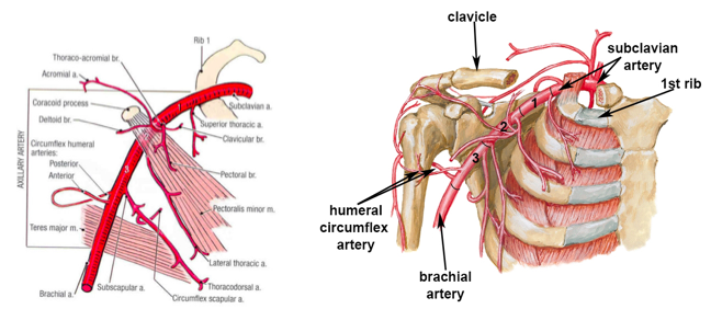
| What are the branches coming of the aortic arch? | On the RHS the brachiocephalic gives rise to the right subclavian and right common carotid. On the LHS the left common carotid comes off next to the brachiocephalic. The the left subclavian. |
| What do the subclavian arteries become? Where does this start? Where does this structure end? What are the three parts? Where does it give branches off to? | 1) Axillary arteries 2) At the lateral border of the first rib 3) The lateral border of the teres major 4) The first part is enclosed in the axillary sheath which is the lateral border of the 1st rib and medial border of the pectoralis minor. It then runs posteriorly to the pectoralis minor. The third part is between the lateral border of the pectoralis minor and the inferior border of the teres major. 5) Branches go off to the chest wall and shoulder girdle (humeral reflex) |
| X | |
| What does the axillary artery become? Where does this structure run? What is the major branch? Where does this structure end? | 1) The brachial artery 2) Anteriorly and superiorly 3) The profunda brachii - deep artery of the arm, runs posteriorly with the radial nerve. 4) 2 cm above the elbow |
| Where is the brachial pulse? | Medial to the distal tendon of the biceps brachii. |
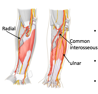
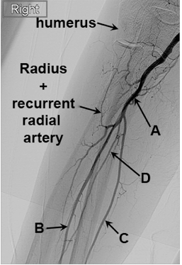
| Where does the ulnar artery run? What does it supply? What is its branch and what doe this supply? | 1) Runs down the medial aspect of the forearm 2) Muscles of the medial forearm 3) Interosseous branch which supplies the deep flexors and extensors of the forearm |
| Where does the radial artery run? What does it supply? | 1) Down the lateral aspect of the forearm 2) Lateral muscles of the forearm |
| X | |
| Where do both the superficial and deep palmar arches arise from? Where are they both located? What arises from the superficial and deep palmar arches? | 1) Both arise from the ulnar and radial arteries - the superficial mainly from the ulnar and the deep arch mainly from the radial. 2) The superficial cross the palm at the level of the extended thumb, the deep lies across the metacarpals at their bases. 3) The metacarpal and digital arteries arise from arches and supply the fingers. |
| Where are the radial and ulnar pulses? Which is more difficult to palpate and why? | 1) The radial pulse is lateral to the tendon of the FCR, the ulnar is lateral to the tendon of the FCU. 2) The ulnar is more difficult to palpate because it runs deeper and lies under the pisiform and the palmar fascia. |
| What is the Allen test? How is it performed? What is a normal filling time? | 1) Determines the patency of the radial and ulnar arteries 2) Step 1 - hand is exsanguinated by putting the hand into a fist and applying pressure on both the ulnar and radial arteries at the distal end of the forearm. Step 2 - Patient opens hand and pressure is released at either the ulnar or the radial artery and capillary refill time is measured 3) Normal filling time is less than 5 seconds |
| X | |
| X | |
| X | |
| X | |
| Describe the venous return in the hand. | There is both deep and superficial drainage. The deep drainage takes the same name as the corresponding artery. The superficial drainage is divided into the basilic and cephalic veins. The cephalic vein is lateral to the basilic vein in the anatomical position. |
| Describe the venous return of the rest of the arm. | Basilic - toward the base, cephalic -toward the head. The cephalic, basilic and deep veins drain into the axillary vein, this drains into the subclavian vein which drains into the brachiocephalic vein and then into the superior vena cava. The median cubital vein crosses the cubital fossa and joins the cephalic and basilic veins. |
| Which vein is most commonly used for venipuncture? | The medial cubital vein |
| At what spinal level does the abdominal aorta end? How does it become the femoral artery? | 1) Ends at L4 where it divides into the common iliac arteries. 2) At the level of the sacroiliac joints the common iliac splits into the external and internal. |
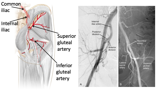
| What does the internal iliac supply? The external iliac? | 1) Pelvic wall and viscera, gluteal region 2) Lower limbs |
| What are the branches of the internal iliac and what do they supply? | The anterior and posterior divisions. Posterior division contains the superior gluteal artery. The anterior division gives rise to the inferior gluteal and the obturator artery. |
| X | |
| What does the obturator artery pass through? What branch does it give off? What branch does this supply? | 1) Obturator artery passes through the obturator foramen 2) The acetabular branch 3) Supplies the hip joint |
| What structure does the femoral artery run underneath? What aspect of the leg does it run down? | 1) The inguinal ligament 2) The anteromedial thigh |
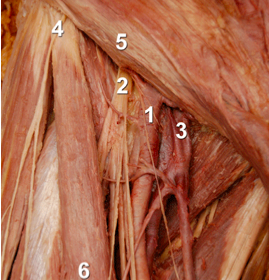
| 1) Femoral artery 2) Femoral nerve 3) Femoral vein 4) ASIS and lateral femoral cutaneous nerve 5) Inguinal ligament 6) Sartorius | |
| Where is the femoral pulse? | Below the inguinal ligament at the mid-inguinal point. Halfway between the pubic symphysis and the anterior superior iliac spine |
| What is the profunda femoris? What does it supply? What branches does it give off and what do these supply? | 1) The deep (and main) artery of the thigh 2) Main arterial supply to the thigh muscles (quadriceps, hamstrings and adductors) 3) Medial and lateral circumflexes - supply the head and neck of the femur. |
| Describe the structure of the medial and lateral circumflexes of the profunda femoris. | The medial circumflex initially runs medially and then passes posteriorly around the neck of the femur. The lateral circumflex passes laterally and anteriorly around the neck of the femur. |
| How does the femoral artery become the popliteal artery? What does the popliteal artery supply? | 1) It passes through a gap in the adductor magnus muscle (adductor hiatus). It then enters the popliteal fossa (back of the knee). 2) Contributes to the anastomosis that supplies the knee region |
| How is the popliteal pulse best felt? | Person prone with knee flexed to relax the hamstrings. Palpate at the inferior aspect of the fossa where the popliteal artery is closer to the tibia. |
| 1) Patella 2) Femoral condyles 3) Sartorius muscle 4) Semitendinosus tendon 5) Medial head of gastrocnemius 6) Popliteal artery 7) Lateral head of gastrocnemius 8) Bicep femoris muscle | |
| What does the anterior tibial artery supply? | The dorsiflexors and extensors of the ankle and foot |
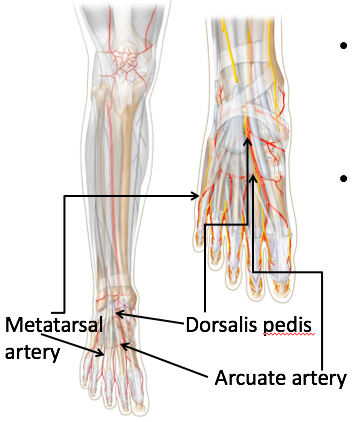
| What is the dorsalis pedis? What does it supply? | 1) Continuation of the anterior tibial artery distal to the inferior extensor retinaculum. 2) Supplies the dorsum of the foot, giving off branches to the arcuate artery from which metatarsals arise |
| X | |
| How does the posterior tibial artery compare to the anterior? Which aspect of the leg does it pass through? What is the deeper branch of the posterior tibial artery? What does the posterior tibial artery (and its branch) supply? | 1) Larger 2) Posteromedial 3) Peritoneal/fibular 4) Posterior (plantarflexor) and lateral muscles of the leg |
| Where is the dorsalis pedis pulse located? Posterior tibial pulse? | 1) Lateral to tendon of extensor hallucis longus 2) Half-way between the medial malleolus and calcaneal tendon |
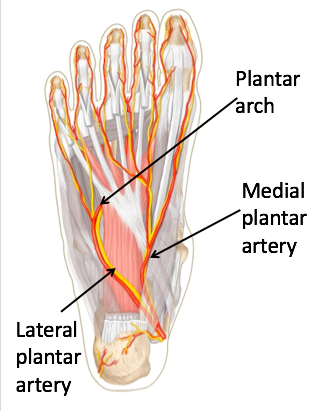
| What does the posterior tibial artery become? What does this supply? What do the digital arteries arise from? | 1) Medial and lateral plantar arteries 2) The sole of the foot 3) The lateral end of the plantAr arch (formed of the lateral plantar artery) |
| Describe the venous return in the leg. | Deep and superficial veins. Deep veins take the same name as the artery. The superficial veins are the small and great saphenous veins. These arise from the dorsal venous arch and run outside the fascia. |
| Where do the small and great saphenous veins drain into? How do the superficial and deep veins relate? | 1) The small saphenous drains into the popliteal vein, the great saphenous (longest vein in the body) drains in the femoral vein at a point just distal to the inguinal ligament 2) Superficial veins have numerous perforating veins that connect with the deep veins - valves ensure that blood only flows from the superficial to the deep. |
| How is venous return controlled? | 1) Movement from superficial to deep veins 2) Respiratory pump 3) Muscular pumps 4) Smooth muscle (venous constriction) 5) Valves |
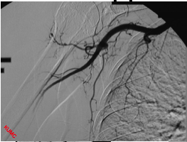
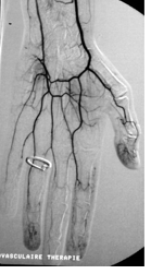
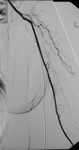
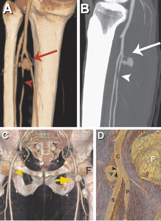
| What is peripheral vascular disease? What are the four most common causes? Describe the pathophysiology. | 1) Occlusive disease of the arteries of the lower extremities 2) Atherosclerosis, arteritis, aneurysm and embolism 3) Arterial narrowing decreases blood flow, pain results when there is insufficient blood flow for demand. |
| The arrow is an aneurysm, the arrowhead is thromboembolism. |
¿Quieres crear tus propias Fichas gratiscon GoConqr? Más información.