8279807
| Pregunta | Respuesta |
| CORE IDEA 1: BIOMOLECULES CHAPTER 4 - ENZYMES | ... |
| Defn of enzymes | Enzymes → biological catalysts that increase the rate of rxn & remain chemically unaltered at the end of the rxn, & thus can be reused. They are effective in small amts. |
| Roles of enzymes in cells | (1) Catalysis → enzymes act as highly specific catalysts that speed up the rate of metabolic rxns (2) Regulation → enzymes provide a means by which indiv. runs can be controlled. The mechanism of these regulatory processes include allosteric control, competitive inhibition, non-competitive inhibition, covalent modification of enzyme & variation in the amt of enzyme synthesised. *Enzymes are vitally impt, w/o them, rxn in cells too slow to sustain life |
| General characteristics in enzymes | (1) Enzymes are mostly globular proteins (2) Enzymes increase rate of rxn (3) Enzymes operate at milder rxn conditns (4) Enzymes exhibit specificity |
| General characteristics (1) Enzymes are mostly globular proteins | -Consists of 1 or more ppd chains coiled tgt & folded into a globular unit - As globular proteins, they are extremely complex molecules w intricate 3D contours & distinct surface geometries - Action of enzymes depend on their 3D conformation - Exceptions: some enzymes composed of RNA |
| (2) Enzymes increase rate of rxn | - Rate of enzymatically catalysed rxn are typically 10^(6) to 10&^(12) times greater than those of correspondingly uncatalysed rxn |
| (3) Enzymes operate at milder rxn conditions | - Enzyme-catalysed rxns occur under relatively milder conditions (temp below 100℃, atmos. pressure & nearly neutral pH conditions encountered in organism) |
| (4) Enzymes exhibit specificity | - An enzyme / particular type of enzymes usually catalyse a specific chem rxn - Absolute specificity → enzyme catalyse a single specific rxn E.g. Maltase catalyses hydrolysis of maltose to 2 glucose - Group specificity → Enzyme attack 1 type of chem bond in a variety of substances (e.g. peptide bond) E.g. Chymotrypsin catalyses conversion of ppd to smaller fragments of ppd |
| Tutorial Qn 2(b) Explain why having the same sequence of amino acids in their active sites allows these two different enzymes to catalyse the same reaction. LO1p [2] Type: Recall Topic: Enzyme specificity Approach: 2 marks = 2 points How does the sequence of amino acids influence the active site? (Sequence determines the way the polypeptide chain folds) What are the important amino acids at the active site and what characteristic must they have to enable proper functioning of the enzyme? | 1. Both enzymes have active sites* of the same shape/conformation and charge* which will be complementary* to the same substrates; 2. The presence of same catalytic R groups will allow the same reactions to be catalyzed; |
| QN (pg 6): What is the critical determinant in the biological function of a protein? | The unique 3D conformation of a protein will determine the fxn of the protein. This 3D conformation in turn is determined by the no & sequence of a.a that make up its primary structure. |
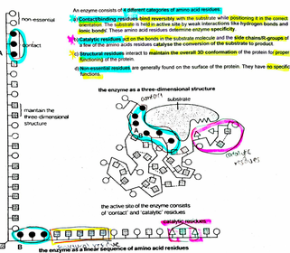
| (p) Explain the mode of action of enzymes in terms of an active site, enzyme-substrate complex, lowering of Ea & enzyme specificity using lock-and-key hypothesis & Induced ft hypotheses. (A) ACTIVE SITE *defn of active site - types of a.a in an enzyme | Active site → enzyme's catalytic center, small region of enzyme which binds w substrate - The 1० structure of enzyme determines its 2०& 3०structure. This in turn specify the overall 3D conformation of enzyme. - There is a precise 3D groove on enzyme at active site which gives it a specific conformatn - Active site consists of 3-12a.a (few). Some are CATALYTIC A.A RESIDUES. Some are CONTACT A.A RESIDUES which interact reversibly w substrate via weak H & ionic bonds, while positioning substrate in correct orientation. |
| (A) ACTIVE SITE - what do catalytic a.a & structural a.a do? * defn of specificity btwn substrate & enzyme | - R grps of catalytic a.a, present within active site catalyse conversion of substrate to pdts - Rest of ppd provide framework that maintains conformation of active site. (maintained by R grp interactions of structural residues) ***Specificity of enzyme attributed to complementary shape & charge btwn substrate & active site - Active site is not rigid. As substances enter it changes shape so that active site fits more snugly arnd substrate to for a more stable structure. *Contact + catalytic + Structural residues = determine complementary in shape & charge btwn substrate & active site |
| (A) ACTIVE SITE - 4 diff categories of aa residues | (1) Contact/binding residues →bind reversibly w substrate while positioning it in correct orientation. Substrate held in active site by weak interactions (H bonds & ionic bonds) . These aa residues determine enzyme specificity. (2) Catalytic residues → act on the bonds in substrate molecules & side chain / R-grps of a few of the a.a residues catalyse conversion of substrate to pdt (3) Structural residues → interact to maintain overall 3D conformatn of protein for proper fxning of protein (4) Non-essential residues → generally foun on surface of protein & hv no specific fxn |
| Tutorial Qn 2(a): Describe how amino acid residues at different positions in the protein may be brought together in the active site when such enzymes are synthesised. [4] Type: Recall Topic: Protein folding Approach: 4 marks = 4 points - Primary structure (with amino acid residues at different positions) must fold to form the secondary structure - What are the types of secondary structure? What are the bonds present? Where do the bonds occur? - Further folding to form the tertiary structure. What are the bonds present? Where do the bonds occur? - How does forming the tertiary structure affect the amino acid residues in question? | 1. The primary structure* of the polypeptide is the no & sequence of amino acids which determines its secondary and tertiary structure; 2. The polypeptide is folded into its secondary structure* such as α-helix* and β-pleated* sheets through hydrogen bonding* between the CO and NH groups of the polypeptide backbone; 3. The secondary structure is further folded into its tertiary structure* via hydrogen bonds*, ionic bonds*, hydrophobic interactions* and disulphide linkages* which are formed between the different R groups* of the amino acids; 4. The folding of polypeptide thus results in a globular structure where the three amino acids are thus brought closer together at the active site; |
| QN (pg 7): How can the 2 a.a, 'A' & 'B' that are ar apart along ppd chain somehow end up next to each other in the active site? | (1) 1० structure is the sequence of aa which determines its 2०& 3० structure. (2) The ppd is folded into its 2० structure, such as alpha-helix & beta-pleated sheets through H bonding btwn the CO & NH groups of the ppd backbone. (3) secondary structure is further folded into its tertiary structure via H bonds, ionic, disulphide bonds & hydrophobic int.s formed btwn the R grps of a.a residues. (4) the folding of the ppd results on a globular structure where the 2 a.a A & B are thus brought closer tgt at the active site. |
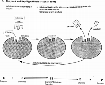
| (B) MODELS OF ENZYME ACTION → illustrates why enzymes are highly specific (1) LOCK-AND-KEY HYPOTHESIS | ***Enzyme active site has a specific surface conformation & charge complementary to the substrate, that is produced by 3D folding of ppd chain - Substrate → key, whose conformation is complementary to active site → lock - When enzyme & substrate collide in the CORRECT ORIENTATION, substrate becomes attached to active site of enzyme (not all collisions results in rxn → must be in correct orientation) - A short-lived E-S complex is formed - Catalysis occurs & pdts formed - Once formed, pdts no longer fit into active site & are released to the surrounding medium, leaving active site free to receive further substrate molecules. - Enzyme specificity arises bcos both enzyme active site & substrate possess SPECIFIC COMPLEMENTARY SHAPE & CHARGE that fit exactly into each other |
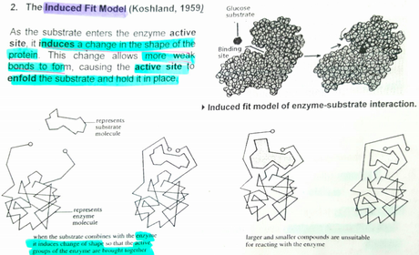
| (B) MODELS OF ENZYME ACTION → illustrates why enzymes are highly specific (II) THE INDUCED FIT MODEL | - As substrate enters enxyme active site, it induces a change in the shape of the protein → allows more weak bonds to form, causing active site to enfold the substrate & hold it in place - Active site of enzymes is complementary in SHAPE but not a perfect fit to the substrates it catalyses - However, when substrates binds to enzyme, it induces a change in the conformation of enzyme & its active site. - Change in conformation allows its active site to be moulded into a more precise fit for the substrate, enabling enzyme to perform its catalytic fxn more effectively |
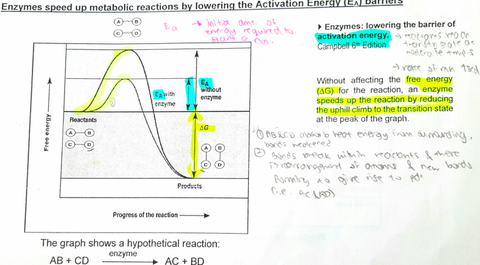
| (C) ENERGY PROFILE OF RXN *defn of Ea - how to reactants absorb energy for rxn? -how are the energy used for rxn? | Enzymes speed up metabolic runs by lowering the Ea barriers. (1) Ea → amt of energy reactants must absorb before a chem rxn will start - Reactants AB & CD absorb energy from the surroundings in the form of heat & absorption of thermal energy: (a) increases KE of reactant molecules for more forceful collisions (b) Increase frequency of collisions of reactants (c) Thermal agitation of atoms in molecules make bonds more likely to break |
| (C) ENERGY PROFILE OF RXN -what happens at peak of graph? - *defn of exergonic rxns | (II) Activation of reactants → peak of graph - At peak, reactant molecules have absorbed sufficient free energy to react & are unstable = transition state - Bonds within the reactants can then break once they have absorbed enough energy to become unstable (III) As bonds break & new bonds form, molecules settle into their new bonding arrangement & energy is released to the surroundings = exergonic rxn *Exergonic rxn → those in which free energy of final state is less than free energy of initial state. |
| (C) ENERGY PROFILE OF RXN How do enzymes enable rxn to take place faster? | Enzymes *lower Ea barrier* enabling reactant molecules to reach transition state at MODERATE temps. - Combi of enzyme + substrate = ES complex, which is equivalent to transition state of reactants in normal rxns. Formation of ES complex require less energy than transition state. *Note: Enzyme cannot change the △G (free energy change) for a rxn, w/o affecting △G for rxn, an enzyme speeds up rxn by reducing the uphill climb for the transition state at the peak of the graph. |
| (C) ENERGY PROFILE OF RXN -Molecular basis of enzyme action: Molecular mechanisms which contribute to a lowering of Ea → enabling a rxn to occur at a lower temp One or more of these mechanisms may work simultaneously in catalysis. | (1) Proximity effects → temp binding of reactants next to each other in enzyme active sites increases the chance of a rxn. Uncatalysed rxns depend on random collisions btwn reactant molecules. (2) Strain effects → slight distortion of reactants as they bind to the enzyme strains the bonds which are to be broken & increases the chances of breakage. (3) Orientation effects → reactants are held by the enzyme in such a way that bonds are exposed to chemical attack (by catalytic r grps) (4) Microenv effects → Hydrophobic aa create a water-free zone in which non-polar reactants may react more easily (5) R-groups of amino acid residues in active site participate in direct catalysis e.g. Acid-base catalysis. Acid-base catalysis → acidic & basic aa in enzyme facilitates catalysis |
| SUMMARY ***NOTE 3 KEY AREAS when answering qns : - BINDING -CATALYSIS - RELEASE | |
| Tutorial Exemplar Q(b) State why an enzyme-catalysed rxn is more likely to occur. [1] | (1) Enzymes lower Ea required for chem rxn to occur. Ea required in an enzyme catalysed rxn is about 2/3 that of uncatalysed rxn (based on graph shown) (2) Increases no of substrate molecules w required energy to cross the Ea barrier so rxn can proceed faster. |
| Tutorial exemplar (c) Describe the ways in which an enzyme interacts w its substrate. [3] - "describe" → give details - "interacts" → how the substrate would bind w enzyme Name the temp bonds btwn substrate & enzyme Specificity of enzyme action must be mentioned | (1) Temporary binding of substrate at the AT site of enzyme forming the ES complex (2) Substrate binds to AT site via H bonds, ionic bonds, hydrophobic int.s (e.g. creates a microenv w a water-free zone in which non-polar reactants may react more easily) *the temp bonds btwn substrate & enzyme* (3) Substrate binds to specific enzyme w AT site that in COMPLEMENTARY in shape & charge to it *specificity of the enzyme* (4) Based on the L&K hypothesis, enzyme → lock, substrate → key OR (4) Based on the induced fit hypothesis, substrate induces a change in shape in enzyme's AT site so that the AT site is a more precise fit for the substrate for effective catalysis. |
| Tutorial Essay Qn 7(a): Describe the mode of action of enzymes. [7] | 3 KEY AREAS: BINDING, CATALYSIS & RELEASE Interaction/binding 1. Enzymes have an specific* active site* which is complementary in shape/conformation and charge* to the substrate; 2. Effective collisions between enzyme and substrate form a temporary enzyme-substrate complex*; 3. Based on the lock and key* hypothesis, enzyme is the lock* and substrate is the key* (lock and key hypothesis) Or 4. Based on the induced fit* hypothesis, substrate induces a change in shape in enzyme active site so that active site is a more precise fit for substrate for effective catalysis. 5. Enzyme-substrate complex held together by weak interactions e.g. hydrogen, ionic bonds, hydrophobic interactions* |
| Tutorial Essay Qn 7(a): Describe the mode of action of enzymes. [7] When stating factors that decrease Ea, state "proximity effects", then explain "temp binding of reactants..." | Catalysis 6. Enzyme lowers the activation energy* barrier by: (max of 2 marks) Aligning substrates next to each other (close proximity) in active site for reaction to occur Strain on bonds to be broken / distorts the substrate and reduces activation energy to achieve transition state Orientates substrate such that its bonds are exposed to attack Provide a favorable microenvironment R-groups of amino acid residues in active site participate in direct catalysis – e.g. Acid-base catalysis Release 7. Products no longer fit active site and are released enzyme is unchanged and can be used again; |
| QN (pg 12): Explain what effect denaturation will have on the shape of the enzyme's active site? | (1) Denaturation results when the H bonds, ionic bonds & other weak interactions that stabilise the 3D conformation are broken (2) The conformation / shape of enzyme is altered including that of the active site (3) Active site is no longer complementary in conformation & charge to the substrate. |
| Enzyme cofactors *defn -examples | *enzyme cofactors → add non-protein substances require by enzymes for catalytic activity (1) Inorganic ions (e.g. zinc in DNA polymerase) - Many enzymes require certain metal ions to change non-functioning active site to functioning - Attachment of ion w the main enzyme (apoenzyme) changes the shape of the enzyme so as to allow ES complex to form more easily (2) Coenzymes - Cofactors that are organic in nature (3) Prosthetic grp (e.g. haem grp of haemoglobin, except haemoglobin not enzyme) - Permanently bound to enzyme |
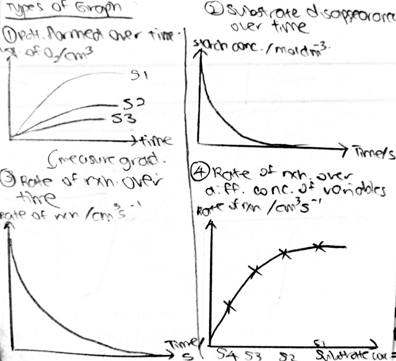
| (q) Investigate & explain effects of temp & pH, enzyme conc & substrate conc of an enzyme-catalysed rxn by measuring rates of formation of products or rate of disappearance of substrate (A) FOLLOWING THE TIME COURSE OF ENZYME-CATALYSED RXN 2 methods to measuring rate of rxn | 2 ways to investigate the effect of diff independent variables on rate: (AI) Measuring product-formation over time - amt of pdt in mixture, how fast pdts appear (AII) Measuring substrate disappearance over time - amt of substrate remaining, how fast substrate disappears |
| (AI) Measuring pdt formation over time E.g Conversion of H2O2 to H2O & O2 by enzyme catalase 2H2O2 → 2H2O + O2 - independent variable? -dependent? -constant conditions? | Independent variable: pH, temp, substrate / enzyme conc. Dependent variable/measurable qty: Volume of O2 (pdt) evolved Constant conditions: Other than independent variable, all other conditions kept constant |
| Experimental set-up For exp: Mention: 1) Rxn 2) Dependent variable 3) Independent variables 4) Constant variables / control set-up 5) Exp set-up & procedure - be able to describe fully | Q: The experiment can be improved by attaching the delivery tube in the above set-up directly to a frictionless gas syringe. Why is this a better approach? - O2 gas may dissolve in H2O resulting in underestimation of gas evolved (lower than actual) |
| Procedure of exp | 1) Add enzyme (catalase) to H2O2, mix & start stopwatch 2) O2 evolved can be measured by downward displacement of H2O in a graduated cylinder OR using frictionless gas syringe 3) Record vol of O2 evolved at fixed time intervals |
| Graph of exp For graphs: Mention (1) Describe trend (measured qty + rate of rxn) (2) Explain trend | -Rate of O2 produced (cm3/s)→ determined by finding gradient of graph - Decrease in rate of O2 pdtn with time → gradient decreases with increasing time |
| Explaining the trend of (1) Vol of O2 increases w time (2) Rate of O2 produced decreases with time | - Both substrate and enzyme molecules move around freely in the soln. When substrate collide w enzyme in correct orientation such that it fits into the enzyme's active site - effective collision - At time 0, rate of rxn max (steepest) → [substrate] highest & chances of effective collision btwn substrate & enzyme highest → higher freq of effective collisions - Thus, rate of formation of ES complex is highest & rate of rxn max - As time progresses, less substrate in soln, [substrate] decreases → less substrate to collide w enzyme molecules - Eventually, all substrates become products → graph shows pdt formation decreasing to horizontal & rate of rxn = 0 |
| Graph of rate of rxn against time | |
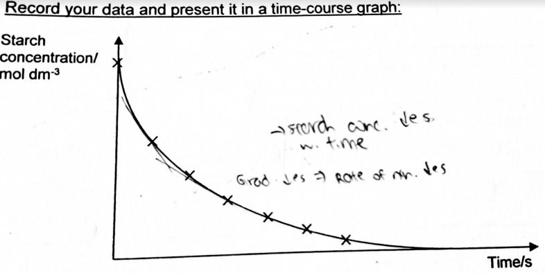
| (AII) Measuring disappearance of substrate over time E.g. of conversion of starch (substrate) to reducing sugars by amylase Starch → mostly maltose (use iodine test for starch) 1) Rxn 2) Dependent variable 3) Independent variables 4) Constant variables / control set-up 5) Exp set-up & procedure - be able to describe fully | - Independent variable: pH, temp, [substrate], [enzyme] - Dependent variable: [starch] - Measured qty: Intensity of blue-black coloration (shows [starch]) - Constant conditions: other than independent variable, rest constant |
| Set-up Procedure | 1) Add amylase to starch & starch stopwatch 2) At fixed time intervals, ALIQUOT out 1cm3 of rxn mixture to test tube & add 3 drops of iodine to rxn mixture 3) In presence of starch, iodine yellowish brown → blue-black. 4) Intensity of blue-black coloration measured using colorimeter & this indicates conc of starch present (to make rxn quantitative: w actual values of starch conc) 5) Use conversion standard to convert colorimeter reading to starch conc |
| Q: Why need to aliquot at fixed time intervals instead of adding I2 directly to rxn mixture & monitoring colour change over time? | Addn of I2 to rxn mixture inhibit digestion of starch to amylase. I2 complexes w starch & may prevent amylase from binding to starch |
| Trend: 1) Intensity of blue-black coloration / conc of starch 2) Rate of starch digestion decreases w time | Explaining the trend: - Both substrate and enzyme molecules move around freely in the soln. When substrate collide w enzyme in correct orientation such that it fits into the enzyme's active site - effective collision - At time 0, rate of rxn max (steepest) → [substrate] highest & chances of effective collision btwn substrate & enzyme highest → higher freq of effective collisions - Thus, rate of formation of ES complex is highest & rate of rxn max - As time progresses, less substrate in soln, [substrate] decreases → less substrate to collide w enzyme molecules - Eventually, all substrates become products → graph shows substrate conc. decreasing to 0 & rate of rxn = 0 |
| Investigating how the rate of enzyme rxn varies w one manipulated variable: [substrate] - Carry out several experiments at diff [S]: [S1] [S2] [S3] [S4] [S5] etc. * must hv at least 5 diff values for independent variable, w other variables kept constant | - Measure the progress of rxns over fixed time intervals & plot graphs (GRAPH A) - Find the INITIAL RATE OF EACH CONC. by finding the grad of tangent at time =0 → initial gradient, steepest & fastest rate of rxn, when there is NO LIMITING FACTOR OTHER THAN SUBSTRATE CONC affecting results Plot graph of rates against [s] (GRAPH B) |
| GRAPHS SUMMARY | |
| (q) Investigate & explain effects of temp & pH, enzyme conc & substrate conc of an enzyme-catalysed rxn by measuring rates of formation of products or rate of disappearance of substrate (B) FACTORS AFFECTING RATE OF ENZYME -CATALYSED RXN (i) Temp | |
| Explanation -when temp increased from low? | At low temp, - Increase in temp results in increase in KE of enzyme & substrate molecules → increase frequency of effective collisions btwn substrate & enzyme active sites Increase KE → (1) Increase rate of formation of ES complex, formation of more ES complexes /unit time (2) Increased no of molecules having energy > Ea / having sufficient energy to overcome Ea barrier to form pdts Thus, rate of rxn increases |
| Exp - temp coefficient? | Temp coeff (Q10) → factor by which rate increases with each 10℃ increase in T (pr how much rate increases up to physiological limit = point in which it denatures) Q10 = Rate of rxn at (X+10) ℃ / Rate of rxn at X ℃ = 2x (doubles every 10 ℃ increase) For enzyme-catalysed rxn btwn 4℃ to optimum temp (physiological T) rate of chem rxn doubles for each 10℃ rise in T, Q10 = 2 for most typical enzyme rxns |
| - what happens at opt T? | - Rxn rate increases w T only until opt T of enzyme is reached. Each enzyme has opt T → rate of enzyme rxn proceeds at max rate - Some enzymes have higher opt T → higher proportion of disulphide bonds (strong cov bonds) OR numerous intramolecular interactions that hold the tertiary structure of enzyme tgt -If the enzyme has many cysteine residues w sulfyhydryl groups → tertiary structure is maintained by many more disulphide bonds formed btwn R grps of cysteine residues. Disulfide bonds are strong cov bonds able to remain intact at higher temp. - Increase in the no of disulfids bonds increases stability of a protein to heat denaturation → holds onto 3D shape v well & retains its function |
| - increase in T beyond opt T? | - Increase in KE at T beyond opt T causes intramolecular (within the enzyme molecule) vibrations to increase → breaks H, ionic & other weak interactions that stabilise the conf / shape→ & disrupts the 2 & 3 structure of the enzyme → denaturation → loss of AT site conf → loss of enzyme activity -Substrate no longer comp to conf/shape of AT site of enzyme → failure of substrate to fit into enzyme AT site → fewer ES complexes formed → lowering rate of rxn |
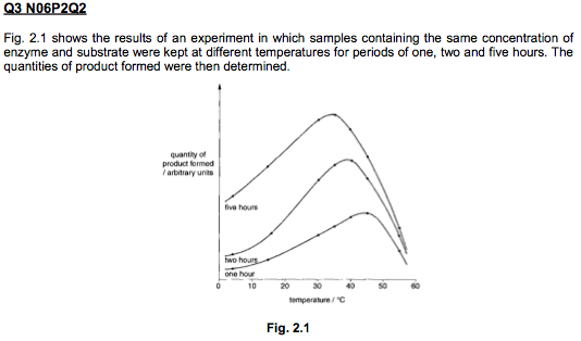
| Tutorial Qn 2.(cii) Explain the effect of increasing temperature on the activity of this enzyme. [4] “Explain” requires details in answer. What is the effect of increasing temperature on the energy of the molecules in the reaction? How does this lead to an increase in the rate of reaction? (think ES complexes) Don’t forget to address the decrease in rate of reaction also as it can be clearly seen in the graph. How does the molecular vibration affect the enzyme? Does this occur from the start or only after optimum temperature is reached? | 1. As temperature increases from 22oC to 100oC, kinetic energy* (K.E.) of the enzyme and the substrate molecules increases; 2. Resulting in an increase in the frequency of effective collisions* between substrate and enzyme active sites and therefore increased chances of forming enzyme-substrate (ES) complex*; 3. Increased number of molecules having sufficient energy to cross the activation energy* barrier to form the products of reaction; 4. Beyond the optimum temperature of 100°C, the higher temperatures cause greater (intramolecular) vibrations of the molecules which causes the bonds that determine enzyme conformation to break, (molecular vibration occurs from the start when temp starts increasing) 5. resulting in denaturation of enzyme where loss of conformation of the enzyme active site results in decrease in rate; |
| (b) Explain why the optimum temperature is lower if the quantity of product formed is measured after five hours rather than one hour. [2] (Optimum temp for 1h is 45°C while increasing duration to 5h decreases optimum temperature to 35°C) | 1. For longer durations of 5h, enzymes would have been exposed to increased temperatures for longer durations; 2. resulting in more weaker bonds, e.g. hydrogen bonds, being broken leading to loss in functional/tertiary conformation which changes the shape of the active site*increased denaturation; |
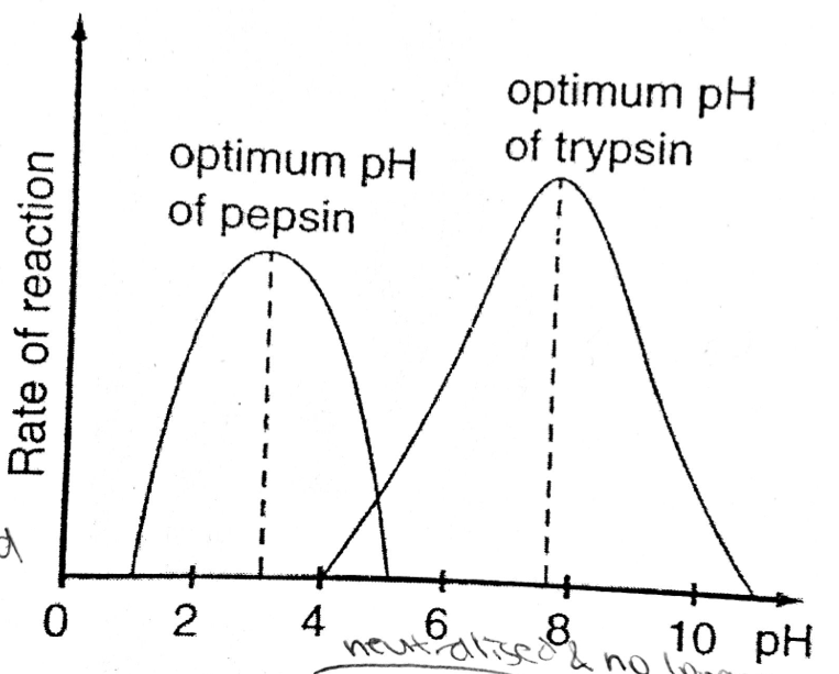
| (B) FACTORS AFFECTING RATE OF ENZYME -CATALYSED RXN (ii) pH | - Each enzyme has an opt pH → rate of rxn max at opt pH - Deviation from opt pH → lowering tf rate of rxn -In acidic/basic env, ionisation state of R-grps of charged a.a altered: Excess H+ /OH- ions affect the ionisation of the R grps of the charged a.a excess H+ → -COO- = -COOH (no longer -vely charged) excess OH- → NH3+ = NH2 (no longer +vely charged) - If these affected a.a are: (1) STRUCTURAL a.a → disruption of ionic & H bond formation which determines the TERTIARY structure of the protein → changes the specific 3D conf of enzyme AT → denatured as shape no longer COMP to substrate (2) CONTACT & CATALYTIC A.A → enzyme - substrate int disrupted & catalysis not take place → NO LONGER COMP IN CHARGE e.g. some catalytic activity require amino group to be in protonated form (NH3+) (3) Part of protein substrate, charges on its residues will change, affects substrate int w enzyme AT site &/or catalysis |
| TUTORIAL ESSAY 8(b) Explain how pH affects the rate of an enzyme catalysed reaction. | |
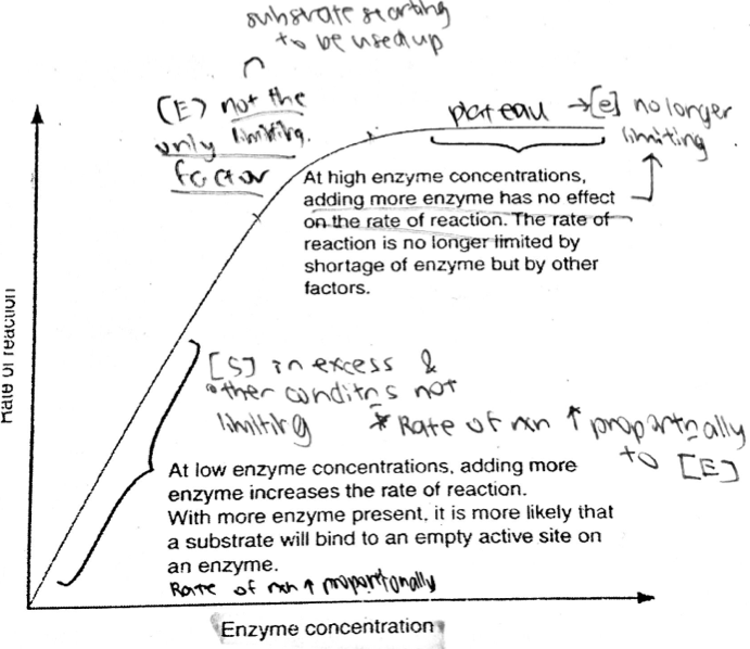
| (B) FACTORS AFFECTING RATE OF ENZYME -CATALYSED RXN (iii) ENZYME CONC | Rate of enzyme-controlled rxn dependent on freq on effective collisions btwn substrate & enzymes - Increased enzyme conc → freq of effective ES collisions increase → increased rate of formation of ES complexes & rxn rate increased (1) At linear portion, enzyme conc limiting → any increase in enzyme conc result in proportional increase in rate of rxn ([S] in excess & other conditions not limiting) (2) At curved portion, enzyme conc not the only limiting factor, some other factor limiting → substrate conc limiting (not enough substrate to occupy all AT sites) (3) At plateau, [E] no longer the limiting factor. Other factors limiting rate of rxn. Increasing [E] no longer increases rate of rxn |
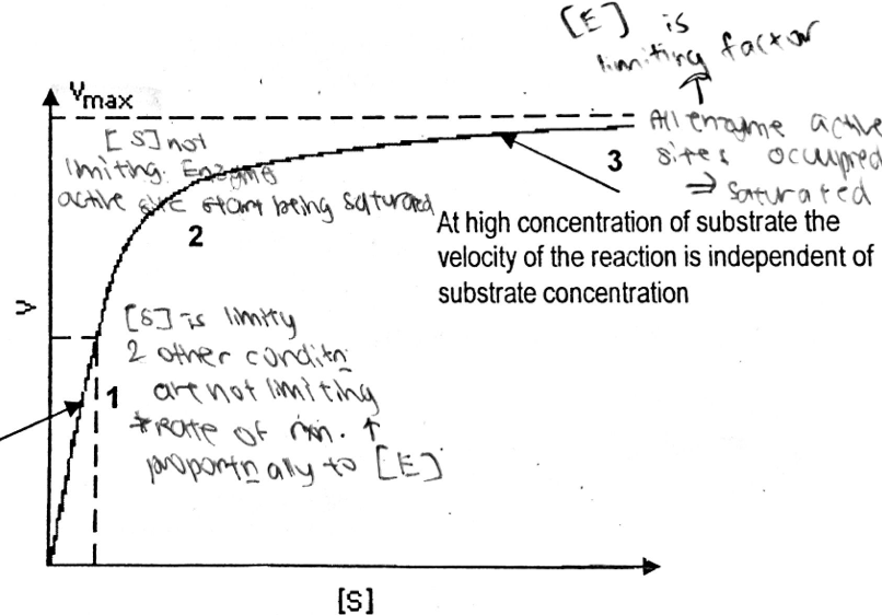
| (B) FACTORS AFFECTING RATE OF ENZYME -CATALYSED RXN (iv) SUBSTRATE CONC | At point (1), rate of rxn increases PROPORTIONALLY w an increase in [S] Freq of effective collision btwn substrate & enzyme molecules increase → Rate of ES complex formation increases → increase in rate of rxn bcos At low [S], AT sites if enzymes are readily available to catalyse rxn → substrate conc limiting. As [S] increases, more AT sites are occupied by substrates. At point (2), enzyme AT sites start being saturated which limits rate of rxn ([E] limiting) At point (3) plateau reached. Enzyme saturation reached → all available AT sites are occupied by substrate molecules. [S] no longer limiting → [E] limiting → further increase in [S] will not cause rate of rxn to increase further → rate of rxn reached max velocity (Vmax) |
| Michaelis constant (Km) *defn | Michaelis constant (Km) → conc of substrate required to make rxn attain half its max rate Km = [S] at 1/2 Vmax → unique for each enzyme -Measure of the affinity of enzyme for its substrate LOW Km → HIGH affinity btwn enzyme &v substrate → low [S] needed to attain half max velocity HIGH Km → LOW AFFINITY → High [S] needed to attain half max velocity |
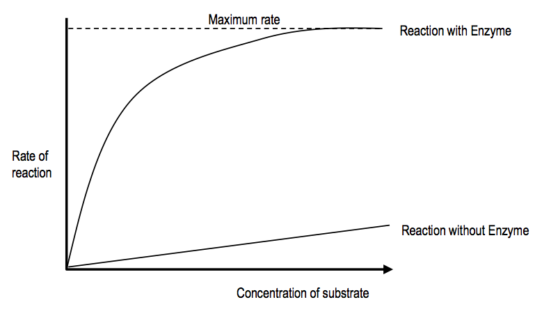
| Tutorial Qn 1(a) With ref to graphs, explain the diff btwn the 2 graphs. [3] Approach: 3 marks 3 points State the difference between the two graphs first. Explain the differences in the magnitude between the two graphs. Explain the differences in the shape between the two graphs. | (1) MAGNITUDE: Across all substrate concentrations, the rate of enzyme catalyzed reaction is higher than the rate of reaction without enzyme; (2) SHAPE: As substrate concentration increases, rate of reaction without enzyme increases linearly whereas, rate of enzyme-catalysed reaction increases at decreasing rate until it plateaus; (Need to state the difference first); |
| (r) describe STRUCTURE of competitive & non-competitive inhibitors wrt to the binding sites of the inhibitor s) explain EFFECTS of comp & non-comp inhibitors (including allosteric inhibitors) on rate of enzyme activity ENZYME INHIBITION types | |
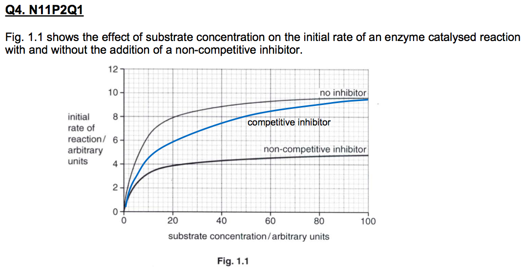
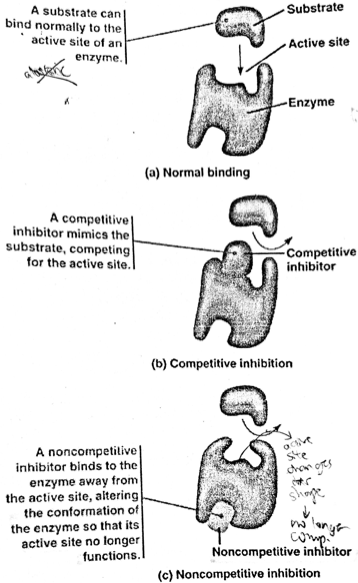
| (I) Competitive inhibition *defn STRUCTURE EFFECTS | Competitive inhibition → when bound to enzyme, prevents substrate molecules from binding to enzyme AT site STRUCTURE: Bear similar shape / conformation & charge to substrate thus competes w substrates for AT site -Thus can bind REVERSIBLY to AT Site → bonds involved are weak, non-cov bonds EFFECTS: Reduces availability of enzyme AT sites for substrate binding thus reduces the rate of rxn *inhibition CAN BE OVERCOME BY INCREASING [S] → increases chances of substrate binding to T site instead of inhibitor binding. At sufficiently high [S] rxn velocity reaches same Vmax (max rate of rxn) observed as in the absence of the inhibitor. |
| Tutorial 1(c) Explain effect of a competitive inhibitor on rate of enzyme catalysed reaction.[3] - How does the competitive inhibitor bind to the enzyme? - Important to qualify that the reaction is reversible (temporary binding) - What does the degree of inhibition depend on? | 1. The competitive inhibitor has similar shape/conformation and charge* to substrate molecule; 2. Competitive inhibitor binds to the active site* thus blocking substrate from binding; 3. With increase in substrate concentration, the degree of inhibition will be reduced; |
| (ii) Explain with reasons the shape of the curve you have drawn. [3] | 1. Competitive inhibitor has the similar charge and shape/conformation* to the substrate 2. allowing it to bind to the enzyme active site* and block substrate binding. 3. The binding of the inhibitor to the active site is not permanent/reversible; 4. Effect of the inhibitor at high substrate concentrations is negligible since the higher proportion of substrate molecules compared to inhibitor molecules can effectively out-compete the inhibitor molecules for the active sites; 5. Hence at very high substrate concentrations, the rate of reaction is the same as that when no inhibitor was present; |
| (II) Non-competitive inhibitor *defn STRUCTURE EFFECTS | Non-competitive inhibitor → bears (STRUCTURE) no structural similarity to substrate molecule, so it binds to A SITE OTHER THAN THE ENZYME AT SITE EFFECT:- Alters the conf/shape of the specific enzyme AT site → substrate cannot bind to AT site in the correct orientation → rate of rxn decreased - Non-competitive inhibitors effectively decreases availability of FUNCTIONING ENZYMES as it forms INACTIVE ENZYME-INHIBITOR COMPLEXES. -Effects CANNOT BE OVERCOME BY HIGH[S] - rate of rxn continue to decrease with increasing inhibit conc. When inhibitor saturation reached (all enzyme other sites alr bound by inhibitors, further increase in [I] does not decrease rate further), rate of rxn almost nil |
| Comparing graphs of comp & non-comp inhibition | |
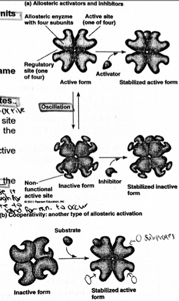
| (III) ALLOSTERIC INHIBITION *defn of allosteric enzymes Characteristics | Allosteric enzymes → regulated by inhibitors & activators Characteristics: (a) Allosteric enzymes usually consists of ⪰2 subunits where each subunit have their own - ACTIVE SITE → binds substrate - ALLOSTERIC (not AT) SITE → binds inhibitor / activators These sites are at diff locations within same subunit (b) Can exist in 2 conformational state (oscillate btwn 2 conf states - active & inactive) - Binding of allosteric activator at allosteric site / binding of substrate at AT site → stabilises the functionally active conformation - Binding of an allosteric inhibitor → stabilises inactive conformation of the enzyme |
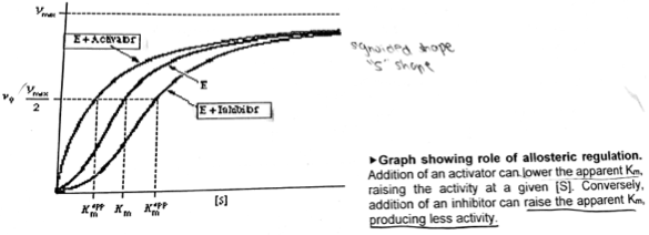
| Characteristics ****S-SHAPED GRAPH IN ENZYME QN → SHOWS ENZYME IS AN ALLOSTERIC ENZYME | (c) A SINGLE ACTIVATOR/INHIBITOR → SUFFICIENT TO ACTIVATE/INHIBIT the activity of the enzyme → NOT permanent bonding (weak bonds only) → stabilise it long enough for substrate to bind for rxn to occur (d) Binding of substrates in allosteric enzymes exhibit COOPERATIVITY → binding of substrate to first subunit changes its conf of the other subunits such that it becomes EASIER to accept subsequent substrates (increases affinity of enzyme for substrates) (e) Rate V against [S] plot, an S-SHAPED SIGMOID CURVE → cooperative binding of substrate to AT site |
| Characteristics | (f) Effect of an allosteric inhibitor on rate of rxn is opposite that of increasing [s]. Hence , in the presence of an allosteric inhibitor, SAME Vmax CAN BE REACHED AT HIGHER SUBSTRATE CONC. Addition of activator → lower Km, raising activity at a given [S] (at the same [S], the rate of added activator rate of rxn is higher than w/o activator rate) → Less [S] required to reach 1/2 Vmax → increased affinity btwn enzyme & substrate Add of inhibitor → higher Km, decreasing activity at a given [S], (at the same [S], the rate of added inhibitor rate of rxn is lower than w/o inhibitor rate) → More [S] needed to reach 1/2 Vmax → decreased affinity btwn enzyme & substrate |
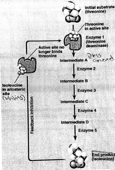
| (IV) Feedback inhibition / End-pdt inhibition | -In end-pdt inhibition, a metabolic pathway inhibited by the BINDING OF AN END PDT of a biochemical pathway TO AN ENZYME (that ACTS EARLY IN THE PATHWAY) - E.g. a.a isoleucine is produced from threonine As isoleucine (END PDT) ACCUMULATES, it inhibits the enzyme threonine deaminase in the first step of the rxn by BINDING TO THE ALLOSTERIC SITE (inhibitor) OF THE ENZYME. Hence the end pdt ALTERS THE CONF/SHAPE OF THE SPECIFIC ENZYME AT SITE → thus substrate cannot bind to AT site in the correct orientation → rate of rxn decreased. -Prevents cell from wasting resources in producing excess isoleucine Q: Why is it impt to inhibit enzyme earlier in the pathway? Don't waste resources, prevent wastage of resources synthesising various intermediates |
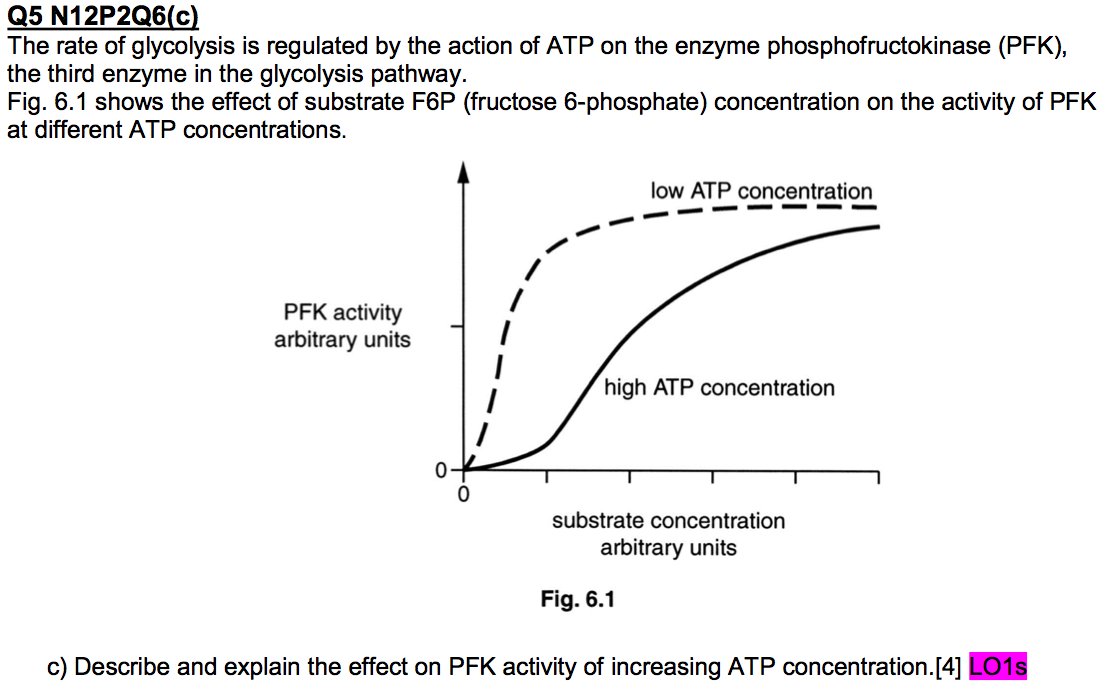
| ATP → inhibitor - At high ATP conc, low [S], why dent? ATP binds to allosteric site of PFK stabilising inactive conf, altering its AT site such that substrate (F6P) cannot bind to AT site → lowering enzyme (PFK) activity At high ATP cons, high [S], substrate binds to AT site & exhibit cooperativity → S-SHAPED GRAPH | 1. High levels of ATP slows down / inhibits PFK activity (as the graph is below the graph represented by low ATP); 2. This inhibition is most pronounced at lower substrate concentrations. This inhibition is overcome by high substrate concentrations. 3. ATP binds to *allosteric site of PFK stabilizing the inactive conformation, altering its active site conformation such that the substrate (F6P) cannot bind to the enzyme active site thus lowering the PFK activity. 4. At high substrate concentration, the substrate binds to the active site exhibiting cooperativity allowing easier binding of subsequent substrate molecules |
| Q(pg 24) How are enzymes regulated by non-competitive inhibition diff from enzymes regulated by allosteric regulation? | |
| Key words defining: (1) 3D conformation (2) AT site (3) Ea (4) Complementary in shape & charge | (1) 3D conf → the 3D arrangement of atoms of a protein (enzyme) molecule (2) AT site → the enzyme's catalytic centre, a small region on the enzyme that binds to the substrate (comp in shape & charge to substrate). Consists of 3 types of a.a, structural, contact & catalytic. Contact a.a determine enzyme specificity (3) Ea → min amt of energy reactants must absorb before a chem rxn can start (4) comp in shape & charge → the enzyme AT site have a specific surface conf/shape (precise 3D groove at AT site) & charge produced from the 3D folding of the ppd chain, that allows the substrate to fit into it well |
| (5) denaturation (6) Effective collisions (7) ES-complex | (5) denaturation → the loss of the specific 3D conf/shape of enzyme AT site, due to the disruption/breaking of weak bonds (H, ionic bonds, hydrophobic int.s) that stabilises the enzyme's conf/shape, as result of high temp / unsuitable pH, causing the AT site of enzyme not being comp to substrate. (6) Effective collisions → collisions btwn enzymes & substrates in the correct orientation, such that the substrate fits into enzyme AT site, forming ES complex (7) ES complex → formed when substrates fits into enzyme AT site, as a result of effective collisions, weak interactions (H bonds & ionic bonds) hold the substrate & enzyme AT site tgt |
| (8) Induced fit (9) KE (10) Lock & Key | (8) Induced fit → binding of substrate to enzyme AT site, inducing a change in conf/shape of the enzyme & its AT site such that AT site is moulded into a more precise fit for substrate, enabling the enzyme to perform its catalytic fxn most effectively. (9) KE → increase in T → increase in KE of substrate & enzyme molecules → increase in freq of effective collisions (10) Lock & Key → "lock" = enzyme "key" = substrate Enzyme & substrate comp in shape & charge to each other such that substrate fits into enzyme AT site |
¿Quieres crear tus propias Fichas gratiscon GoConqr? Más información.