9560734
Descripción
Fichas por Marissa Alvarez, actualizado hace más de 1 año
|
|
Creado por Marissa Alvarez
hace más de 7 años
|
|
| Pregunta | Respuesta |
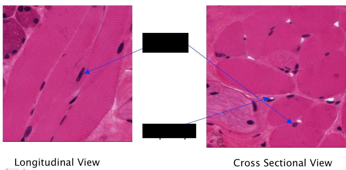
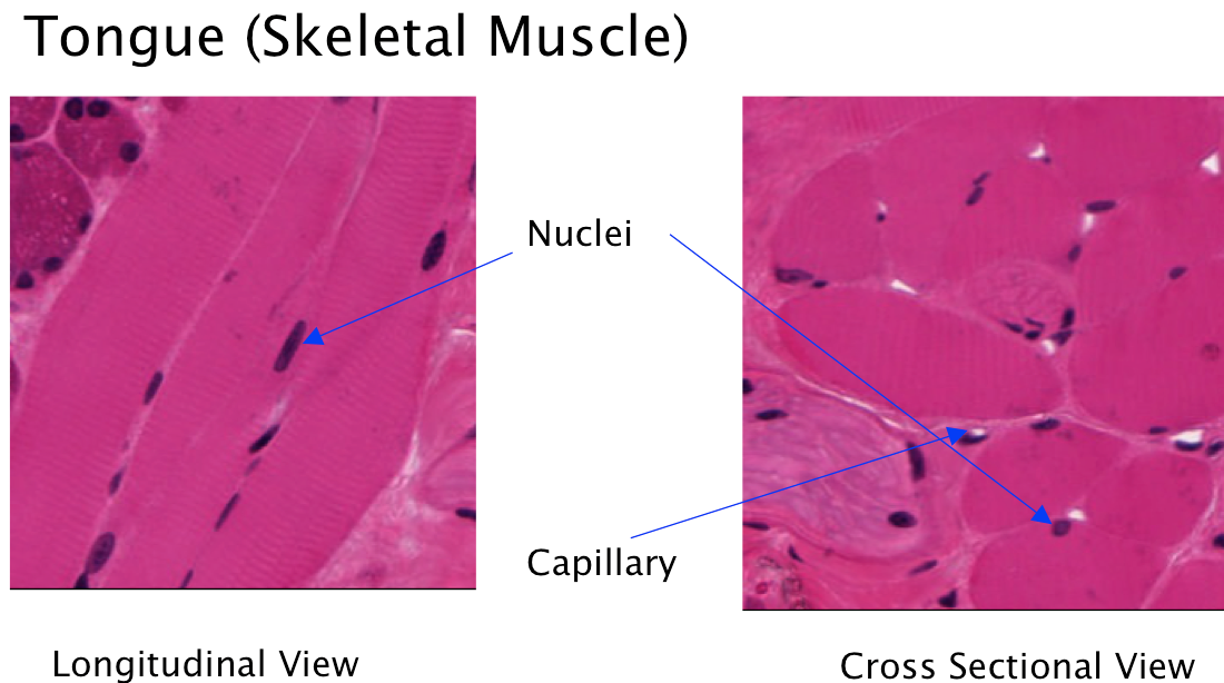
| What type of muscle tissue is this? What are the arrows pointing at? | Note the distribution of nuclei at the PERIPHERY of the fibers and numerous capillaries especially obvious in the cross-sectioned muscle. The distribution of muscle bundles is consistent with VOLUNTARY control of complex movements of the tongue, and skeletal muscle bundles are NOT attached to tendons. |
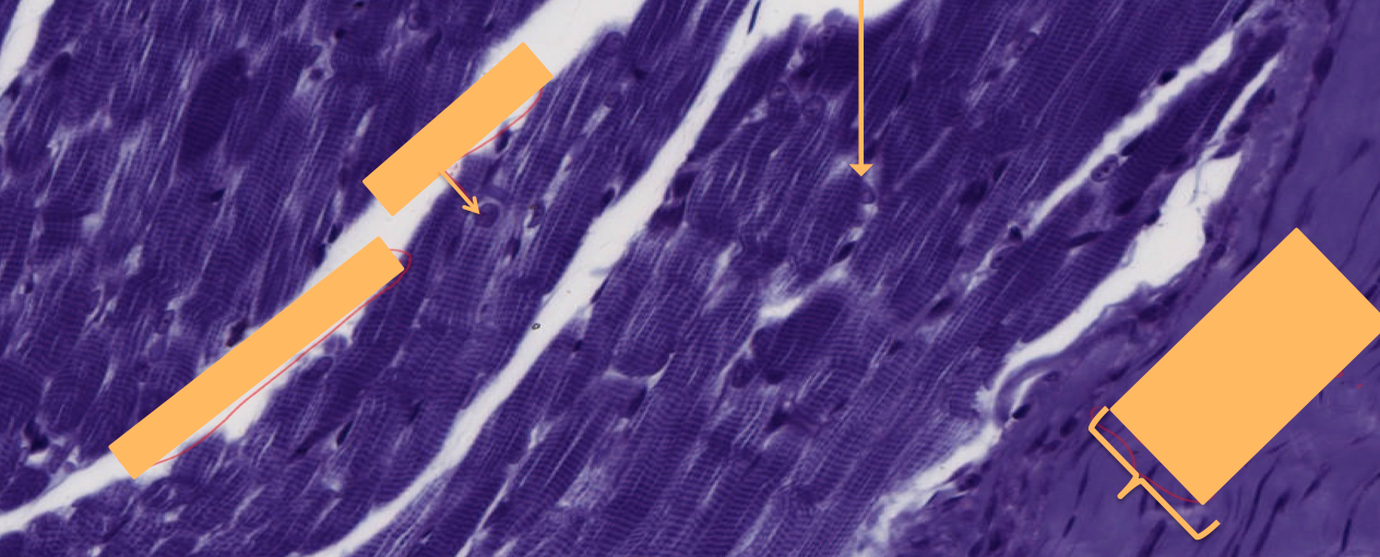
| Which type of muscle tissue is shown? What are the arrows/boxes indicating? | Overview of SKELETAL muscle (i.e., STRIATED VOLUNTARY MUSCLE) Note that the fibers may be quite LONG & ALL the fibers have STRIATIONS in contrast to the smooth tendon (shown where Dense regular tissue CT is located). The numerous open spaces between muscle fibers are artifacts of preparation. Normally the fibers are closely packed. |
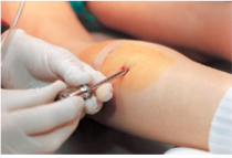
| Clinical Point: Skeletal muscle biopsy | a valuable clinical procedure for diagnosis and management of many neuromuscular diseases: is removal of a small piece of muscle tissue under local anesthesia via open biopsy (surgical excision) or by less invasive percutaneous needle followed by microscopic evaluation |
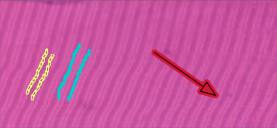
| Identify the regions labeled in the image. What type of muscle tissue is this? | Skeletal Muscle Tissue: Longitudinal Section Identification of the Z line (in the middle of light portion, I band) The A band is outlined in the yellow and is characterized by dArk outline The I bind is outlined in the blue and is characterized by the LIght portion |
| Question: Of the following regions of a sarcomere (A band, I band, and H band), which one(s) shorten(s) during muscle contraction? | During contraction, the actin and myosin filaments interact. The actin are pulled toward the center of the myosin filaments. As a result the sarcomeres shorten. In the fully contracted muscle, the ends of actin myofilaments overlap, the H zones disappear, and the I band becomes very narrow. *A band remains unchanged |
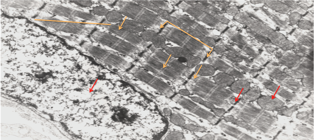
| Identify the following: M, H, A, I, Z, Sarcomere, Mitochondria, T tubule system, Triad Which type of muscle tissue is shown in the image? | Skeletal Muscle Tissue (Electron micrograph) (could not find T tubule system or Triad, waiting on Julianna to find answer!) |
| Question: What is the difference between a fiber, fibril, and filament in muscle tissue? | Muscle Fiber: contains both myofibrils & is a elongated, multinucleated cylindrical fiber (cell), exhibits striations Myofibril: contains filaments, is a long, cylindrical contractile element within muscle fiber; as long as the muscle fiber itself; exhibits striations Myofilament: contains short contractile proteins such as myosin (thick) and actin (thin) |
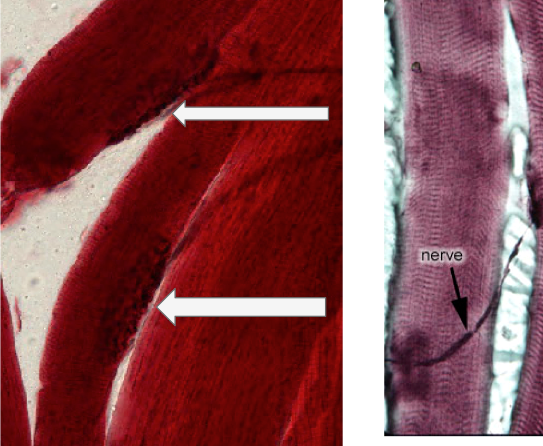
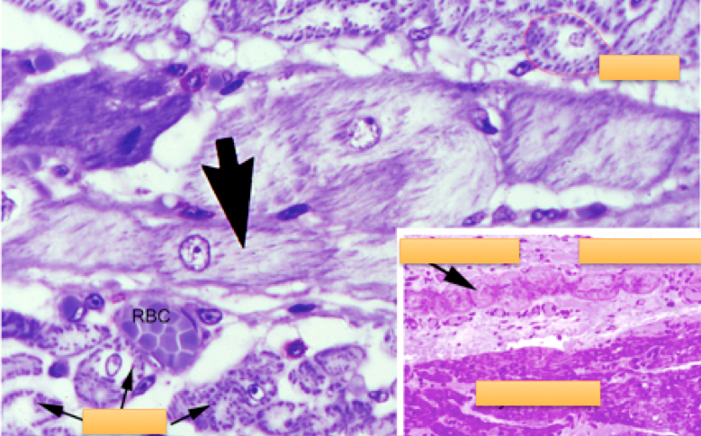
| What is being depicted in the image above? | Neuromuscular Junction! Note the large cylindrical skeletal muscle fibers. Each fiber is innervated by a neuron that terminates in a neuromuscular junction (the darkly stained and speckled bulb like projection on end of long axon string). |
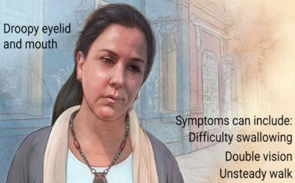
| Clinical Point: Myasthenia Gravis | the most common hereditary disorder of neuromuscular transmission but this autoimmune disease most often results from an acquired immunologic abnormality Symptom onset > age of 30 yo in women and later in men. Muscle weakness often fluctuates but is usually progressive. In the acquired disorder, a distortion of the postsynaptic sarcolemmal membrane of the neuromuscular junction is accompanied by a reduction in the concentration of acetylcholine receptors. Antibodies are attached to the postsynaptic membrane, which makes it less sensitive to acetylcholine and leads to a reduced muscle action potential in response to a nerve impulse. |
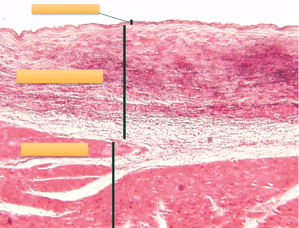
| Endocardium: inner surface of heart (near lumen) Myocardium: the middle muscular layer of the heart wall Epicardium: (not shown in image): outer surface of heart *hard to see but rows of Purkinje fibers but they lie b/w endocardium and myocardium (in sub-endocardium) | |
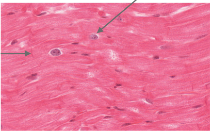
| What are the arrow pointing at and what type of muscle tissue is shown? | Heart: Cardiac Muscle Tissue |
| Epicardium (outside surface) *Note the unilocular fat cells -blood vessel indicates the outside → vascularizing the heart | |
| Endocardium (inside surface) -towards the lumen - layer on internal portion - possible epithelial lining | |
| Cardiac myocytes and Purkinje fibers: Their main role is to conduct a depolarization impulse along the fibers through gap junctions (electrical synapses) into the myocardium | |
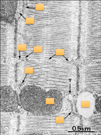
| What type of muscle is depicted in the image? What are the arrows indicating? | Electron micrograph: Cardiac muscle w/ intercalated disk Note the mitochondria (M), glycogen (G), T tubules (T), sarcoplasmic reticulum (SR), Z-lines, & striated appearance of actin and myosin. Identify the intercalated disk and its components: fascia adherens (F), desmosomes (D), and gap junctions (N, nexus). |
| What type of muscle tissue is shown? What do each of the arrows indicate? | Electron micrograph: cardiac muscle w/ a dyad at Z-line Note the lipid droplet (L), mitochondria (M), glycogen (G), T tubules (T), sarcoplasmic reticulum (SR), Z lines, & striated appearance of actin and myosin. In cardiac muscle, one T tubule and single SR cisterna form a DYAD, which is located at or near the Z-line. (This is in contrast to skeletal muscle where one T tubule and 2 SR cisternae formed a triad at the A-I junction.) |
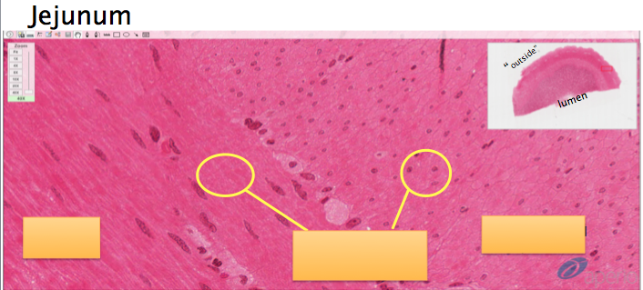
| Jejunum Smooth muscle tissue | |
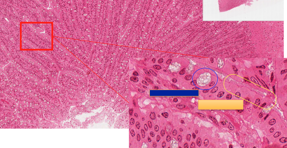
| What is indicated by the blue and yellow areas? | Intestinal villi are small, finger-like projections (yellow area) that extend into the lumen of the small intestine. Each villus is approximately 0.5–1.6 mm in length, and has many microvilli projecting from the enterocytes of its epithelium which collectively form the striated or brush border. Goblet cell (blue area) |
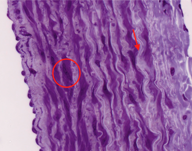
| What type of tissue is depicted in the image? What is the arrow indicating? The circle? | Smooth muscle: the muscle structure that helps retain and manipulate the artery’s shape. When contracted, the nuclei take on a “corkscrew” appearance. The smooth muscle also is able to make Elastic cartilage. The elastic cartilage is visible BETWEEN the smooth muscle cells and has a lighter, “rubber band-like” appearance. |
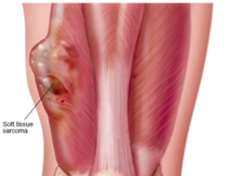
| Clinical Point: Rhabdomyosarcoma | Most common soft tissue sarcoma in children and adolescents. Sarcoma = malignant tumor of connective or other nonepithelial tissue Two histologic subtypes of this skeletal muscle neoplasm: the more common embryonal and less frequent alveolar Neoplasm: a new and abnormal growth of tissue in some part of the body, especially as a characteristic of cancer. Even though both are fast growing and malignant, long-term survival rates show improvement with advances in surgery combined with high-dose chemotherapy, stem cell rescue, and radiation. Tumor cells may arise directly from SATELLITE cells or MESENCHYMAL STEM cells with capacity to become striated muscle; they resemble preinnervated fetal skeletal muscle. |
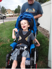
| Clinical point: Duchenne muscular Dystrophy | Is a genetic disorder caused by a deficiency in dystrophin Characterized by rapid progression of skeletal muscle degeneration occurring early in life. Dystrophin maintains mechanical integrity of the cell during contraction by anchoring cytoskeletal elements. Women are carriers but men are actually affected by the disorder (X-linked) -mainly children are affected specifically (young boys) Symptoms: muscle weakness & wasting, heart involvement, worsens with age. |
¿Quieres crear tus propias Fichas gratiscon GoConqr? Más información.