Cerrar
Thoracic Skeleton consists of:
Sternum
12 pairs of ribs and the associated costal cartilages
12 thoracic vertebrae and their associated intervertebral (IV) discs
Intervertebral (IV) discs are joints located b/w the vertebrae
STERNUM: Flat elongated bone that forms the anterior and middle portion of the thoracic cage
Composed of three parts:
1. Manubrium
2. Body
3. Xiphoid Process
**Sternal angle (Angle of Louis) is the articulation b/w the manubrium & the body
**The sternal angle:
marks the 2nd rib w/associated costal cartilage is opposite of ~T4/T5 thoracic vertebrae (highlighted by yellow box)
STERNUM:
Jugular Notch:
easily palpable concave notch along superior border of the manubrium
COSTAL CARTILAGES
Prolong the ribs ANTERIORLY
Contribute to the elasticity of the thoracic wall
First 7 generally articulate DIRECTLY w/the sternum
The 8th, 9th and 10th articulate with the cartilages just SUPERIOR to them
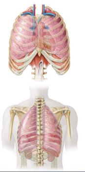
Thoracic Apertures
The thoracic cage has superior and inferior openings (apertures)
Superior Thoracic Aperture:
doorway b/w thoracic cavity and the neck
Clinicians generally
refer to the superior thoracic
aperture as the “thoracic outlet”
Inferior Thoracic Aperture:
the muscular diaphragm closes the aperture, separating the thoracic cavity from the abdominal cavity
Functions of the Thoracic Cage
General Features of Thoracic Vertebrae:
Like most all vertebrae, a typical thoracic vertebra consists of three major features:
1. Vertebral body: for weight bearing
2. Vertebral (neural) ARCH consisting of pedicles and laminae:
protection of the spinal cord
3. Numerous (7) processes for:
muscular attachment and joint surfaces
VERTEBRAL BODIES ARE heart-shaped and have costal facets for articulation with ribs
SPINOUS PROCESSES are long and slant inferiorly
TRANSVERSE PROCESSES are long and slender and have a transverse costal facet for articulation with the tubercle of a rib
ARTICULAR FACETS (4) for articulation with adjacent vertebrae (i.e. facet joints)
Three types of RIBS:
1. Vertebrocostal (True):
articulates directly with sternum via its own costal cartilage (1-7)
2. Vertebrochondral (False):
indirect articulation with sternum via costal cartilage of the superior rib (8-10)
3. Vertebral (Floating):
do not articulate with sternum, end in the posterior abdominal wall (11-12)
Characteristics of typical Ribs:
Typical ribs are considered ribs 3-9
HEAD:
has two facets (superior and inferior) for articulation with two vertebrae.
**There is a crest b/w these two facets
Neck:
slightly constricted area just distal to head
Tubercle:
located at junction of neck and shaft, has a smooth facet for articulation with corresponding transverse process of the vertebra
Shaft:
thin, flat and curved.
Has a costal angle where rib turns anterolateral and a COSTAL GROOVE inferiorly for neurovascular structures
Typical Rib ARTICULATIONS:
Understand that the head of typical ribs articulates with TWO vertebrae:
1. The body of the numerically corresponding vertebra and also 2. the body of the vertebra superior to it
Example: Rib 5 articulates with the body of T5 and T4
Understand that the tubercle of a typical rib articulates with the numerically corresponding transverse process
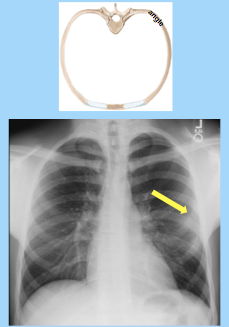
In the Clinic:
RIB FRACTURES
JOINTS of the Thoracic Skeleton
The thoracic skeleton consists 3 major types of articulations (joints):
1. Costochondral
2. Sternocostal
3. Costovertebral
All VERY STRONG articulations aimed to protect underlying structures!
JOINTS of the Thoracic Skeleton Cont'd...
Costochondral = fibrous joints
Sternocostal = synovial joints
Costovertebral = synovial joints
MUSCLES of the Thoracic Wall
Several EXTRINSIC muscles of the upper limb cover the thoracic wall but are primarily muscles of the upper limb
These include:
Pectoralis Major
Pectoralis Minor
Serrratus Anterior
These muscles, although primarily move and stabilize the upper limb, they can also assist in respiration by moving the ribs and thoracic cage
PECTORALIS MAJOR
Large fan-shaped muscle in superior thorax
Two heads of origin: clavicular and sternocostal
Inserts into the humerus
Powerful ADDUCTOR; flexor, and medial (internal) rotator of the arm
***Innervated by the medial & lateral pectoral nerves
PECTORALIS MINOR
Originates from anterior surface of the 3rd - 5th ribs
Inserts into the coracoid process of the scapula
Stabilizes the scapula against thoracic wall (accessory respiratory muscle also)
Innervated primarily by the MEDIAL pectoral nerve (receives a small contribution by the lateral pectoral nerve)
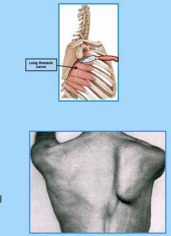
SERRATUS ANTERIOR MUSCLE
Originates on the surface of the 1st - 8th ribs
Inserts into the medial border of the scapula
Stabilizes and protracts the SCAPULA (shoulder blade)
**Innervated by LONG thoracic nerve
In The Clinic: WINGED SCAPULA
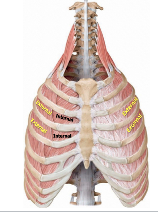
INTRINSIC MUSCLES of the Thorax
External Intercostals
Internal Intercostals
Innermost Intercostals
Intercostal muscles are all accessory muscles of respiration
Innervated by intercostal nerves
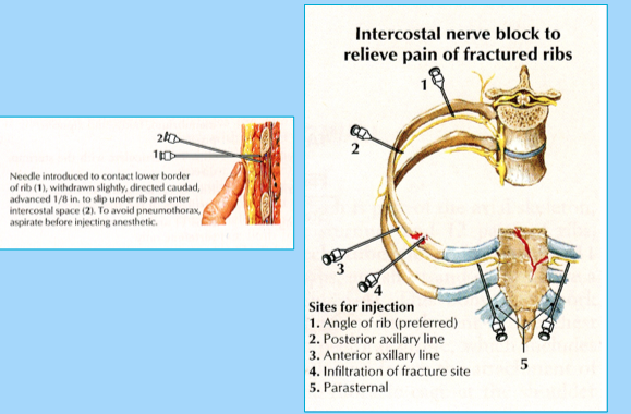
Note the order “VAN” of neurovascular structures within the costal groove:
Intercostal Vein
Intercostal Artery
Intercostal Nerve
Small collateral branches… (minor significance, "angel hairs")
In The Clinic:
INTERCOSTAL NERVE BLOCK
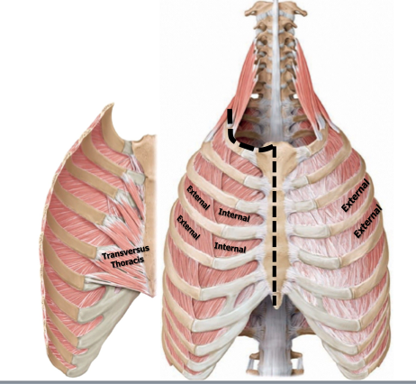
INTERCOSTAL MUSCLES
INNERMOST Intercostal
Muscles
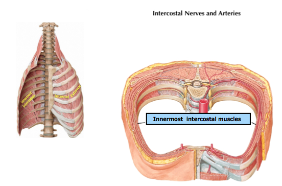
INNERmost Intercostal Muscles
ONE IN PARTICULAR:
(TTM)
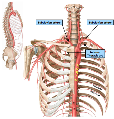
VASCULATURE of the Thoracic Wall
VASCULATURE of the Thoracic Wall
Cont'd...
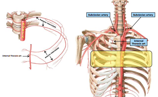
Vasculature of the Thoracic Wall
Cont'd......
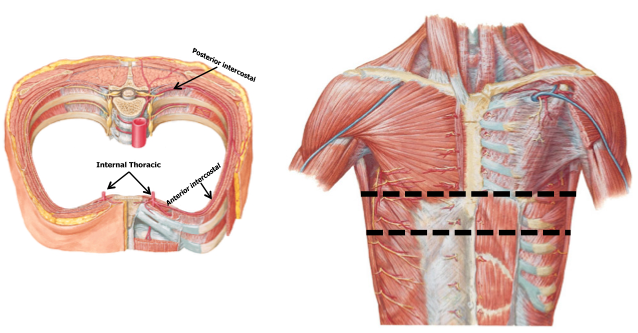
If we removed the anterior aspect of the thoracic wall….
Note the internal thoracic arteries divides into the MUSCULOPHRENIC and SUPERIOR EPIGASTRIC arteries
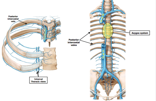
Venous Drainage of the Thoracic Wall
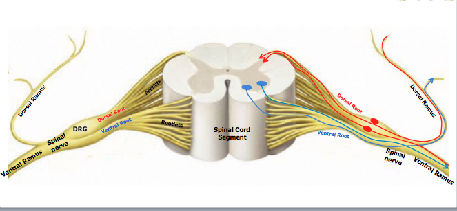
Nerves of the Thoracic Wall
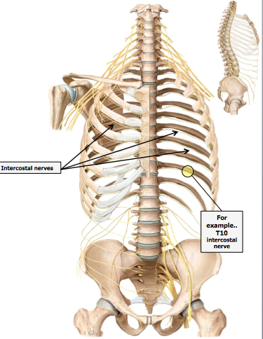
Spinal Nerves
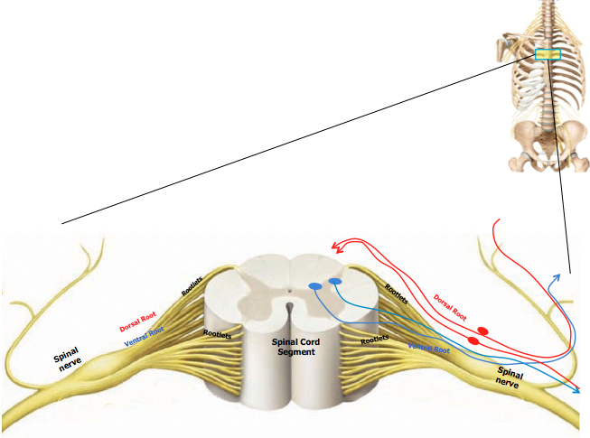
Spinal nerves divide into two branches (or rami)
Nerves of the Thoracic Wall
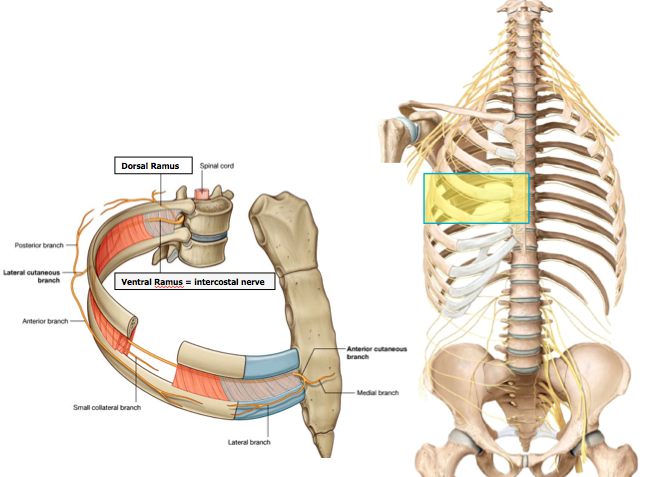
Nerves of the Thoracic Wall Cont'd...
This segmental innervation pattern is reflected on the surface of the body
Note in this dermatome map, the thoracic nerves innervate successive segments along the body wall
Every sensory nerve innervates a specific skin area
These “dermatomes” then correspond to a specific spinal cord segment
The dermatomes are arranged in a “band-like pattern
Note that the T4 dermatome corresponds to the level of the nipples
Note that the T10 dermatome corresponds to the level of the umbilicus
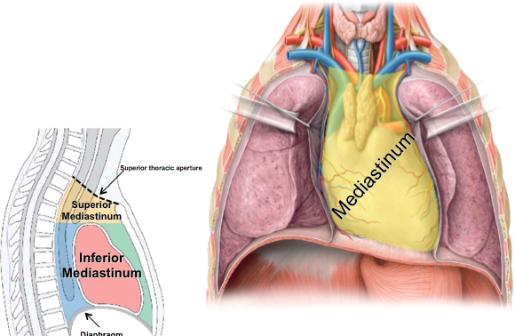
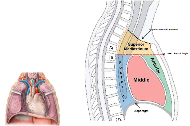
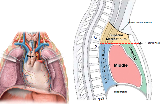
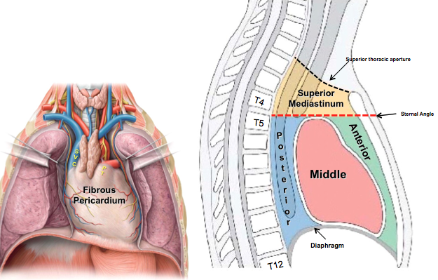
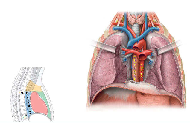
DIVISON of the Thoracic Cavity
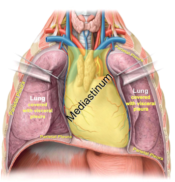
DIVISON of the Mediastinum
Lateral Chest X-Ray
ANTERIOR Mediastinum
MIDDLE Mediastinum
BOTTOM LINE CONCEPTS
ACC
Anatomical Connection to the Clinic:
65 yo male, falls in bathroom, hits chest on bathtub, left side chest pain, pain worsens when breathing deeply
Severe point tenderness over the lateral aspect of his chest wall (w/superficial swelling & bruising)
Need chest x-ray
Diagnosis?
Possible complications?


