10663916
Description
Flashcards by Dolu Falowo, updated more than 1 year ago
|
|
Created by Dolu Falowo
over 8 years ago
|
|
| Question | Answer |
| What happens to a tissue for it to be viewed? | 1) Fixed using a chemical/physical methods to prevent autolytic degradation or putrefaction of the tissue 2) Embedded in wax (blocks) before being cut into sections 3) Placed on microscope slide and wax is removed 4) Stained using H&E, Masson's trichrome or elastic van Gieson. |
| What are the types of microscopes used? | -Bright-field or light microscope -Fluorescence microscope -Transmission electromicroscopy (TEM) |
| Main types of tissue? | Epithelial Connective/support Muscle Nervous |
| What is the function of epithelia? | -covers body surfaces, lines tracts/tubes, forms glands -covers surfaces that communicate with the exterior of the body (form a barrier) -selective diffusion, absorption or secretion (simple epithelia) -physical protection (stratified) -in urinary tract (transitional) -transport of particles out of airways (pseudostratified) -avascular |
| Shapes of epithelial cells? | -squamous (flat cell/nucleus) -columnar (elongated cell/nucleus) -cuboidal (round/square cell with a spherical nucleus) |
| Types of intracellular junctions? | Occluding/tight Adherins Desmosomes Hemidesmosomes Gap |
| Describe: Occluding/tight Gap Adherins | Lie immediately underneath apical surface. Prevent leakage of molecules between cells and maintain cellular polarity Forms channels that allow small molecules (ie. ions) to pass between cells. Helps communication between cells. Connect actin filament bundles in adjacent cells |
| Describe: Desmosomes Hemidesmosomes | Connect intermediate filaments in adjacent cells Anchor intermediate filaments in a cell to the basal lamina (prevent cells sliding off) |
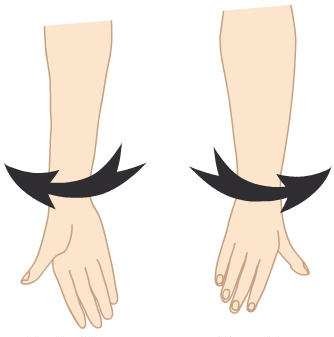
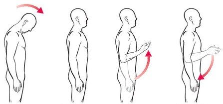
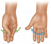
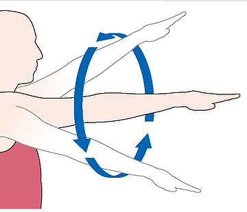
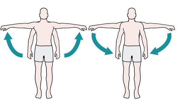
| Label: | |
| Supination Pronation | |
| Flexion Extension Flexion Extension | |
| Opposition Reposition | |
| Circumduction | |
| Abduction Adduction | |
| Dorsiflexion Plantarflexion | |
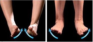
| Inversion Eversion | |
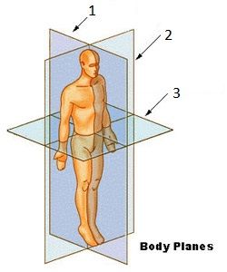
| 1. Sagittal plane 2. Coronal plane 3. Transverse/horizontal | |
| Anterior Posterior Lateral Medial Para-sagittal plane Dorsal | Front Back Further away from medial plane Closer to medial plane Parallel to medial plane Back of hand |
| Ipsilateral Contralateral Inferior Superior Supine Prone | Same side Opposite side Towards feet Towards crown of head Lying horizontally on their back Lying horizontally on their front |
| Define: Protraction Retraction | Movement of body part in the anterior direction. Movement of body part in the posterior direction. |
| What is connective tissue? | Tissue of mesodermal origin which gives structural and metabolic support for other tissues and organs throughout the body. It fills volume in the body |
| What are the 2 types of connective tissue and what is their function? | Loose connective tissue surrounds blood vessels and underlies the basement membrane of epithelia. It is the site of metabolic exchange between capillary and the tissue it supplies. Dense connective tissue provides tough physical support and protection |
| What are the 3 components of connective tissue? | Fibres Ground substance (collectively the ECM) Cells |
| What are the 2 types of fibres and their properties? | Collagen fibres are flexible but have a high tensile strength. Elastin fibres provide connective tissue with the ability to stretch and recoil back to its original shape. |
| What is ground substance composed of? | Proteoglycans which are highly hydrophilic molecules that trap water within the matrix. |
| What are fibroblasts? Shape? | They produce the collagen and elastin fibres and ground substance Cigar-shaped (elongated with triangles cut out of the sides) |
| Difference between loose/dense connective tissue | Loose is composed largely of hydrated ground substance with few fibres and fibroblasts. Dense is composed largely of fibres with sparse ground substance and fibroblasts. Dense regular has collagen fibres in an organised fashion while dense irregular has collage fibres arranged randomly. |
| What type of cell stores fat? What are the 2 types and their function? Where is its nucleus? How does it appear on histology slides? | Adipose tissue/adipocytes White adipose is used for long term storage of fat for energy. It has 1 large cytoplasmic lipid droplet. Brown adipose is used to release heat to warm the blood. It has many lipid droplets with many mitochondria. Its nucleus is pushed to 1 side in white adipose but in the middle in brown adipose. It appears white on slides as the lipid is dissolved by the solvent used. |
| What are reticular fibres? | -Made up of type III collagen fibres -Form a network where lymph and cells can pass |
| What other cells are present? | -Fibroblasts -Macrophages (derived from monocytes in the blood) -Mast cells -Adipocytes -Plasma cells -Other leukocytes |
Want to create your own Flashcards for free with GoConqr? Learn more.