13726799
Description
Flashcards by Sophie Byrne, updated more than 1 year ago
|
|
Created by Sophie Byrne
almost 8 years ago
|
|
| Question | Answer |
| Briefly outline the 3 main mechanisms by which membrane-bound organelles import proteins. | 1. Proteins and RNA move b/w nucleus and cytosol through nuclear pore complexes 2. Transmembrane protein translocators directly translocate proteins across a membrane from cytosol to ER and mitochondria 3. Secretory pathway - vesicles loaded w/ cargo |
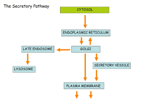
| Summarise the secretory pathway using a diagram | |
| What is the first step of the secretory pathway, using the example of the ER? | Synthesis and transport of nascent protein into ER from cytosol. - Ribosome directed to secretory pathway using signal sequences/peptides - SS depends on where protein is going to end up e.g. the ER |
| What is the structure of the ER signal sequence? | 16-30 AAs, 1 or more +vely charged AA Followed by 6-12 hydrophobic AAs |
| What is the SRP and what does it do in the first stage of the secretory pathway? | Signal recognition particle. Wraps around large ribosomal unit, 1 end bound to signal sequence as it emerges from the ribosome |
| How does the SRP direct the ribosome complex to the ER membrane? | - Other end of SRP binds elongation factor binding site at the interface b/w small and large ribosomal units - Stops protein synthesis as soon as signal peptide emerges from ribosome - Gives ribosome enough time to bind to SRP receptor in rough ER membrane before the polypeptide chain is completed - SRP detaches from ribosome complex - Cycle repeats |
| Why is it important that the ribosome has enough time to bind to the ER membrane before the polypeptide chain is completed? | 1. Stops lysosome enzymes being released into the cytosol 2. Avoids large portions of the protein folding into a compact structure before reaching the ER translocator |
| What is the ER translocator? | Channel that facilitates insertion of newly-forming protein into the ER |
| What is the human translocon also known as, and what does it consist of? | Sec61 Made up of 3 subunits |
| How is a soluble protein translocated into the ER once the ribosome complex has docked into the ER? | - Docking w/ Sec61 (translocon) - Signal sequence's run of hydrophobic AAs act as a 'stop transfer sequence' which is held in the translocon - Synthesis of the polypeptide resumes and polypep is pushed thry translocon into ER lumen til protein synth is complete - Signal peptidase cleaves polypep chain from signal sequence - Protein released into ER lumen where protein folding into tertiary sturcture occurs - Signal seq moved out of translocon - Cycle repeats |
| Where is signal peptidase located? | In the membrane of the ER |
| How are transmembrane proteins translocated? | - Signal sequence (START-transfer sequence) held in translocon - Protein synthesised and pushed into ER lumen so part of it is in the translocon and partly in the lumen - Signal peptidase cleaves away signal sequence - N-terminus is in the ER lumen; C-terminus in cytosol; transmembrane domain inside the translocon - Protein moves by lateral diffusion out of translocon so ot sits in the ER membrane - Further p[rocessing occurs and protein is trabsported to elsewhere in the cell |
| What happens if there are multiple transmembrane domains? | Same principle but there are multiple internal stop-transfer sequences |
| What is N-linked glycosylation? | Addition of oligosaccharides to asparagine in a protein, occuring in the ER lumen. |
| Where does each of the 2 stages occur? | 1. Cytoplasmic face of ER membrane - core oligosaccharide synthesis 2. Exoplasmic face of ER membrane |
| Describe the process of N-linked glycosylation. | Core oligosaccharide synthesis on cytoplasmic face Sequential adding of sugars to dolichol - the carbohydrate side is flipped from the cytosol side to the lumen side Oligosaccharide is to asparagine of a protein by oligosaccharide transferase |
| Why is N-linked glycosylation a cotranslational process? | It occurs at the same time as translation of the polypeptide chain |
| What happens after N-linked glycosylation? | Procesisng of ologosaccharide continues as the glycoprotein moves through the Golgi |
| Describe the principle of vesicular transport. | Budding off from donor compartment (ER membrane) containing cargo Vesicle trafficked to target organelle Recognition Fusion w/ target compartment and release of cargo into target compartment vesicle |
| How does budding occur? | - Vesicles have protein coats surrounding phospholipid bilayer - |
| What is clathrin? | - Component of vesicle protein coat - Consists of havy chain and light chain - Triskelion shapes interlock to create cage-like structure around bilayer |
| How does clathrin drive vesicle formatio budding? | 1. Protein coat assembly - clathrin clusters together on Golgi surface, associates w/ adaptor proteins which are inserted into bilayer 2. Cargo receptors bind to cargo molecules (sol. proteins) and associate w/ c;lathrin complex which causes curvature of membrane 3. More and more curvature occurs til 'lollipop' structure occurs where a vesicle is almost formed 4. 'Neck' of vesicle (lollipop stick) pinches off and vesicle leaves donor membrane 5. Clathrin complex dissassembles and leaves vesicle --> results in naked transport vesicle |
| How does 'pinching off' of the vesicle occur? | Dynamin wraps around vesicle neck and Conformational changes occur which squeezes off vesicle neck Vesicle forms as membrane fuses |
| How does the vesicle fuse with the target compartment/organelle? | - v-SNARE inserted into vesicle bilayer - Rab associates w/ vesicle - Rab recognised by Rab effector (tethering protein) on target membrane and docking occurs - v-SNARE and t-SNARE intertwine and pull vesicle into close proximity to target membrane - Fusion of transport vesicle and target membranes occurs and enters lumen - SNAREs unravel - v-SNAREs repackaged and leave; t-SNAREs remain on target membrane |
| How are soluble ER-resident proteins retaines within the ER? | KDEL sequences (Lys-Asp-Glu-Leu) at C-terminus Retains protein in ER |
| Describe the mechanism for protein retention in the ER. | - KDEL-containing protein trafficked to the Golgi in the forward pathway - KDEL receptor in the Golgi recognises and binds to the KDEL sequence in the protein - This KDEL complex is packaged into a retrieval vesicle w/ a COPI coat - Vesicle trafficked back to ER and KDEL-containing protein is released back into the ER lumen |
| What is another example of a retrieval mechanism for rwsident ER proteins? | KKXX (lysine-lysine-anything-anything) |
| What is exocytosis? | Membrane-bound vesicle carrying cargo fuses w/ membrane and releases cargo into outside world |
| Example of exocytosis? | Histamine release by mast cells |
| What are the 2 types of exocytosis/ | Regulated Constitutive |
| How does trafficking to a lysosome occur? | Lysosomal enzymes are transported: N-glycosylation in ER and early Golgi Phosphorylation of mannose residues on carbohydrate side of enzyme (mannose-6-phosphate forms) M6PRs on transgolgi network bind to enzymes containing mannose-6-phosphate Enzymes bound to M6PR packaged into transport vesicles and delivered to late endosome Enzymes dissociate from M6PR due to lower pH than in lumen (5.5 vs 6.4) |
| Describe receptor-mediated endocytosis. | 1. Binding 2. Lateral diffusion 3. Invagination 4. Vesicle formation 5. Uncoating 6. Fusion with early endosome and release of ligand 7a. Transport to late endosome and then to lysosome for digestion 7b. Transport of receptors to cell surface for recycling 7c. Transcytosis |
| What is familial hypercholesterolaemia? | Inherited disease where patients have abnormally high LDL levels in the blood Increased incidence of CHD at a young age Most common mutation involves LDLR mutation |
| What is different between normal and mutant LDL receptors? | LDL receptor can't bind to adaptor proteins (isn't recognised) LDL ligand not incorporated into vesicle so less LDL is broken down Leads to higher LDL levels in blood as it remains in circulation |
Want to create your own Flashcards for free with GoConqr? Learn more.