1744771
Description
Flashcards by Faith Adams, updated more than 1 year ago
|
|
Created by Faith Adams
about 11 years ago
|
|
| Question | Answer |
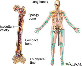
| Functions | 1. support 2. storage of minerals (calcium) and lipids (yellow marrow) 3. Blood cell production (red marrow) 4. Protection: ribs, vertebra, skull, pelvis 5. Leverage: force of motion (movements) |
|
Image:
9582 (image/jpg)
|
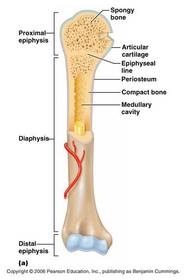
Long Bone Long and thin. Found in the arms legs, hands, feet, fingers and toes. e.g. the femur which is the longest bone of the thigh. |
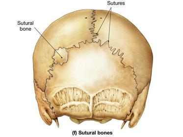
| Sutural Bone Small, irregular bones between flat bones of the skull. Aka Wormian bones | |
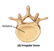
| Irregular Bone Complex shapes such as spinal vertebrae and pelvic bones, skull bones with short, flat notched or ridged surfaces. | |
|
Image:
7707 (image/jpg)
|
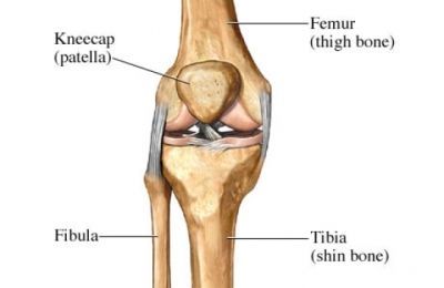
Sesamoid Bone Small, flat bones that develop inside tendons near joints of knees, hands and feet. e.g. patella |
|
Image:
9889 (image/jpg)
|
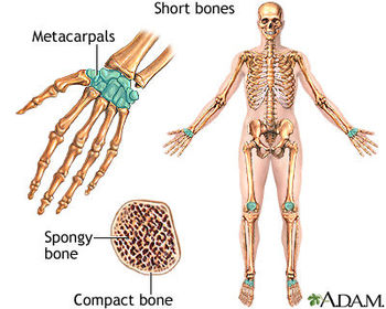
Short Bone Small and thick, including ankle and wrist bones. (tarsal and carpal) |
|
Image:
landmarks0001 (image/jpg)
|
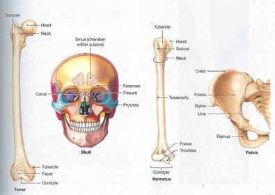
Bone Markings: Elevations and Projections 1. Process: projection or bump 2. Ramus: extension of a bone making an angle with the rest of the structure |
|
Image:
landmarks0001 (image/jpg)
|
Bone Markings: Tendons and Ligaments 1. Trochanter: large, rough projection 2. Tuberosity: smaller, rough projection 3. Tubercle: small, rounded projection 4. cCrest: prominent ridge 5. Line: low ridge 6. Spine: pointed or narrow process |
|
Image:
landmarks0001 (image/jpg)
|
Bone Markings: Articulation with adjacent bones 1. Head: extended end of epiphysis separated from shaft by neck 2.Neck: connection between the epiphysis and diaphysis 3. Condyle: smooth, rounded articular process 4. Trochlea: smooth, grooved articular process shaped like a pulley 5. Facet: small, flat articular surface |
|
Image:
landmarks0001 (image/jpg)
|
Bone Markings: Depressions 1. Fossa: Shallow depression 2. Sulcus: narrow groove |
|
Image:
landmarks0001 (image/jpg)
|
Bone Markings: Openings 1. Foramen: rounded passageway for nerves and blood vessels 2. Canal: duct or channel 3. Meatus: passageway through a bone 4. Fissure: elongated cleft or slit 5. Sinus: chamber within a bone |
| Flat Bone Thin with parallel surface and are found in the skull, sternum, ribs and scapula. Provides protection for underlying soft tissue and extensive surface area for muscle attachment. | |
| Diaphysis | Extended tubular shaft |
| Epiphysis | Wide part at each end, where the femur articulates with other bones |
| Metaphysis | Area where the epiphysis and diaphysis connect. |
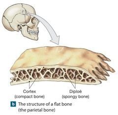
| Structure of Flat Bone Parietal Bone | Diploe: layer of spongy bone between the compact bone within the cranium. |
| Osseous Tissue | Dense supportive tissue containing specialized cells. The matrix of the bone tissue is solid because of the calcium salts deposited around protein fibers in its ground substance. |
| Characteristics of Bone Tissue | 1. Dense matrix containing deposits of calcium salts. 2. Matrix contains bone cells within the lacunae (spaces that chondrocytes occupy) which are organized around blood vessels. 3. Canaliculi (narrow passageways thru matrix) that form pathways for blood vessels to exchange nutrients and wastes. 4. Outer surfaces of bones are covered by periosteum consisting of outer fibrous and inner cellular layers. |
| Bone Matrix | 2/3 of bone matrix is made up of calcium phosphate and reacts with calcium hydroxide to form hydroxyapatite which incorporates with other calcium salts like calcium bicarbonate and ions and crystallizes. Calcium phosphate is hard but brittle and flexible. (withstands compression) Collagen fibers are strong when subjected to pull and are stronger than steel. They are flexible and tolerates twisting and bending. |
| Bone Cells Osteocytes | 1. Mature bone cells 2. Each live in a lacunae 3. Canaliculi through the lamellae allow osteocytes to connect at gap junctions between cytoplasmic extensions. 4. Osteocytes do not divide. Main Functions -maintain protein and mineral content -repair damaged bone (reverts to osteoblasts or osteoprogenitor) |
| Bone Cells Osteoblasts | 1. Immature blood cells 2. Secrete the matrix by process of osteogenesis: secretion of proteins and other inorganic compounds of the matrix 3. Osteoid: the matrix before calcium salts are deposited. 4. When surrounded by bone they become osteocytes. |
| Bone Cells Osteoprogenitor Cells | 1. Mesenchymal stem cells that divide to produce osteoblasts 2. Located in the inner, cellular layer of periosteum (endosteum) 3. Assists in repairing bone structures. |
| Osteoclasts | 1. Giant, multinucleate cells 2. Secrete acids and protein digesting enzymes which dissolve bone matrix and release stored minerals (process of osteolysis) 3. Derived from stem cells that produce macrophages. |
| Bone building | Bone building and recycling must be kept in balance. When osteoclats break down bone faster than osteocytes build bone, bones become weak. Bones get stronger with exercise, which causes osteocytes to build bone. |
|
Image:
Slide9.JPG (image/JPG)
|
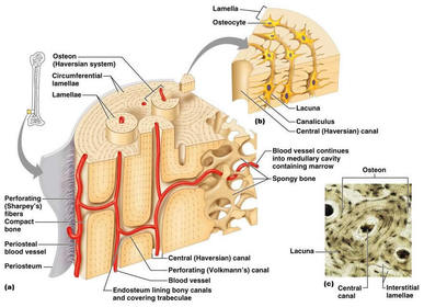
1. Basic unit of mature compact bone is the osteon (Haversian system) 2. Haversian canal: cirucular channel (concetric lamellae/circles) running longitudinal in the center of the osteon containing lymph and blood vessels (capillary and venule) 3. Volkmann's Canal: minute passageway by means of of which blood vessels and nerves from the periosteum of bone penetrate into compact bone. (perpendicular to the HC) |
|
Image:
Slide9.JPG (image/JPG)
|
4. Concentric lamellae: Rings of calcified bone matrix surrounding the HC of compact bone. 5. Lacunae: little lake, small hollow space w/in the bone matrix where osteocytes reside. Located in the CL. 6. Canaliculus: Small channel or canal connecting 2 lacunae in CB. Each contains a cell process of osteocyte. 7. interstitial lamellae: fragments of older CB found between newer osteons. Are partially destroyed during bone replacement. All osteons run the length of the bone and strengthen the bone in that direction. |
|
Spongy Bone
Image:
image006 (image/jpg)
|
1. Contains no osteons 2. Matrix forms a trabeculae which contains no blood vessels 3. Trabeculae is filled with red bone marrow which has BVs and supplies nutrients to osteocytes and yellow bone marrow which stores fat. |
| Periosteum | 1. Covers all bone except parts of joint enclosed w/in the joint capsule. 2. Includes an outer fibrous layer and inner cell layer 3. Collagen fibers here connect the CF of the bone w/ those of joint capsules, attached tendons and ligaments (perforating fibers) |
| Periosteum Functions | 1. isolates bone from surrounding tissues 2. provides route for circulatory and nervous supply 3. participates in growth and repair |
| Endosteum | 1. incomplete cellular layer covering the trabeculae of SB in marrow cavity and line central canals 2. contains osteoblasts, osteoprogenitor cells and osteoclasts 3. Active in bone repair |
| Intramembranous ossification | 1. Dermal ossification 2. Occurs in the dermis, produces dermal bones e.g. mandible and clavicle |
| Steps of IMO | 1. Mesenchymal cells collect, differentiate into osteoblasts and begin ossification at the ossification center where developing bones grow out in projections called spicules. 2. BVs grow into the area to supply the osteoblasts. Spicules connect, trapping BVs inside the bone. 3. SB develops which can be remodeled into osteons of CB, periosteum or marrow cavity. |
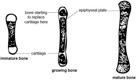
| Endochondral ossification | 1. Bones originate as hyaline cartilage which then becomes ossified. |
| Ossification occurs in 6 steps | 1. Chondrocytes in the center of the HC enlarge and form struts which calcify. Enlarged chondrocytes die, leaving cavities in the cartilage. 2. BVs grow around the edges of the cartilage. Cells in the perichondrium change to osteoblasts, producing a layer of superficial bone around the shaft which will then become CB. 3. BVs enter cartilage bringing fibroblasts that become osteoblasts. SB develops at primary ossification center. 4. Remodeling creates marrow cavity. Bone replaces cartilage at the metaphyses. 5. Capillaries and osteoblasts enter the epiphyses creating secondary ossification centers. 6. Epiphyses fill w/ SB. The cavity remaining w/in the joint cavity is the articulation cartilage. The cartilage at the metpahysis is the epiphyseal cartilage. |
| Epiphyseal line | When the long bone stops growing, after pubert, the epiphyseal cartilage disappears but is visible as the EL. |
| Appositional Growth | Growth of compact bone on the surface (periosteum) of the bone, continues to thicken and strengthen the long bone w/ layers of circumferential lamellae. |
| Blood and Nerve Supplies | As the long bone matures, osteoclasts enlarge and marrow cavity and osteons form around BVs in the CB. |
| Blood Vessels Nutrient artery and vein | single pair of large blood vessels that enter the diaphysis through the nutrient foramen. (femur has more than 1 pair) |
| Blood Vessels Metaphyseal vessels | Supply the epiphyseal cartilage where bone growth occurs |
| Periosteal vessels | Provide blood to the superficial osteons and the secondary ossification centers. |
| Bone Remodeling | 1. Cycle that recycles and renews bone matrix. 2. Involves osteocytes, osteoblasts and osteoclasts. 3. When deposition is more than removal bone is stronger and when removal is faster bone is weaker. |
| Exercise and Bones | 1. Mineral recycling allows bones to adapt to stress. Heavily stressed bones become thicker and stronger. 2. Degenerative changes in the skeleton occur after short periods of inactivity. Up to 1/3 bone mass can be loss in a few weeks without stress. |
Want to create your own Flashcards for free with GoConqr? Learn more.