3969239
Description
Flashcards by Josie Capolingua, updated more than 1 year ago
|
|
Created by Josie Capolingua
about 9 years ago
|
|
| Question | Answer |
| What are the (7) Functions of Blood? | Transports oxygen. Transports CO2. Transports nutrients. Transports hormones. Maintains pH. Distributes heat. Protects against disease. |
| What are the (4) Components of Blood? | Plasma: liquid part of blood. Erythrocytes: red blood cells, carry oxygen. Leukocytes: white blood cells, fight infection. Thrombocytes: platelets, clot blood. |
| How is oxygen transported? | Carried in red blood cells (erythrocytes). Oxygen combines with haemoglobin to form oxyhaemoglobin. |
| How are Erythrocytes suited to their function? | Contain haemoglobin. Have no nucleus and therefore more room for oxygen. Bi-concave in shape and therefore have a increased surface area. |
| How is CO2 transported? | 8% dissolved in plasma. 22% combine with haemoglobin. 70% carried in plasma as bicarbonate ions. |
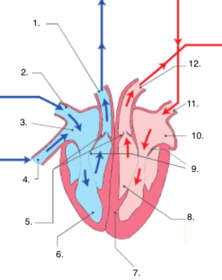
| Label the Heart. | 1. Pulmonary Artery 2. Superior Vena Cava 3. Right Atrium 4. Inferior Vena Cava 5. Semi-Lunar Valves 6. Right Ventricle 7. Septum 8. Left Ventricle 9. Bicuspid Valve (left) and Tricuspid Valve (right) 10. Left Atrium 11. Pulmonary Vein 12. Aorta |
| List the Steps of Pulmonary Circulation. | 1. Deoxygenated blood (DB) enters the right atrium through vena cavae. 2. DB pumps through the tricuspid valve and into the right ventricle. 3. Ventricle pumps DB through the semi-lunar valve to the pulmonary artery. 4. Pulmonary artery takes blood to the lungs to be oxygenated. |
| List the Steps of Systemic Circulation. | 1. Oxygenated blood (OB) returns from the lungs via the pulmonary vein. 2. OB enters the left atrium and is pumped through the bicuspid valve into the left ventricle. 3. OB is pumped through the semi-lunar valve. 4. OB exits the heart via the aorta and goes to the body. |
| The (3) Steps of the Cardiac Cycle. | 1. Diastole: the atria and ventricles fill with blood. 2. Atrial Systole: the atrium contracts, blood moves to ventricle. 3. Ventricular Systole: ventricles contract, blood moves into arteries. |
| Define Heart Rate. | How fast the heart is beating. How many beats it does per minute. |
| Define Stroke Volume. | The volume (amount) of blood forced from the ventricles. |
| Define Cardiac Output. How is it regulated? | The amount of blood leaving the ventricles every minute. SA Node in the wall of the right atrium regulates the cardiac cycle and therefore output. |
| Explain Arteries. | Carry blood away from the heart to the body. Contain oxygenated blood. Thick, muscular, walls, containing elastic fibres. Large arteries branch into smaller arterioles. |
| Explain Vasoconstriction. | The walls of the arteries retract in size. Reduces blood flow to an area. |
| Explain Vasodilation. | The walls of the arteries relax. Increases blood flow to an area. |
| Explain Capillaries. | Connect the arteries and the veins. Site of diffusion. Very thin walls. |
| Explain Veins. | Carry deoxygenated blood to the heart from the body. Thin, inelastic, walls with no muscle. Valves that push blood along. |
| What is Blood Flow? | The amount of blood flowing through an organ or blood vessel. Can be altered by hormones, exercise or adrenaline. |
| Why does blood clot? | To prevent pathogens from entering and stop blood vessels after damage has occurred. |
| (3) Steps of Blood Clotting. | 1. Vasoconstriction to reduce blood flow. 2. Platelets stick to roughened area and create a plug (clot), reduces blood flow and acts as a barrier to pathogens. 3. Vasoconstrictors released by platelets. |
| (5) Steps of Clot Formation. | 1. Blood releases clotting factors. 2. A meshwork forms from fibrin, the product of reactions. 3. Clot retraction pulls the edges of damaged skin together. 4. Serum (fluid) is squeezed out. 5. Clot dries (scab). |
| What is the Lymphatic System? What is its function? | A one way system of capillaries joined to larger lymph vessels. Lymph nodes located along lymph vessels. Valves and skeletal muscles move fluids up. Function: collect remaining fluids from tissues and fight infection. |
| Role of Lymph Vessels. | Return excess fluid back to the circulatory system. Contain infection fighting cells. |
| Where are Lymph Nodes found? What do the contain? | Found at intervals along the lymphatic system. Contain lymphoid tissue and cells that fight infection. (Lymphocytes, Macrophages, Plasma Cells) |
| How does the Lymphatic System defend against disease? | When unfamiliar particles are detected, they are trapped, engulfed and destroyed. |
| List the (4) ABO Blood Groups. | A, B, AB, O. A has antigen A. B has antigen B. AB has antigen A and B. O has neither antigen A nor B. |
| What are RH antigens? | Antigens that occur on the surface of red blood cells (along with A and B). RH positive: has RH antigens. RH negative: does not have RH antigens. |
| What criteria must be fulfilled in a Blood Transfusion? | The ABO blood type of the donor must match that of the recipient. The RH grouping of the donor must match the recipient Failure to match blood types will result in agglutination (clumping of blood). |
Want to create your own Flashcards for free with GoConqr? Learn more.