
Flashcards
Perio Assessment, PSR and recording
Cards in this deck (116)
- 1periodontal probingaids in the diagnosis of gingival and periodontal diseases, should be completed on every adult patient
- 2characteristics of a periodontal probeslender, blunt end, steel or plastic, working end can be tapered, straight, curved, round, flat or rectangular
- 3nabers probefor measuring into furcations
- 4perio probes examinedepth and topography of gingival sulcus
- 5Novatech probesright angled, facilitate examination of the sulcus/pocket of the direct distal or posterior teeth
- 6measuring purpose of probessulcus, pocket, recession, width attached gingiva, size or lesions
- 7other purposes of probesdetection, consistency of gingival tissue, evaluate bleed, post-treatment evaluation
- 8who probescreening probe, ball on the end =.5mm, from the end to the beginning of the black band is 3.5mm, the end of the black band is 5.5
- 9unc 12 probeused for complete/comprehensice periodontal examination. each thing black band 1mm, first wider black band 4-5mm, second wider band 9-10mm
- 10clinician probing techniqueprobe tip placed in contact w/tooth surface, length of the probe is positioned parallel to long axis of tooth, probe slightly angled under the contact to access the col
- 11bobbing strokeaka walking stroke, vertical stroke in 1mm steps
- 12probing pressurebetween 10-20 grams, you put pressure till you see the tissue slightly blanch and feel resistance
- 13JE in healthfeels firm and resilient
- 14JE in diseaseprob may pass through the JE and encounter physical resistance from the underlying CT
- 15parallelismposition probe as parallel as possible to long axis of the tooth, probe must be parallel in the M-D direction and in the F-L dimension
- 16probing interproximallyprobe parallel to the proximal surface until it touches the contact area, slant the probe slightly so the tip reaches under the contact, probe into the JE
- 17adaptionthe tip of the probe should be kept in contact with the tooth surface
- 18walking strokecover the circumference of the base of the sulcus or pocket, walk the probe through the sulcus while keeping the probe tip against the tooth surface, move stroke up and down in short bobbing strokes and forward 1mm. probe to JE w/each stroke
- 19comprehensive chartingeach tooth measured in 6 areas, (3F & 3L), measure from the base of the sulcus/pocket to the height of the gingival margin in mm measurements. record deepest reading in each area.
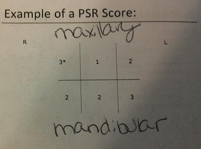
- 20periodontal screening and recordingaka psr, easy to use system for detecting PDZ & streamline record keeping. usually specially designed probe (screen or who probe), .5mm balled end an a colored band extending 3.5-5.5 from tip. this is a screening technique
- 21PSR codeqdetermined by how much of the colored band is visible on the who probe when placed in the gingival crevice
- 22PSR scoredated and recording in each patients chart
- 23when do you perform the PSRall new patients (NP) as part of the periodontal assessment, and for every recall (REC) patient. not needed for the return patient
- 24PSR code 0colored area of the probe completely visible-pocket depth <3.5, no calculus or defective margins, no BOP. treat with appropriate preventive care
- 25PSR criteria code 1colored area of probe completely visible- pocket depth <3.5mm no calculus or defective margins, BOP. treat with OHI, appropriate therapy
- 26PSR criteria code 3colored area of probe partially visible-pockets between 3.5- 5.5mm. if 2 or more sextants score code 3 complete periodontal charting
- 27PSR criteria code 4colored area of probe completely disappears, indicates probing depth >5.5mm. if one 4 in any sextant: complete periodontal charting
- 28PSR critera * (asterisk)added to the PSR when there is any of the following: furcation involvement, mobility, recession, mucogingival involvement
- 29PSR code 0, 1, or 2 with an *the clinician should make a specific notation and/or treatment for that condition as needed
- 30PSR code 3 or 4 with an *a comprehensive periodontal examination and charting are necessary to determine an appropriate care plan
- 31Example of a PSR score
- 32when do you perform the PSRafter medical history, I/E exam, dental charting, periodontal assessment, prior to treatment planning, every NP and REC pt. Use PSR to determine if complete perio charting is necessary.
- 33Microbial plaqueprimary etiologic factor in PDZ
- 34contributing factors of PDZhost response, plaque retentive calculus, rough surface on teeth/restorations, tooth contour or position, smoking/chew, poor POH, genetics (interlukin 1), iatrogenic
- 35signs and effects of inflammationgingival changes, bleeding & exudate (BOP), mucogingival involvement, attachment levels, furcation involvement, plaque & calculus, mobility, radiographic evidence
- 36symptoms of PDZbleeding gums when brushing or flossing, sensitivity to hold and cold, tenderness or pain while eating, bad breath or taste, loose teeth
- 37tissue changes of the inflammatory response are ignited bypresence of specific microorganisms modified by the host
- 38during tissue changes of the inflammatory response bacterial endotoxinsbreakdown the intercellular substance of the sulcular epithelium, causes ulceration
- 39the inflammatory response which causes tissue changes is initiated byirritants passing into the CT
- 40during an inflammatory response erythema isincreased blood flow to the area, increase in # of capillaries, increased vasodilatation, venous stasis-megenta, bluish-red tissues
- 41increased permeability of capillaries during inflammatory responseedema, increased collection of defense cells and fluid in gingival CT, tissue will be edematous (soft and spongy)
- 42tissue will be fibrotic during inflammatory response ifattempt to repair dominates the inflammatory response due to increase collagen production and hyperkeratosis
- 43localizedgingiva around a single tooth or group of teeth <30%
- 44generalizedgingiva around all or nearly all of the teeth (maxilla, mandible, or both) >30%
- 45marginalconfined to free or marginal gingiva (localized or generalized)
- 46papillaryinvolves the papilla but not the rest of the gingiva (localized or generalized)
- 47diffuseinvolves attached gingiva and free gingiva-vertical extension, most often localized, rarely generalized
- 48gingival examinationrecord tissues vertically (free to attached) describe, color, size, shape, consistence, surface texture, position, mucogingival junction, bleeding and exudate
- 49color of healthy gingival tissuepale pink-coral pink, darling in people w/darker complexions, influenced by vascular supply, thickness of epithelium, degree of keratinization, melanin pigmentation. pink tissue isn't always healthy
- 50gingival tissue color in diseaseerythema, marginal redness, papillary redness, magenta, bluish-red, bluish-pink
- 51shape (size/form/contour) of free gingiva in healthy gingival tissueflat, knife-edged margin or slightly rounded on the facial and lingual, fits snugly around the tooth
- 52shape (size/form/contour) of papillae in healthy gingival tissuefills the embrasure space, pyramidal-anterior, saddle shaped-posterior, flat-where teeth are not in contact/diastema
- 53shape (size/form/contour) of attached gingiva in healthy gingival tissuewidth varies among patients and among teeth, 1-9mm wide, wider in maxilla, broadest zone usually involves incisors, narrowest at cuspid and premolar regions
- 54shape (size/form/contour) of free gingiva in diseased gingival tissuelocalized or generalized, rounded, rolled festooned (McCall's festoon-really really rolled), cleft (still mans cleft, floss cleft)
- 55shape (size/form/contour) of papilla in diseased gingival tissuelocalized or generalized, bulbous, cratered (NUG characterized by "punched out" papillae), blunted, enlarged (col deepens as the papillae increase in size
- 56shape (size/form/contour) of papillae in diseased gingival tissue caused by med side effectscaused by gingival hyperplasia, drugs included phenytoin, cyclosporine, calcium channel blockers
- 57shape (size/form/contour) of attached gingiva in diseased gingival tissuedecreases in width as the periodontal pocket deepens, lack of adequate attached gingiva (mucogingival problem, disease, congenital or malpositioned teeth)
- 58consistency of healthy gingival tissuegingival margin and papillae firm when palpated with side of probe, attached tissue firmly bound to underlying bone
- 59consistency of gingival margin of diseased gingival tissuesoft and spiny, edematous, dents when pressed with side of probe, margin may hang slightly away from tooth or be easily displaced by a blast of air, gingival fibers destroyed, acute or chronic inflammation
- 60consistency of disease gingival tissue with chronic inflammationtissue may become hard and fibrotic due to increased fibrosis (excessive collagen formation)
- 61relationship of color and consistency of diseased gingival tissuetissue may be pink and well stippled, bleeding occurs in deep part of the pocket, tissue could also be erythematous, magenta, bluish-red and still be fibrotic
- 62surface texture of free gingiva margin in healthy gingival tissuesmooth
- 63surface texture of interdental gingiva in healthy gingival tissuemay or may not be stippled, stippling-orange peel effect as a result of the rate pegs extending into the underlying CT
- 64surface texture of attached gingival in healthy gingival tissuemay or may not be stippled
- 65surface texture of free gingival margin in diseased gingival tissueloss of stippling with smooth shiny surface (edema), hyperkeratosis (may result in leathery hard or nodular surface), attached tissue may or may not be stippled
- 66surface texture in chronic disease of gingival tissuetissue may be hard and fibrotic with pink coloring and deep stippling
- 67actual position of gingival epitheliumactual position is not directly visible, it is where the JE is located, determined by probing
- 68apparent position of gingival epitheliumlevel of the gingival margin or gingival crest of the free gingiva, seen by direct observation
- 69position of apparent position in healthin a fully erupted permanent tooth the apparent position of gingiva is 1-2mm above the CEJ
- 70position of actual position in healthJE is located at or just below the CEJ, in health 1mm long
- 71position in disease with hyperplasia or hypertrophygingival margin is higher on the enamel, partly or nearly covering the anatomic crown
- 72position in disease with gingival recessionexposure of the root surface as a result of apical migration of the junctional epithelium, actual verses visible, periodontal pocked with or without visible recession
- 73apparent recession AKA visible recessionexposed root surface that is visible clinically, measured from gingival margin to CEJ, may be localized, wide, narrow, deep, shallow, may extend to or through JE, measure with UNC probe
- 74actual recessionaka clinical attachment loss (CAL)-pocket depth (gingival margin to JE) + visible recession (gingival margin to CEJ) or total attachment loss, measured from CEJ to base of the sulcus/pocket (JE)
- 75bleeding in healthno bleeding upon probing, no spontaneous bleeding
- 76bleeding in diseaseBOP, may have spontaneous bleeding, sulcular spithemlium become pocket epithelium, extensions of rete pegs into the pocket/sulcular epithelium, bleeding comes from underlying CT
- 77exudatecomposed of fluid, cells and cellular debris that have escaped from blood vessels and are deposited in tissues. result of inflammation and exulting increase in vascular permeability, no exudate in health
- 78exudate suppurationformation or secretion of pus
- 79exudate on gingival exammy appear at entrance to pocket or squeezed out by light finger pressure, amount related to severity of inflammation, not depth of pocket. increase in volume of GCF is considered an inflammatory exudate.
- 80gingiva of the primary dentition in healthpink or slightly red, thick rounded or rolled, firm but not tightly adapted to tooth, more shiny than stippled
- 81healthy gingiva in mixed dentitionconstant state of change related to exfoliation and eruption
- 82diseased gingiva of young childrengingivitis occurs frequently in children, periodontitis relatively rare, mucogingival problem can occur in children can be significant issue for children who need orthodontic treatment
- 83eight warning sings of PDZgums that bleed when brushing your teeth, are red swollen or tender, that have pulled away from teeth, pus between teeth &gums when pressed, permanent teeth that are loose or separating, any change in the way teeth fit together when you bite, any changes in the fit of your partial denture, bad breath
- 84gingivitisinflammation of gingival tissue
- 85initially lesionw/in 2-4days, migration & infiltration of WBC to JE and gingival sulcus, increased flow of sulcular fluid, early breakdown of collagen, fluid fills the spaces in CT (edema) may not see any changes clinically in earliest phase
- 86early lesiondays 7-14 older & thicker plaque, inflammatory response continues, breakdown of collagen fiber support in gingival margin, gingival enlargement, early gingivitis is reversible
- 87the established lesion in gingivitis15-21 days, fluid increases (edema) predominate inflammatory cells are plasma cells, formation of pocket epithelium, continued loss of collagen and CT support, these lesions may remain stable for long period of time or progress to periodontal lesion
- 88formation of pocket epithelium in established lesion of gingivititsJE and sulcular epithelium proliferate, areas of ulceration, gingival pocket results
- 89periodontitisinflammation in the periodontium, gingival tissues, PDL, cementum and bone. bacteria from supra gingival plaque proliferate apically and become source for sub gingival plaque
- 90extension of inflammationexdontoxin (LPS) production by microorangisms, inflammation continues through CT, inflammation enters the bone through small vessel channels in alveolar crest, spreads though the bone marrow & out into PDL
- 91progressive destruction of CT in PDZct fivers below the JE are destroyed, apical migration of JE exposed cementum is altered by inflammatory products of microorganisms, w/out treatment pocket deepens
- 92advanced lesion of PDZdevelopment of periodontal pocket, calculus formation progressive, period of inactivity and activity, can be site specific can have varying degrees of destruction in on mouth
- 93gingival pocketgingival enlargement without the apical migration of the JE, inner wall is enamel, fully erupted tooth the base of the sulcus is at or near the CEJ, all gingival pockets are supra bony, the base of the pocket is coronal to the crest of the alveolar bone
- 94periodontal pocketformed as the result of apical migration of the JE, involves attachment apparatus, inner wall is C or partly C & partly E, base of pocket is on C, may be supra boney or intrabony (infra bony), intrabony: base of pocket is apical to crest of alveolar bone
- 95mucogingival involvementlack of adequate attached gingival tissue, attach tissues supports the marginal gingiva, w/stands the frictional stresses of mastication & toothbrushing, provides a solid base for the moveable alveolar mucosa, may be the result of disease, genetics. BARRIER TO THE PASSAGE OF INFLAMMATION
- 96tobacco usesmoking is a major risk factor for PDZ, increased # of microorganisms, impairs host immune response, inhibits function of fibroblasts, impaired bone cell function;mineral content of bone reduced
- 97variables of smoking on PDZtype of tobacco products, length of year used, number of daily exposures to tobacco product
- 98clinical effect of smokingfibrotic gingiva, minimal redness, bone loss, pocketing in maxillary anterior lingual area, gingival recession above anterior teeth, increased temperature in pocket
- 99tobacco use in response to treatmentresistance to conventional therapy, dental implant have greater risk for infection, delayed healing after surgical and non-surgical procedures
- 100treatment plan-preventive phaseeducation- POH, FL
- 101Treatment plan-preparatory phasedental hygiene treatment, initial treatment of extensive carious lesions, endodontic therapy, oral surgery is necessary
- 102treatment plan-treatment phaseperiodontal surgery if necessary, restorative treatment, prosthetic treatment, orthodontic treatment, tissue maintenance
- 103treatment plan-maintenance phasePOH instruction, dental hygiene recall, re-evaluation
- 104periodontal case typeAAP classification system
- 105Periodontal case type 0indicates periodontal and gingival health
- 106Periodontal case type 1gingival disease, inflammation of the gingiva, characterized by change in color, contour, position, consistency and presence of bleeding &/or exudate, no bone loss, no apical migration of JE
- 107Periodontal case type IIearly periodontitis, progression of gingival inflammation into the deeper periodontal structures & alveolar bone crest w/slight bone loss, usually slight loss of CT attachment & alveolar bone
- 108Periodontal case type IIImoderate periodontitis, more advanced stage of preceding condition, increased destruction of the periodontal structures, noticeable loss of bone support, possible tooth mobility, may be furcation involvement
- 109Periodontal case type IVadvanced periodontitis, further progression of periodontitis w/major loss of alveolar bone support, usually accompanied by increased tooth mobility, furcation involvement likely
- 110Periodontal case type Vrefectory periodontitis, multiple disease sites that continue to demonstrate attachment loss after appropriate therapy, these sites presumably continue to be infected by periodontal pathogens despite excellent POH & frequent maintenance, disease recurs at a single site or multiple sites
- 111mineralized case typeamount of calculus & stain present, not a universal classification system (unique to VTC), method for tracking the types of patients that you treat in terms of amount of plaque retentive calculus and stain
- 112mineralized case type slight (SL)no deposits present or slight deposits in localized areas, L of man anteriors, B of maxillary molars
- 113mineralized case type light (LT)light deposits, subgingivally and or supragingivally in several areas through out the dentition
- 114mineralized case type moderate (MD)moderate deposits, located both sub and supra gingivally which are generalized throughout the dentition
- 115mineralized case type heavy (HV)heavy tenacious deposits throughout the dentition
- 116generally house many appts to scheduleI, II, III, IV, SL:1appt. I, II, III, IV, LT:1-2appt. I,II,III,IV,V MD: 5-6 appt. IV,V, HV: 7-8 appt
Information
- Description
- Flashcards on Perio Assessment, PSR and recording, created by angela.dennis22 on 10/12/2013.
- Created by
- angela.dennis22
- Views
- 70
- Rating