9844613
Description
Flashcards by Christianna Ziccardi, updated more than 1 year ago
|
|
Created by Christianna Ziccardi
over 7 years ago
|
|
| Question | Answer |
| Gross Anatomy | Study of structures visible to the unaided eye |
| Topographical anatomy | Anatomy of one part of the body in relation to another |
| Applied Anatomy | applied to disease |
| Microscopic anatomy | study of tissue with a microscope |
| Ultra Structural Anatomy | study of tissues with electron microscope |
| What type of plane? | Median |
| What is a sagittal plane? | Any plane parallel to the median plane |
| What type of plane? | Dorsal plane (any level that is perpendicular to the median plane |
| What type of plane? | Transverse plane |
| Cranial vs Caudal | Closer to head=cranial Closer to tail=caudal Can also be used to directly compare parts (ie the forelimb cranial to the hind limb) |
| Where is rostral vs caudal used? | The head region |
| Rostral vs caudal | Rostral= toward the nose (nostral) Caudal= toward the tail |
| Dorsal vs ventral | Dorsal=closer to top Ventral=closer to bottom (think V) |
| Axial vs Abaxial | Axial= Facing or towards the central line or axis Abaxial= facing away or farther from the central line or axis |
| Medial vs Lateral | Medial- closer to median plane Lateral- farther away from median plane |
| Proximal vs distal | Proximal- closer to the trunk (body) Distal- farther from the trunk |
| Which three terms are used distal to the carpal bones? | Dorsal and Palmar/Plantar |
| Dorsal and Palmar and Plantar | Dorsal- top Palmar- bottom (palm) forelimb Plantar- bottom (palm) hindlimb |
| Superficial vs deep | Superficial- near the surface of the body deep- near the center (inside the trunk) of the body |
| 5 general functions of bones | 1) Provide form and support 2) Protect soft tissues (i.e lungs) 3) act as levers to facilitate locomotion 4) have a role in blood cell formation 5) maintain mineral homeostasis |
| Two classes of skeleton and what they include | Axial skeleton- forms and supports the body trunk Appendicular skeleton- forms the limbs |
| 7 classes of bones | 1) Long bones 2) Irregular bones 3) Short bones 4) flat bones 5) sesamoid (round) bones 6) pneumatic bones 7) Heterotropic or splanchnic bones |
| What class of bone? | Long |
| What class of bone? | Irregular |
| What class of bone? | Short |
| What class of bone? | Flat |
| Form and function of sesamoid (round) bones | small, nodular, embedded within tendons and adjacent to joints where the tendons are compressed |
| Pneumatic bones | air filled bones |
| Heterotrophic or splanchnic bones (3 types) | -os penis (dog) -os cordis (cattle) os rostrum (pigs) |
|
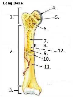
#2?
What is it composed of?
Image:
O7owhx (binary/octet-stream)
|
Diaphysis, composed of compact bone |
| What structure is the diaphysis covered by? | periosteum |
|
#1?
Composed of what?
Image:
O7owhx (binary/octet-stream)
|
Epiphysis, made up of spongy bone covered by compact bone |
|
#3/#4
Image:
O7owhx (binary/octet-stream)
|
Articular cartilage (AKA hyaline cartilage) |
|
#5, function?
Image:
O7owhx (binary/octet-stream)
|
Epiphyseal line, thin strip of bone marking the fusion of epiphysis to diaphysis |
| What is the difference between epiphyscal growth plate and the epiphyseal line | Area actively growing in bones is called epiphyscal growth plate. It is replaced by the epiphyseal line when the bone stops growing |
|
#6
Image:
O7owhx (binary/octet-stream)
|
Spongy bone |
|
#7?
Image:
O7owhx (binary/octet-stream)
|
Medullary cavity |
| Where is metaphysis on a long bone and what is it made of? | Metaphysis is part of the diaphysis that borders the epiphyseal plate/line and consists of spongy bone |
| Blue? | Articular cartilage (hyaline cartilage) |
| Light purple (top) | Epiphyseal plate or line |
| Orangish pink? | Medullary cavity/spongy bone |
| Green (outer covering) | Periosteum |
| Yellow? (middle) | Diaphysis (compact bone) |
| Dark purple (inner) | Endosteum |
| Almost mature animals have an open or closed growth plate? | Closed |
| number and locations of ossification centers in long bones | (3), one diaphyseal center (middle of the bone) and two epiphyseal centers (top and bottom) |
| Number of ossification centers in a short bone | one |
| Which two bone classifications have variable numbers of ossification centers? | Flat and irregular |
| Where does the nutrient artery attach? | in the middle of the diaphysis |
| What is the nutrient foramen? | A gap in the bone that allows arteries through |
| Three properties of muscles that allow them to produce movement | 1) Contractility 2) Excitability 3) Extensibility |
| Why do muscles differ in shape? | Based on the amount of work they do (shortening depends on fiber length, power depends on cross sectional area) |
| Two ways muscles attach to bones and their characteristics | Tendons- discrete band of tissue Aponeurosis- flat, wide, thin sheet of fiberous connective tissue |
| 2 Different muscle attachments and their characteristics | Origin- more fixed (least movable), usually proximal attachment Insertion- more movable, usually distal attachment |
| Muscle belly | fleshy contractile part of muscle |
| Retinaculum |
Band of dense regular tissue than binds down muscle tendons as they pass over bone surfaces
Image:
1 (binary/octet-stream)
|
| When is the retinaculum generally broad? | When it passes over many tendons (ex dorsal aspect of carpus) |
| Function of synovial bursa? | Permits frictionless movement of a tendon over bone |
| Differences between synovial bursa and tendon sheath? |
Same function, tendon sheath (4) normally lays under a retinaculum (3) and wraps completely around the tendon (1)
Image:
11 (binary/octet-stream)
|
| How does the synovial sheath relate to the tendon sheath | The synovial sheath is one of two layers that surrounds a tendon sheath |
Want to create your own Flashcards for free with GoConqr? Learn more.