15925215
Description
Mind Map by Moza Rashid, updated more than 1 year ago
|
|
Created by Moza Rashid
about 7 years ago
|
|
Tumor
- Signs and Symptoms
- Headaches
- Nausea
- Difficulty with balance
- Speech difficulties
- Types
- Apraxia of speech
- Cluttering
- Dysarthria
- Dysprosody
- Muteness
- Speech Sound Disorders
- Stuttering
- Voice Disorders
- Apraxia of speech
- Types
- Confusion
- Seizures
- Hearing problems
- Behavior changes
- Limb Weakness
- Vomiting
- Causes
- Central nervous system
- Closed head injury
- Increased ICP
- Migraine
- Seizures
- Vestibular
- Closed head injury
- Gastrointestinal
- Functional disorders
- Obstruction
- Organic disorders
- Functional disorders
- Infectious
- Medications and Toxins
- Metabolic
- Miscellaneous
- Acute glaucoma
- Psychiatric disorders
- Acute MI
- Nephrolithiasis
- Pain
- Acute glaucoma
- Central nervous system
- Causes
- Loss of consciousness
- Headaches
- Eye
- Normal
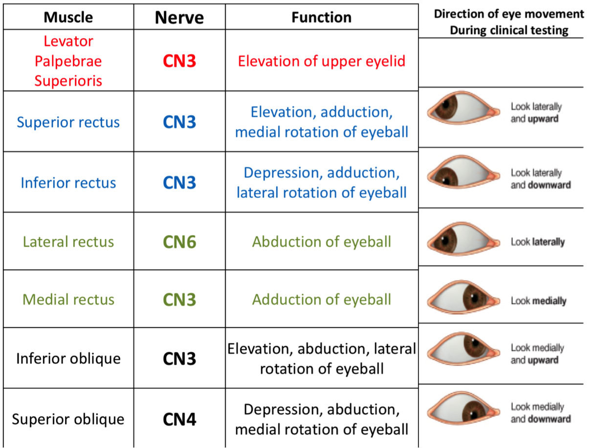
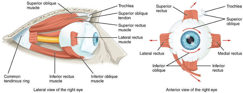
- Muscles
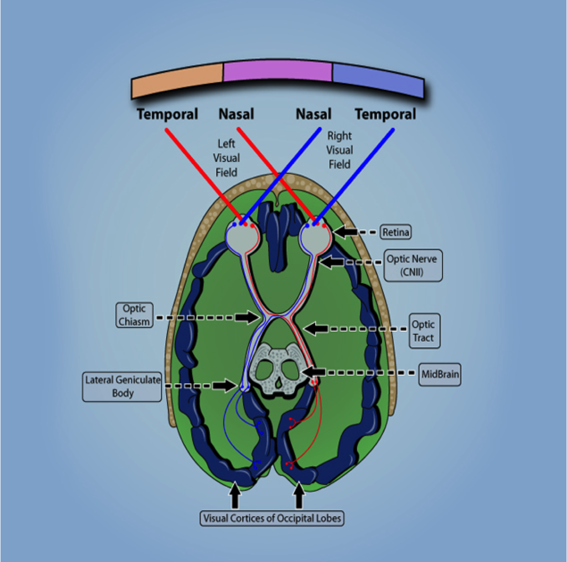
- Visual Pathway
- 1- Retina
- 2- Optic nerve
- 3- Optic chiasma
- 4- Optic tract
- 5- Lateral Geniculate Body (L.G.B.) - Diencephalon
- 6- Optic radiation
- 7- Primary visual cortex (visual cortex, area 17)
- 7- Primary visual cortex (visual cortex, area 17)
- 6- Optic radiation
- 5- Lateral Geniculate Body (L.G.B.) - Diencephalon
- 4- Optic tract
- 3- Optic chiasma
- 2- Optic nerve
- 1- Retina
- Muscles
- Abnormal
- Double Vision
- Causes
- Brain problems
- Migraine
- Increased ICP
- Migraine
- Nerve problem
- Diabetes
- Myasthenia gravis
- MS
- Guillain Barre syndrome
- Diabetes
- Cornea Problems
- Astigmatism
- Dry eyes syndrome
- Infections
- Astigmatism
- Lens Problems
- Cataract
- Cataract
- Eye Muscle Problems
- Strabismus
- Graves disease
- Strabismus
- Brain problems
- Causes
- Double Vision
- Normal
- Risk factors
- Age
- Home & Work Exposures
- Gender
- Family History
- Exposure to infections, viruses and allergens
- Electromagnetic fields
- Race and ethnicity
- Ionizing Radiation
- Head Injury & Seizures
- Age
- Metastatic (50%) / Primary (50%)
- Primary tumors
- In adults are usually supra-tentorial
- In children are usually infra-tentorial
- Rarely metastasize outside the CNS
- Classification
- Tumors of neuroepithelial tissue
- Gliomas
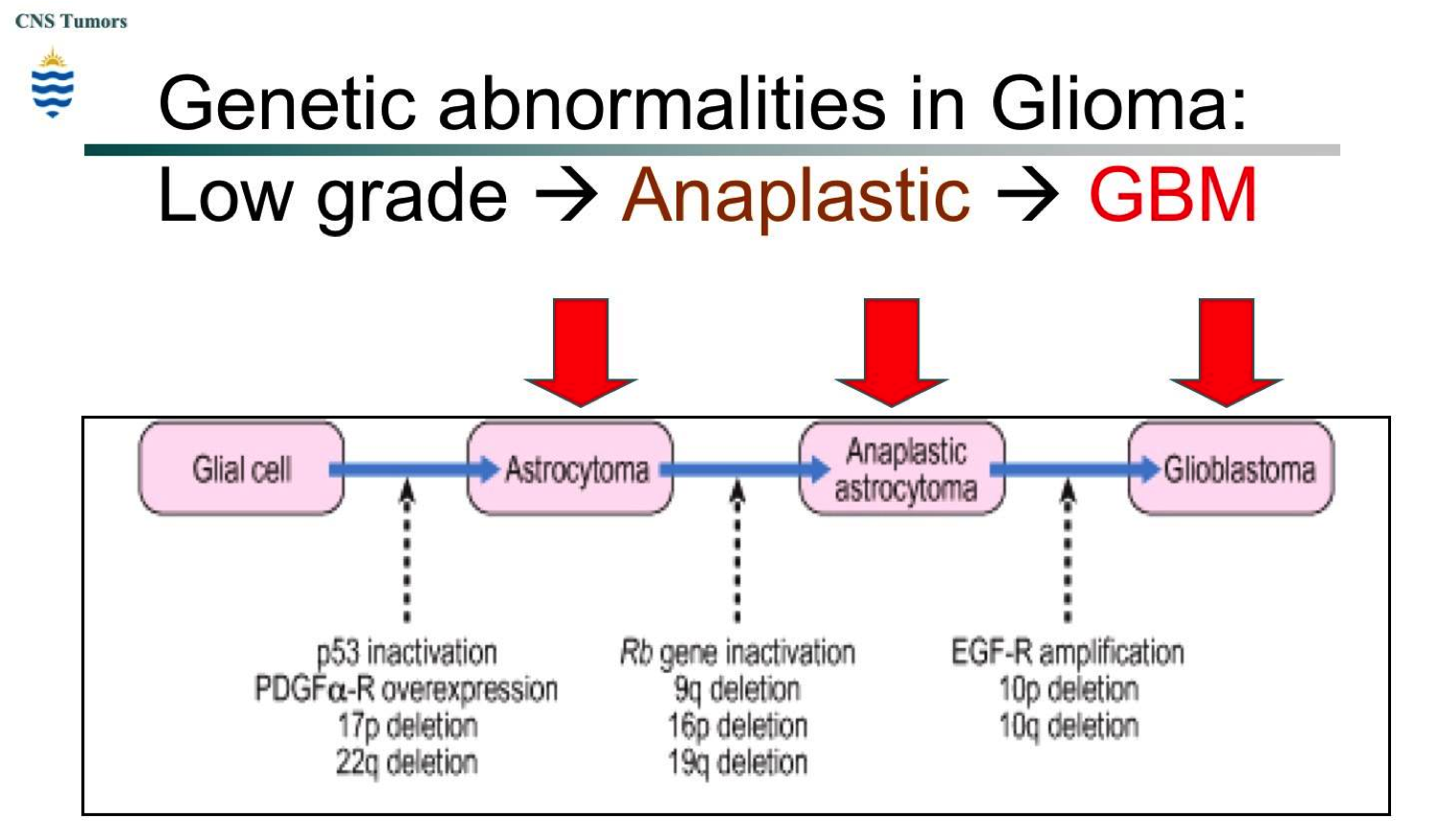
- Astrocytoma
- Infiltrating astrocytic tumors
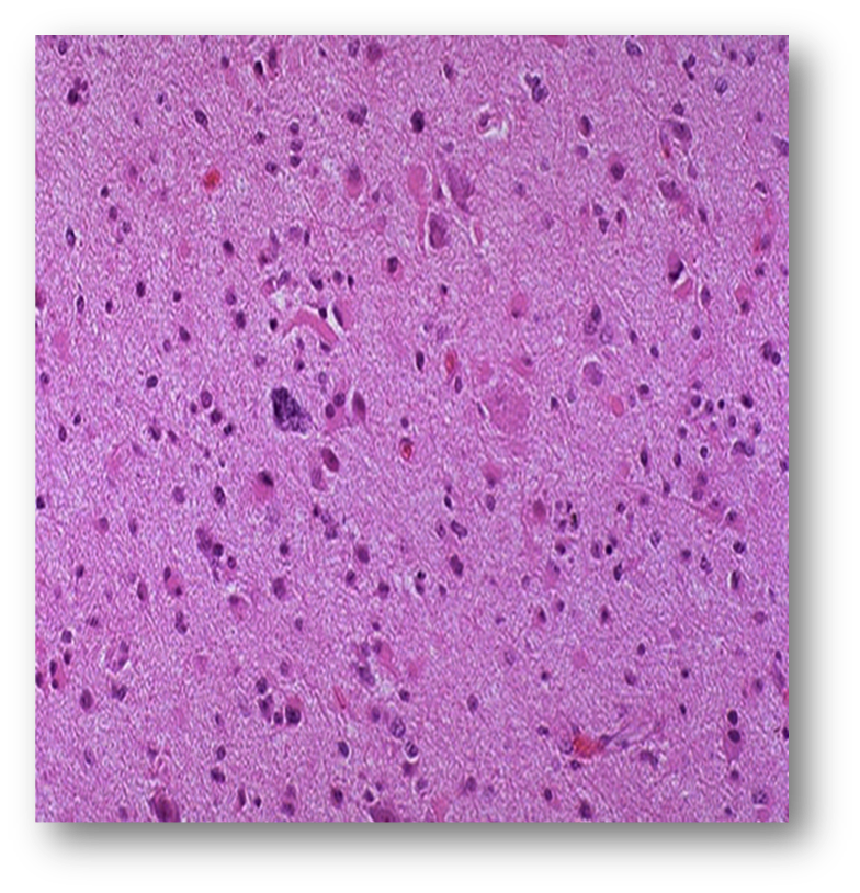
- Diffuse astrocytoma, grade II
- Moderate cellularity / No anaplasia
- Mild to moderate increase in the
number of glial cell nuclei /
Variable nuclear pleomorphism /
Branching fibrillary processes
- Moderate cellularity / No anaplasia
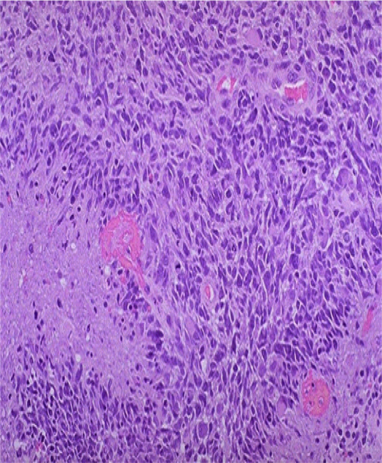
- Anaplastic astrocytoma, grade III
- Cellularity, anaplasia, mitoses
- More cellular areas / Greater
nuclear pleomorphism /
Mitotically active cells are often
- Cellularity, anaplasia, mitoses
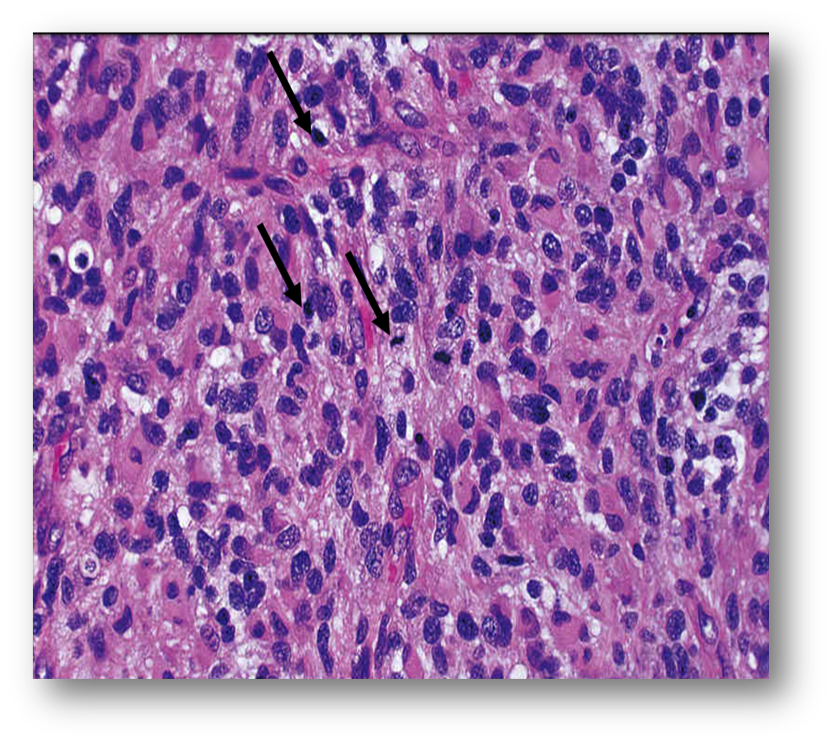
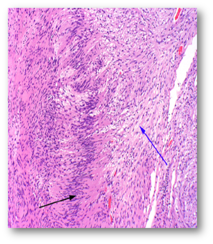
- Glioblastoma, grade IV
- Most common primary malignant
tumor in adults usually arises in
the cerebral hemisphere
- Malignant, high-grade tumor
- Characteristically crosses
the corpus callosum
(butterfly lesion)
- Tumor cells are GFAP positive
- Poor prognosis, death in
less than one year
- Marked anaplasia and pleomorphism / Pronounced
vascular changes with endothelial hyperplasia
(glomeruloid appearance / Areas of necrosis
surrounded by tumor cells (pseudopalisading)
- Most common primary malignant
tumor in adults usually arises in
the cerebral hemisphere
- Diffuse astrocytoma, grade II
- Localized astrocytic tumors
- Pilocytic astrocytoma, grade I
- Benign cytological features
- Benign cytological features
- Pilocytic astrocytoma, grade I
- Associated with a
variety of acquired
mutations
- Mean survival 6-8 years
- Aggressive, affect older patients
- Infiltrating astrocytic tumors
- Oligodendroglioma
- Closely packed cells with large round nuclei
surrounded by a clear halo of cytoplasm
“Fried egg” appearance of cells"
- Long survival (5-10 yrs)
- Closely packed cells with large round nuclei
surrounded by a clear halo of cytoplasm
“Fried egg” appearance of cells"
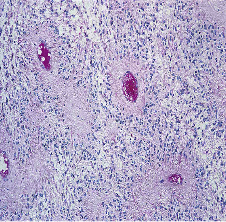
- Ependymoma
- Tubules, rosettes with cells encircling vessels or
pointing towards a central lumen / Perivascular
pseudo-rosettes are a characteristic finding on biopsy
- Recur after surgery, acquire more aggressiveness
- Tubules, rosettes with cells encircling vessels or
pointing towards a central lumen / Perivascular
pseudo-rosettes are a characteristic finding on biopsy
- Astrocytoma
- Choroid plexus tumors
- Neuronal tumors
- Tumors of the pineal region
- Gliomas
- Embryonal tumors
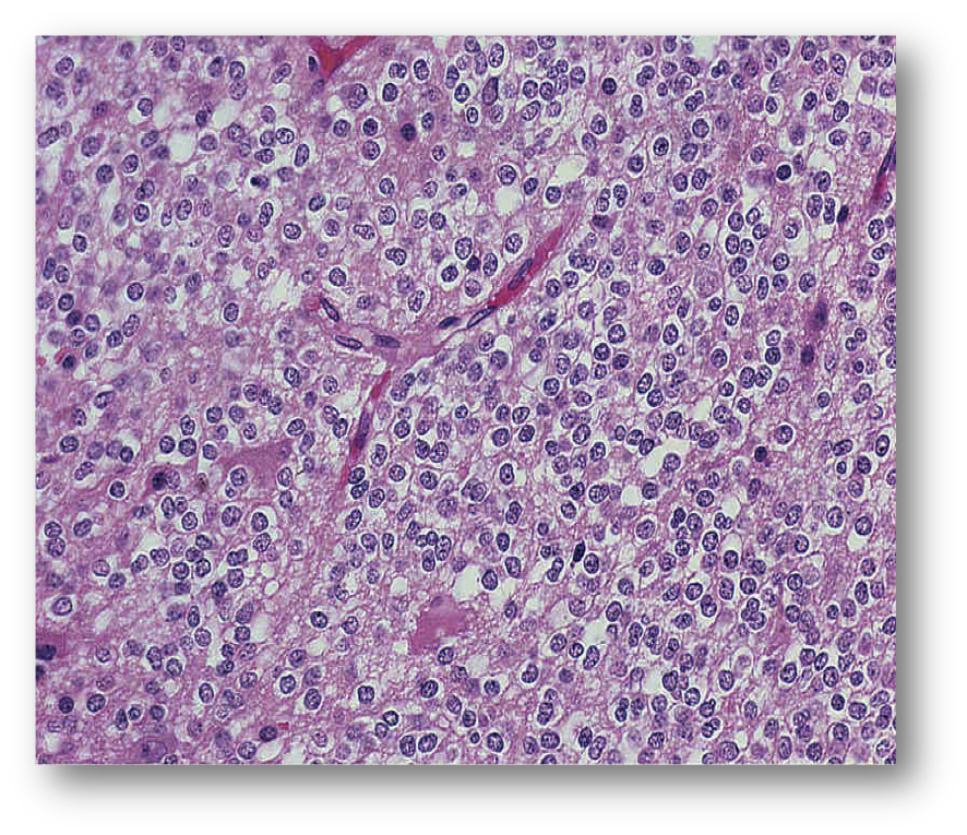
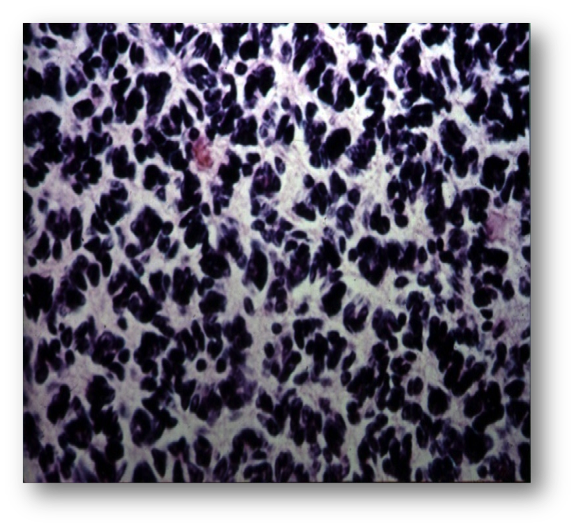
- Medulloblastoma, grade IV
- Histology reveals small round blue cells,
homer- wright rosettes present
- Histology reveals small round blue cells,
homer- wright rosettes present
- Craniopharyngioma
- Medulloblastoma, grade IV
- Tumors of cranial nerves
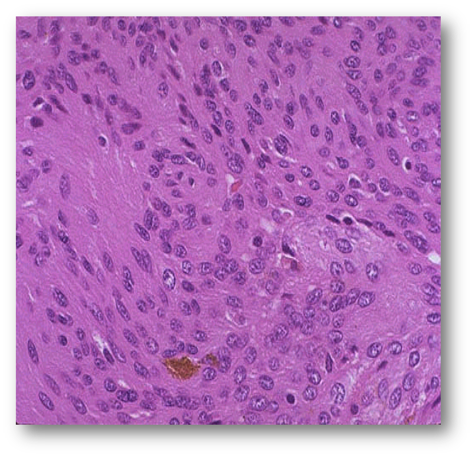
- Schwannoma
- Two patterns (Antoni A interlacing bundles of
elongated cells with palisading of nuclei / Antoni
B looser, less cellular pattern than Antoni A)
- Two patterns (Antoni A interlacing bundles of
elongated cells with palisading of nuclei / Antoni
B looser, less cellular pattern than Antoni A)
- Neurofibroma
- MPNST
- Schwannoma
- Tumors of the meninges
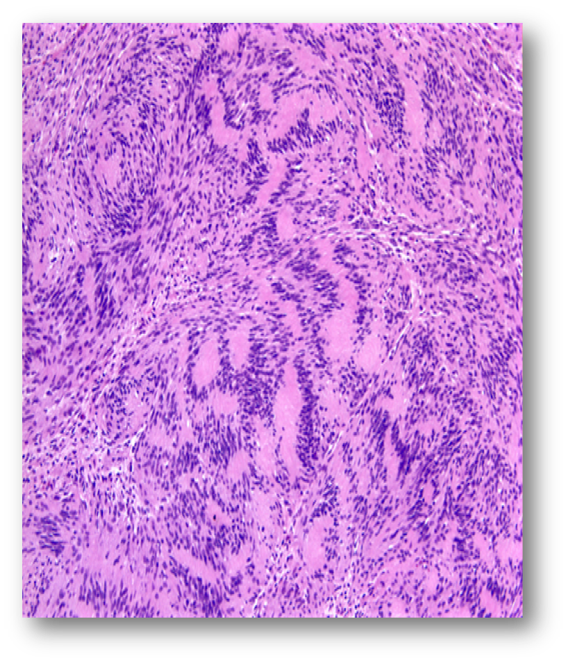
- Meningioma
- Good prognosis
- Meningioma
- Tumors of the sellar region
- Pituitary adenma
- Pituitary adenma
- Metastatic
- Mostly carcinomas
- Common primary sites (Breast / Lung / Skin / Kidney / GIT)
- Single or multiple lesions
- May be the first presentation of the primary
- Mostly carcinomas
- Tumors of neuroepithelial tissue
- In adults are usually supra-tentorial
- Primary tumors
- Investigations
- Neurological examinations
- Blood tests
- X-ray (Skull and Chest)
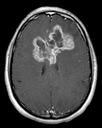
- Brain scans
- CT) / Contrast enhancement
- MRI / Contrast enhancement
- Positron emission tomography (PET)
- CT) / Contrast enhancement
- Single photon emission computed tomography (SPECT)
- Ultrasound imaging (Neurosonography)
- Angiography
- CSF analysis
- EEG
- Evoked potentials
- Biopsy
- Eye Examination
- Cranial nerve
- Optic II (vision)
- Visual acuity - Eye chart /
Visual field – confrontation /
Visual reflexes (2-3) &
accommodation reflex /
Colour vision / Fundoscopy
- Visual acuity - Eye chart /
Visual field – confrontation /
Visual reflexes (2-3) &
accommodation reflex /
Colour vision / Fundoscopy
- III, IV and VI (Ocular
gaze and posture)
- Following finger /
Smooth pursuit
- Following finger /
Smooth pursuit
- Optic II (vision)
- Cranial nerve
- Neurological examinations
- Management
- Treat/Manage Symptoms
- Surgery
- Risks of Surgery
- Allergic reaction to anesthesia / Bleeding in
the brain / Blood clot / Brain swelling /
Coma / Impaired speech, vision,
coordination, or balance / Infection /
Memory problems / Seizures / Stroke
- Allergic reaction to anesthesia / Bleeding in
the brain / Blood clot / Brain swelling /
Coma / Impaired speech, vision,
coordination, or balance / Infection /
Memory problems / Seizures / Stroke
- Risks of Surgery
- Chemotherapy
- Radiation
- Targeted therapy
- Clinical trials
- Palliative treatment
- Treat/Manage Symptoms
Media attachments
Want to create your own Mind Maps for free with GoConqr? Learn more.