
Mind map
Cardio Vascular Overview
Map content
Cardio Vascular Overview
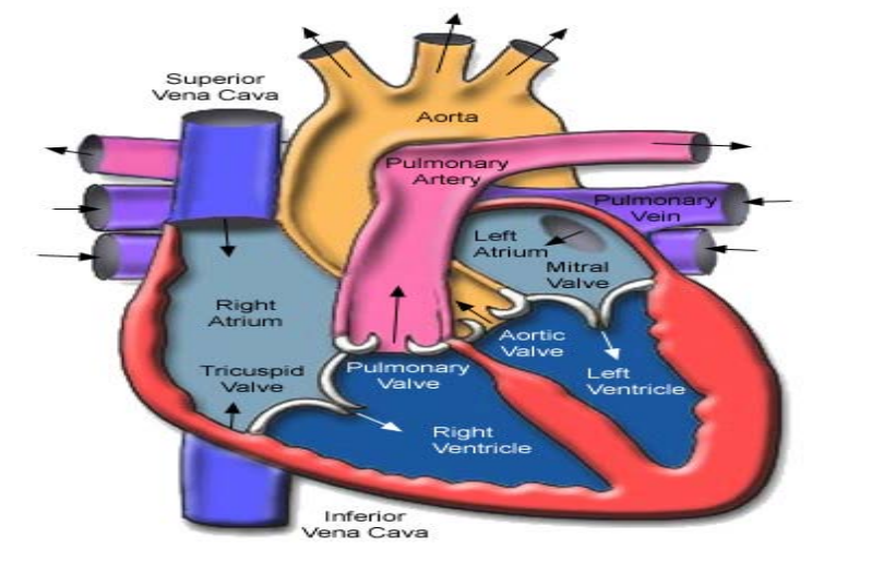
Heart anatomy
- blood flow
- RIGHT ventricle
- Pulmonary artery (deoxygenated)
- lungs
- pulmonary vein
- left aorta. etc...
- pulmonary vein
- lungs
- Pulmonary artery (deoxygenated)
- RIGHT ventricle
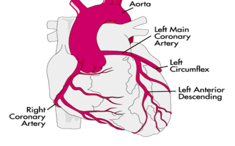
- coronary arteries
- left coronary (main)
- circumflex
- extends from left coronary and wraps around the heart
- left anterior descending
- circumflex
- right coronary
- left coronary (main)
- arteries
- tunica externa
- loose connective tissue
- outermost layer
- tunica media
- middle layer
- smooth muscle
- tunica intima
- inner most
- simple squamous epithelium and elastic fibers
- vs. veins...
- arteries have more muscle
- arteries are more round
- veins collapse if not filled
- valves in veins, not arteries
- tunica externa
- blood flow
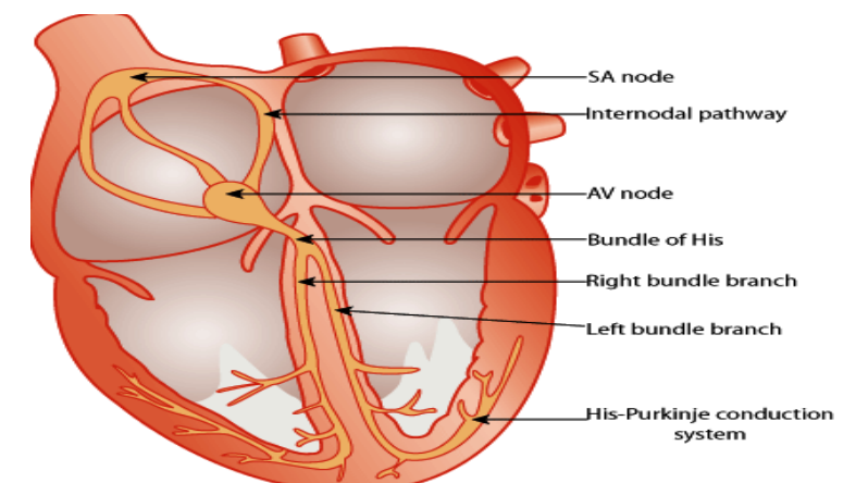
electrical activity of the heart
- sinoatrial node
- usually = HR
- AV node
- bundle of His
- right or left bundle branch
- purkinje fibers
- large conducting cells, most of ventricles
- purkinje fibers
- right or left bundle branch
- bundle of His
- sinoatrial node
RAAS (Renin Angiotensin Aldosterone System)
- angiotensinogen (kidneys)
- angiotensin I (Renin)
- angiotensin II (ACE)
- AT 1 (Type 1)
- ↑ CO
- ↑ aldosterone
- → myocardial fibrosis
- left ventricular hypertrophy
- sodium retention
- potassium loss
- hypertension
- sympathetic activation
- ↑ GFR
- ↑ ADH + thirst
- AT 2 (Type 2)
- vasodilation
- anti-proliferation
- cell differentiation
- tissue repair
- AT 1 (Type 1)
- angiotensin II (ACE)
- angiotensin I (Renin)
- angiotensinogen (kidneys)
hemodynamic definitions
- preload
- pressure stretching the ventrical after diastole (also equals end diastolic volume)
- afterload
- end systolic volume
- tension or pressure used to eject blood out ventricles
- stroke volume
- end diastolic volume - end systolic volume (preload - afterload)
- cardiac output
- CO = HR * SV
- amount of blood pumped / time
- cardiac index
- CO adjusted for BSA
- CI = CO/BSA
- ejection fraction
- fraction of blood ejected by left ventricle during contraction or ejection
- EF = SV/EDV * 100 %
- amount ejected after systole
- mean arterial pressure
- CO * SVR + CVP
- or [(2x diastolic pressure) + systolic pressure]/3
- pulmonary capiullary wedge pressure (PCWP)
- provides estimate of diastolic filling and left ventricular end diastolic volume (when mitral valve is open)
- preload
forrester's classification
- subset 1 (no hemodynamic compromise)
- cardiac index (> 2.2 L/min/m^2)
- PCWP (0-18 mm Hg)
- subset II (pulmonary congestion)
- cardiac index > 2.2
- PCWP > 18
- subset IV (pulmonary congestion and hyperfusion
- high PCWP, low CI
- subset IV (pulmonary congestion and hyperfusion
- subset III (hyperfusion)
- PCWP < 18
- CI < 2.2
- subset 1 (no hemodynamic compromise)
hemodynamic clinical assessment
- preload
- hepatomegaly (megaliver)
- daily weight
- pulmonary crackles
- peripheral edema
- S3 heart sound
- mucous membrane and skin turgor
- afterload
- vascular diastolic
- pulse pressure
- pulses
- preload
Images and files in the map
Information
- Description
- Pharmacotherapy 2 Mind Map on Cardio Vascular Overview, created by owen proctor on 21/08/2017.
- Created by
- owen proctor
- Views
- 11
- Rating