33973957
Descrição
FlashCards por Greg MacPherson, atualizado 5 meses atrás
|
|
Criado por Greg MacPherson
aproximadamente 3 anos atrás
|
|
| Questão | Responda |
| Unit Two - Module 12 The Brain: Brain Regions and Structures | The Brain: Brain Regions and Structures |
| hindbrain | Located at the lower back part of the brain, it includes most of the brainstem and the cerebellum. |
| midbrain | The midbrain is the topmost part of the brainstem, the connection central between the brain and the spinal cord. It controls some motor movement and transmits auditory and visual information. |
| forebrain | By far the largest region of your brain which contains the entire cerebrum and the limbic system. |
| brainstem | The brainstem is one of the most important parts of the entire central nervous system, because it connects the brain to the spinal cord and coordinates many vital functions, such as breathing and heartbeat. It includes the midbrain, the pons, and the medulla. |
| medulla (medulla oblongata) | Located in the hindbrain at the base of the brainstem. It controls heartbeat, breathing, and swallowing. |
| pons | The large swelling between the medulla and the midbrain. It is involved in a number of functions including the coordination of movement of the left and right side of the body. It is also involved in sleep. |
| reticular formation | A network of neurons that runs through the medulla and pons up into the thalamus. It is involved in our state of alertness, the sleep-wake cycle, and filtering sensory information. |
| cerebellum | Located at the rear of the brainstem. This "little brain" has functions that include processing sensory input, coordinating movement output and balance, and enabling nonverbal learning and memory. |
| limbic system | A group of structures in the forebrain (thalamus, hypothalamus, amygdala, hippocampus) that sits under the cerebral hemispheres. It is the part of the brain involved in our behavioural and emotional responses, especially when it comes to behaviours we need for survival: feeding, reproduction and caring for our young, and fight or flight responses. |
| thalamus | A pair of egg-shaped structures that act as the brain's sensory control centre for the sorting of sensory information before it connects on to the cortex. It receives information from all the senses - except smell - and routes that information to the higher brain regions that deal with seeing, hearing, tasting, and touching. The thalamus also receives some of the higher brain's replies and relays them to the medulla and cerebellum. |
| hypothalamus | A very small but very important structure of the limbic system, located below and in front of the thalamus. It plays an important role in body regulation. This includes the regulation of body temperature, thirst, hunger, biological rhythms of sleeping and waking, sexual arousal, and emotions. |
| hippocampus | Two armlike structures that surround the thalamus. The name comes from the Greek word for "seahorse" given its shape. It is instrumental in processing long-term memories that are then sent to other locations in the cerebral cortex for permanent storage. Damage to this structure leads to an inability to form new memories of facts and events. |
| amygdala | Two small , almond shaped structures located in front of the hippocampus. It is vital to our experiences of emotion. In other words, it organizes response patterns to emotional stimuli. This is particularly true of negative emotional stimuli such as surprise, fear, anger, sadness, and disgust. Research in animals and humans has shown that damaged or missing amygdala results in no fear response. |
| cerebrum | The largest part of the brain, forming most of the forebrain. It consists of two cerebral hemispheres bridged by the corpus callosum. Each hemisphere is divided into four main lobes. |
| corpus callosum | A large tract of nerve fibres running across the longitudinal fissure of the brain and connecting the cerebral hemispheres. It is the principal connection between the two sides of the brain. |
| cerebral cortex | The layer of grey matter that covers the outside of the cerebral hemispheres in the brain and is associated with higher cognitive functions, such as language, learning, perception, and planning. |
| frontal lobe | One of the four main lobes of each cerebral hemisphere of the brain, lying in front of the central sulcus. It is concerned with motor and higher order executive functions. |
| parietal lobe | One of the four main lobes of each cerebral hemisphere, lying behind the frontal lobe. Parts of the parietal lobe participate in somatosensory activities, such as the discrimination of size, shape, and texture of objects; awareness of sensation such as touch, pain, and temperature, and spatial awareness of the body. |
| temporal lobe | One of the four main lobes of each cerebral hemisphere, lying to the side and below the frontal and parietal lobes. Parts of the temporal lobe are responsible for auditory activities while other areas are involved in language and in smell. |
| occipital lobe | One of the four main lobes of each cerebral hemisphere, lying to the back of the cerebral cortex. Primarily responsible for receiving and processing visual stimuli. |
| prefrontal cortex | The most anterior (forward) part of the cerebral cortex of each frontal lobe in the brain. It functions in attention, planning, working memory, and the expression of emotions and appropriate social behaviours; its development in humans parallels improvement in cognitive control and behavioural inhibition as an individual grows into adulthood. |
| executive functions | Higher level cognitive processes of planning, decision making, problem solving, organization, and inhibition of competing impulses, among others. Associated with neural networks that include the frontal lobe, particularly the prefrontal cortex. |
| motor cortex | The region of the frontal lobe of the brain responsible for the control of voluntary movement. It is divided into two parts. The primary motor cortex responsible for voluntary muscle movement and the motor association cortex responsible for planning upcoming movements and learning new movements. |
| somatosensory cortex | The region of the parietal lobe of the brain responsible for somatic sensation. It is divided into two parts. The primary somatosensory cortex is responsible for awareness of sensation. The somatosensory association cortex is responsible for recognizing, analyzing, and memory of sensations. |
| auditory cortex | The region of the temporal lobe of the brain responsible for auditory stimuli. It is divided into two parts. The primary auditory cortex is responsible for the awareness of auditory stimuli and the auditory association cortex which is responsible for recognizing, analyzing, and memory of sounds. |
| visual cortex | The region of the occipital lobe of the brain responsible for visual stimuli. It is divided into two parts. The primary visual cortex is responsible for awareness of visual stimuli and the visual association cortex which is responsible for recognizing, analyzing, and memory of visual stimuli. |
| association areas | Areas of the cerebral cortex that are not involve in primary motor or sensory functions. They are involved in higher mental functions such as learning, remembering, thinking, and speaking. |
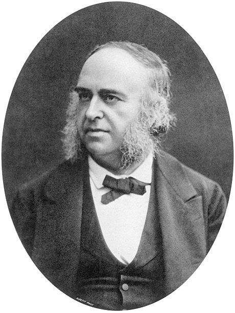
| Paul Broca | French physician who is most famous for his discovery of the speech production centre of the brain located in the frontal lobes (now known as the Broca's area). |
| Broca's area | A region of the frontal lobe that is associated with the production of speech. It is located on the left hemisphere of right-handed and of most left-handed individuals. (discovered in the 1860s and studied and researched by Paul Broca). |
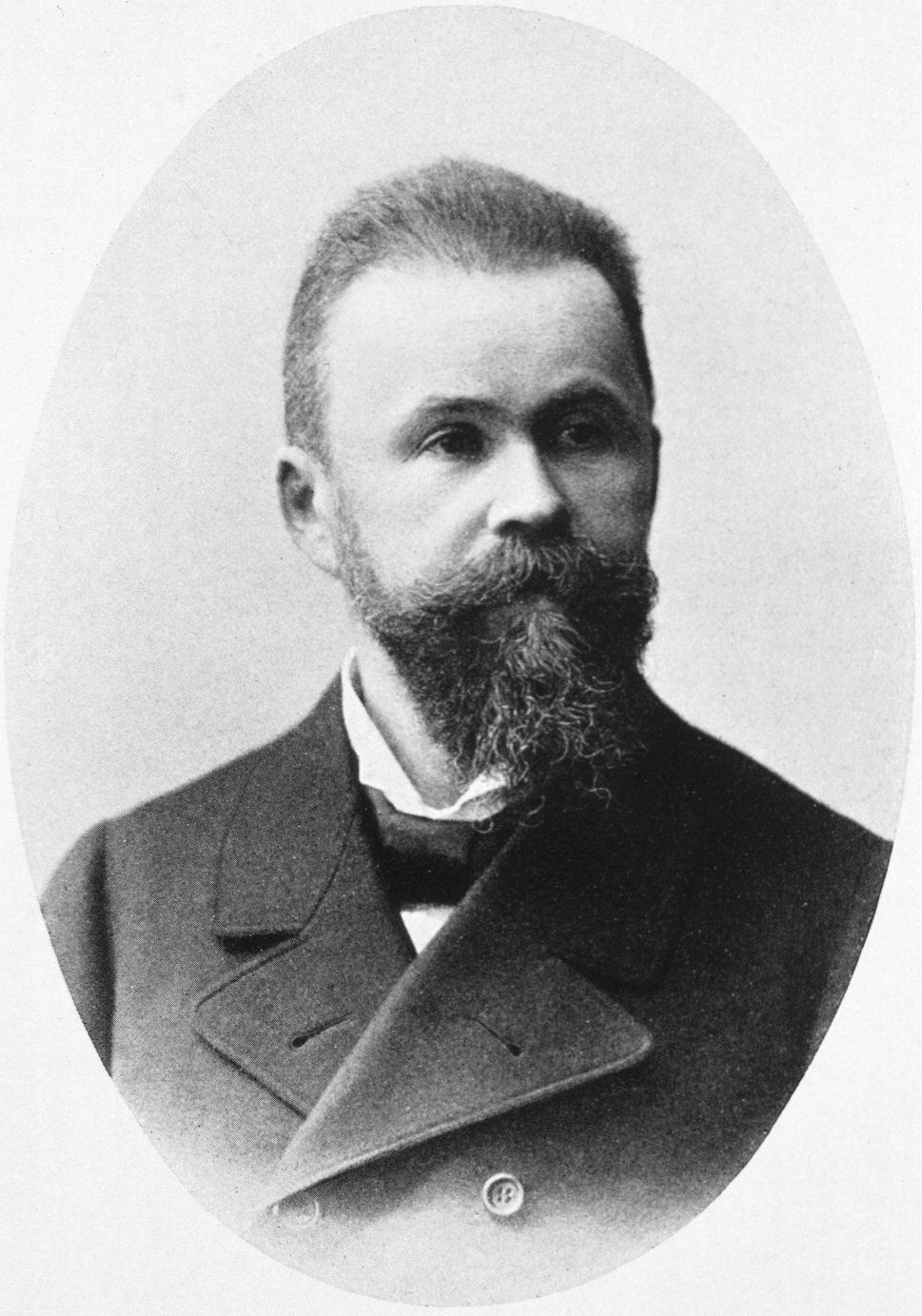
| Carl Wernicke | A German physician whose work led to our understanding of the areas of the brain responsible for the understanding and production of meaningful speech. |
| Wernicke's Area | A region toward the back of the temporal lobe of the left hemisphere of the cerebrum containing nerve tissue associated with the language comprehension and expression. |
| cognitive neural prosthetics | Cognitive neural prosthetics record activity related to higher level cognitive processes that organize behaviour. Recordings of neural activity are used to decode the state of the subject, their goals and the expected value they place on those goals. Decoding these and other cognitive processes directly means patients can have new ways to control their prosthetic device and their control can be more flexible. |
Quer criar seus próprios Flashcards gratuitos com GoConqr? Saiba mais.