4269788

| Questão | Responda |
| 3.1 Biological molecules | N/A |
| 3.1.1 Monomers and polymers | N/A |
| What are monomers? | Smaller unit molecules that join to make larger molecules |
| What are polymers? | Larger molecules make from smaller molecules joined together? |
| What are some examples of monomers? | - Monosaccharides - Amino acids - Nucleotides |
| What is a condensation reaction? | Reaction releasing water and forming a chemical bond between two molecules |
| What does a condensation reaction look like? | |
| What is a hydrolysis reaction? | Reaction taking in water in order to break up a chemical bond between two molecules |
| 3.1.2 Carbohydrates | N/A |
| What are monosaccharides? | Small sugar molecules from which carbohydrates are made |
| What are some examples of monosaccharides? | - Glucose - Galactose - Fructose |
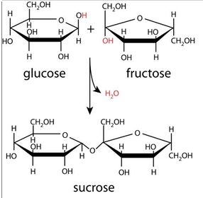
| How are monosaccharides joined together? | Condensation reaction, releasing water and forming a glycosidic bond between two molecules |
| What are disaccharides? | Molecules made from two monosaccharides joined by a glycosidic bond |
| What are some examples of disaccharides? | - Maltose - Sucrose - Lactose |
| What is maltose made from? | Two α glucose molecules |
| What is sucrose made from? | An α glucose molecule and a fructose molecule |
| What is lactose made from? | An α glucose molecule and a galactose molecule |
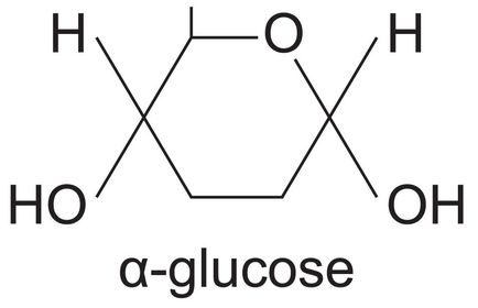
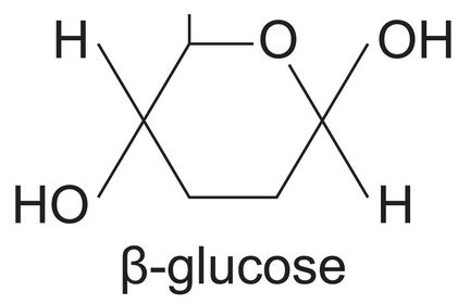
| What are the two isomers of glucose? | - α glucose - β glucose |
| What does the structure of α glucose look like? | |
| What does the structure of β glucose look like? | |
| What are polysaccharides? | Long-chain molecules formed from many monosaccharides joined together by glycosidic bonds |
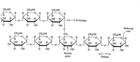
| What is glycogen? | Polysaccharide formed from many α glucose molecules joined by glycosidic bonds |
| What is the structure of glycogen like? | - Long chain - Branches off frequently - Multiple ends |
| What does the structure of glycogen look like? | |
| What is the function of glycogen in cells? | Long-term energy storage molecule in animal cells, especially liver |
| How does the structure of glycogen relate to its function? | - Branching ends allow for enzymes to act on multiple sites, breaking the molecule down faster - Compact, taking up less space in cells - Insoluble, does not affect water potential |
| What is starch? | Polysaccharide formed from many α glucose molecules joined by glycosidic bonds |
| What is the structure of starch like? | - Long chain - No branching - Helix structure |
| What does the structure of starch look like? | |
| What is the function of starch in cells? | Long-term energy storage molecule in plant cells |
| How does the structure of starch relate to its function? | - Helix structure, compact molecule reducing area used for storage - Unbranched, cannot be easily broken down by enzymes - Insoluble, does not affect water potential |
| What is cellulose? | Polysaccharide formed from many β glucose molecules joined by glycosidic bonds |
| What is the structure of cellulose like? | - Long chain - Straight, unbranched - Layers interlinked with hydrogen bonds |
| What does the structure of cellulose look like? | |
| What is the function of cellulose in cells? | Makes up cell walls in plants |
| How does the structure of cellulose relate to its function? | - Unbranched chain, difficult for enzymes to break down - Long chain interlinked with hydrogen bonds provides cell wall with mechanical strength - Insoluble, does not affect water potential |
| What are the steps involved in the Benedict's test for reducing sugars? | - Add equal volume of sample solution and Benedict's solution to test tube - Mix - Leave in water bath for 5 minutes - Observe colour changes |
| What results are expected from the Benedict's test for reducing sugars? | - Colour change from blue to brick red, indicating the presence and quantity of reducing sugars - If there is no colour change, reducing sugars are not present |
| What are the steps involved in the Benedict's test for non-reducing sugars? | - Add 1cm^3 of sample solution and 1cm^3 dilute hydrochloric acid - Boil for one minute - Allow tube to cool - Neutralise acid with sodium hydrogencarbonate - Re-test for reducing sugars - Observe colour changes |
| What results are expected from the Benedict's test for non-reducing sugars? | - Colour change to brown/brick red, indicating that non-reducing sugars were present |
| What are the steps involved in the iodine test for starch? | - Add 10cm^3 of sample solution to test tube - Add 5 drops of iodine/potassium iodide - Observe colour changes |
| What results are expected from the iodine test for starch? | - Colour change from orange to black/dark blue, indicating the presence of starch |
| 3.1.3 Lipids | N/A |
| What is a lipid? | Any molecule that is a fatty acid or derivative of one |
| What does the structure of a fatty acid look like? | |
| What does it mean if a fatty acid is unsaturated? | It contains a double bond besides the one in the (R-C=O-OH) carboxylic acid group - therefore it has a double bond in the R group |
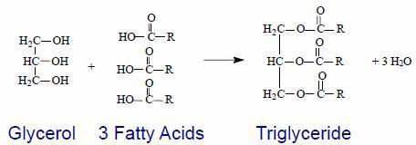
| What is a triglyceride? | A lipid molecule formed by the condensation of three fatty acid molecules and a glyceride molecule, held together by ester bonds |
| What does the structure of a triglyceride look like? | |
| What is the function of triglycerides in animals? | - Good source of energy - Source of metabolic water - Temperature insulator - Storage molecule - Provide buoancy |
| How does the structure of triglycerides relate to their functions? | - High ratio of energy storing carbon-hydrogen bonds to carbon atoms, good source of energy - Produce water molecules on oxidation, source of metabolic water - Poor heat conductor, temperature insulator - Insoluble, can be stored without affecting water potential - Less dense than water, provide buoancy |
| What is a phospholipid | A lipid molecule formed by the condensation of two fatty acids, glycerol and a phosphate 'head', held together by esther bonds |
| What does the structure of a phospholipid look like? | |
| What is the function of phospholipids in cells? | Make up the lipid bilayer of the cell membrane |
| How does the structure of a phospholipid relate to its function? | - Phosphate 'head' is hydrophillic, fatty acid 'tails' are hydrophobic, causing phospholipid molecules to line up in a double layer with their tails facing inwards, making up the lipid bilayer |
| What are the steps involved in the emulsion test for lipids? | - Add a few drops of the sample solution to a test tube - Add 2cm^3 ethanol and shake it thoroughly - Add 2cm^3 of ionised water - Observe formation of precipitate |
| What results are expected from the emulsion test for lipids? | - If a layer of white emulsion forms, lipids are present |
| 3.1.4 Proteins | N/A |
| 3.1.4.1 General properties of proteins | N/A |
| What are amino acids? | The monomers from which proteins are made |
| What does the structure of an amino acid look like? | |
| How many differing R groups are common in all organisms? | 20 |
| What are dipeptides? | Two amino acids joined together in a condensation reaction by a peptide bond |
| What are polypeptides? | Multiple amino acids joined together in a condensation reaction by peptide bonds |
| How many polypeptides are contained in a functional protein? | One or more |
| What are the functions of proteins? | - Making/repairing structural tissue - Hormones - Enzymes - Antibodies |
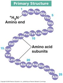
| What are the structures of protein? | - Primary - Secondary - Tertiary - Quaternary |
| What is the primary structure of protein? | Long chain of amino acid 'units' |
| What does the primary structure of protein look like? | |
| What is the secondary structure of protein? | 3D folded structure caused by weak hydrogen bonds between twisting amino acids resulting in a coiling/folding - Alpha helix - Beta pleated sheet |
| What does the secondary structure of protein look like? | |
| What is the tertiary structure of protein? | 3D structure caused by further twisting of polypeptide chain and formation of strong disulphide bonds, weak ionic bonds that hold the geometric shape - Determines protein function e.g. enzyme with active site - Contains numerous easily-broken hydrogen bonds that can deform shape if PH or temperature too high |
| What does the tertiary structure of protein look like? | |
| What is the quaternary structure of protein? | 3D structure made up of multiple polypeptides with tertiary structure coiled together - Often contains non-protein molecules that aid function e.g. heme groups |
| What does the quaternary structure of protein look like? | |
| What are two types of protein? | - Fibrous - structural e.g. cartillage - Globular - metabolic functions e.g. haemoglobin |
| What are the steps involved in the Biuret test for proteins? | - Add few drops of strong base (e.g. sodium hydroxide then few drops of aqueous copper (II) sulphate OR Biuret Reagent - Observe colour changes |
| What results are expected from the Biuret test for proteins? | If proteins are present, solution will change colour from blue to purple |
| 3.1.4.2 Many proteins are enzymes | N/A |
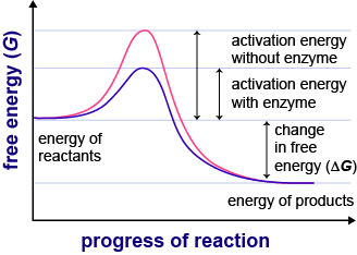
| What effect do enzymes have on the activation energy of a reaction? | Each enzyme lowers the activation energy of the reaction catalysed |
| What does the effect of enzymes on the activation energy of a reaction look like? | |
| What is the induced-fit model of enzyme action? | Enzymes are able to change the shape of their active site slightly to fit a specific substrate |
| What does the induced fit model of enzyme action look like? | |
| To what do the properties of an enzyme relate? | - Tertiary structure of active site - Ability to combine with substrate and form enzyme-substrate complexes |
| What is enzyme specificity? | Enzymes are specific to a single type of substrate due to the shape of their active site |
| What is the effect of enzyme concentration on the rate of an enzyme-controlled reaction? | Increasing enzyme concentration increases the rate of reaction until the plateau when concentration of substrate is exceeded, as there will be no more substrate to form complexes with excess enzymes |
| What is the effect of substrate concentration on the rate of an enzyme-controlled reaction? | Increasing enzyme concentration increases the rate of reaction until the plateau when concentration of enzymes is exceeded, as there will be no more available active sites to form complexes with excess substrate |
| What is the effect of PH on the rate of an enzyme-controlled reaction? | All enzymes have an optimum PH at which the rate of reaction is highest - PH that is too low can cause enzymes to denature by altering the charge of the amino acids that make up the active site, changing its shape so that it can no longer accommodate a substrate, or by breaking down the bonds in the tertiary structure, warping the active site |
| What is the effect of temperature on the rate of an enzyme-controlled reaction? | Rate of reaction increases as temperature increases as the substrates gain more energy, moving faster and thus increasing the chance of successful collisions with the active sites, up until the optimum temperature is reached, after which point the temperature is too great and the bonds making up the active site break down, warping it |
| What are competitive inhibitors? | Inhibitor that blocks the active site of the enzyme |
| What does competitive inhibition look like? | |
| What are non-competitive inhibitors? | Inhibitor that enters another part of the enzyme and changes its structure, changing the shape of the active site so the substrate will no longer fit |
| What does non-competitive inhibition look like? | |
| 3.1.5 Nucleic acids are important information-carrying molecules | N/A |
| 3.1.5.1 Structure of DNA and RNA | N/A |
| What is DNA? | Deoxyribonucleic acid - Holds genetic material - Found in nucleus of cells |
| What is RNA? | Ribonucleic acid - Transports genetic material to ribosomes - Found in cells |
| What are ribosomes formed from? | RNA and proteins |
| What are nucleic acids made up of? | Multiple nucleotides joined together by phosphodiester bonds |
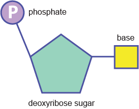
| What are nucleotides made up of? | Backbone: - Phosphate group - Pentose sugar Attached to pentose sugar: - Nitrogenous base |
| What does the structure of a nucleotide look like? | |
| What is the pentose sugar in DNA? | Deoxyribose sugar |
| What is the pentose sugar in RNA? | Ribose sugar |
| What are the nitrogenous bases in DNA? | - Pyrimidines - T & C - Purines - A & G |
| What are the nitrogenous bases in DNA? | - Pyrimidines - U & C - Purines - A & G |
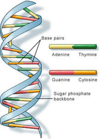
| What is the structure of a DNA molecule? | - Two long DNA strands joined together by hydrogen bonds between complimentry paired bases (T&A, C&G) - Double helix structure |
| What does the structure of a DNA molecule look like? | |
| What is the structure of an RNA molecule? | - Single relatively short RNA strand - Single helix structure |
| What does the structure of an RNA molecule look like? | |
| 3.1.5.2 DNA replication | N/A |
| What does it mean that DNA replication is 'semi-conservative'? | One of the two DNA strands in the new molecule is conserved from the original |
| What are the steps involved in DNA replication? | - DNA strands 'unwind' - DNA helicase catalyses breakdown of hydrogen bonds, strands split - Free nucleotides in the nucleus bind to complimentary base pairs - DNA polymerase catalyses condensation of strong chemical bonds between nucleotides, forming a complete strand |
| 3.1.6 ATP | N/A |
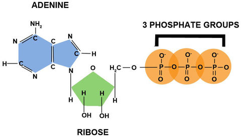
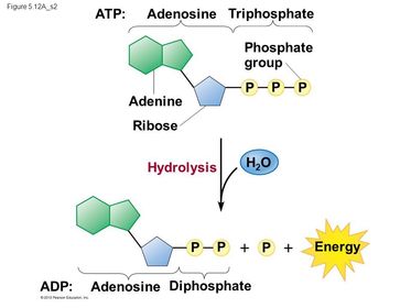
| What is ATP? | A single molecule of a nucleotide derivative |
| What is ATP made up of? | - Adenine base - Ribose sugar - Three phosphate groups |
| What does the structure of ATP look like? | |
| What happens during hydrolysis of ATP? | Bonds between phosphate group furthest from ribose sugar break down - Inorganic phosphate (Pi) and energy released |
| What does the hydrolysis of ATP look like? | |
| What happens during ATP resynthesis? | Bonds between ADP and Pi catalysed by ATP synthase - Happens during photosynthesis and respiration |
| How is ATP used in cells? | Primary short-term energy source in cells - active transport - cell division - muscle contractions - protein synthesis - DNA synthesis |
| 3.1.7 Water | N/A |
| What are the functions of water in the body? | - Metabolite in digestion reactions e.g. hydrolysis - Solvent in which metabolic reactions occur - Temperature buffer - Cooling internal temperatures - Photosynthesis in plants - Transport of waste and nutrients |
| What are the properties of water? | - Can break down large molecules - Solvent - High heat capacity - Large latent heat of vaporisation - Cohesion between molecules |
| How do the properties of water relate to its function? | - Can break down large molecules - able to hydrolyse molecules during digestion - Solvent - able to contain reactants that can only react when dissolved - High heat capacity - buffer against external temperature change as water absorbs lots of heat - aquatic environments change temperature very slowly - Large latent heat of vaporisation - animals and plants can effectively cool themselves by transferring energy to surroundings through evaporation (sweat and stomata) - Cohesion between molecules - polar so attract each other - causes surface tension so small things can live on water surface and transpiration can occur |
| 3.1.8 Inorganic ions | N/A |
| What are inorganic ions? | Also known as electrolytes - Ions used in vital cellular processes |
| What are some examples of inorganic ions? | - Hydrogen ions - Iron ions - Sodium ions - Phosphate ions |
| Where are inorganic ions found? | - Cytoplasm - Bodily fluids - Some in high concentrations and some in low concentrations |
| What is the role of hydrogen ions in maintaining PH? | - greater concentration of hydrogen ions = greater PH - optimum PH for enzymes |
| What is the role of iron ions in haemoglobin? | - Heme group contains iron atom, responsible for association with oxygen - Gives haemoglobin red colour |
| What is the role of sodium in co-transport of amino acids and glucose? | Sodium ion and amino acid/glucose pass through protein channel called symport simultaneously - Sodium moving along concentration gradient drives transport of acid/glucose moving against it - Both simultaneously bind to symport |
| What is the role of phosphate ions in DNA and ATP? | - Makes up phosphate-sugar backbone of DNA - Breaking of bonds between phosphates in ATP is the main immediate energy source in cells |
Quer criar seus próprios Flashcards gratuitos com GoConqr? Saiba mais.