20476076
Descrição
Quiz por Charlotte Jakes, atualizado more than 1 year ago
|
|
Criado por Charlotte Jakes
aproximadamente 5 anos atrás
|
|
Questão 1
Questão
The first cleavage of a zygote occurs immediately after [blank_start]fertilisation[blank_end]. Before the zygote is implanted into the uterine wall at day 7-10, it undergoes a series of subdivisions without [blank_start]growth[blank_end]. The cleavage generates a [blank_start]blastocyst[blank_end] - a hollow ball of cells consisiting of an outer layer of cells known as the [blank_start]trophoblast[blank_end]. This outer layer will interact with the [blank_start]uterine[blank_end] wall. Inside the [blank_start]blastocyst[blank_end] is the [blank_start]inner cell mass[blank_end] which will develop into the embryo.
Responda
-
fertilisation
-
growth
-
blastocyst
-
trophoblast
-
uterine
-
blastocyst
-
inner cell mass
Questão 2
Questão
After implantation, the cells of the inner cell mass in the zygote rearrange to form what?
Responda
-
Blastodisc
-
Epiblast
-
Hypoblast
-
Trophoblast
Questão 3
Questão
In what structure of the embryo does the primitive streak develop?
Responda
-
Trophoblast
-
Epiblast
-
Hypoblast
-
Mesoderm
Questão 4
Questão
What is gastrulation?
Responda
-
The organisation of the cells to generate the body plan by differentiation into the primary germ layers
-
The folding of the neural plate to form the neural tube
-
The process by which the embryo implants into the uterine wall
-
The pre-implantation divisions of the zygote
Questão 5
Questão
Fill in the blanks below to describe the process of gastrulation.
1. The primitive streak develops in the [blank_start]epiblast[blank_end].
2. The end of the primitive streak expands to form the [blank_start]primitive node[blank_end].
3. Cells of the epiblast migrate inwards towards the streak, detach from their layer and migrate [blank_start]beneath[blank_end] it.
4. The first cells of the epiblast to migrate invade the [blank_start]hypoblast[blank_end] layer, displacing its cells.
5. Eventually, the hypoblast cells will be entirely replaced by a new layer. This layer is the definitive [blank_start]endoderm[blank_end].
6. Some of the invaginated epiblast cells remain in the space between the layers. This layer is the [blank_start]mesoderm[blank_end].
7. The remaining cells of the epiblast form the definitive [blank_start]ectoderm[blank_end].
Responda
-
epiblast
-
primitive node
-
beneath
-
hypoblast
-
endoderm
-
mesoderm
-
ectoderm
Questão 6
Questão
The epiblast cells no longer migrate after the definitive germ layers have formed.
Responda
- True
- False
Questão 7
Questão
What is secondary neurulation?
Responda
-
The formation of a tube by the hollowing out of a solid precursor's interior
-
The formation of a tube by the infolding of its wall
-
The formation of a tube by the joining of two hollow structures
-
The formation of a tube by the layering of rings of cells
Questão 8
Questão
What name is given to the formation of the neural tube in the embryo?
Responda
-
Neurulation
-
Neurogenesis
-
Gastrulation
-
Invagination
Questão 9
Questão
Using the image, fill in the blanks to describe each stage of neurulation.
1. Neuroectodermal cells differentiate to form the [blank_start]neural plate[blank_end] with a border to separate it from regular ectoderm.
2. The neural plate folds upwards with its apex extending [blank_start]dorsally[blank_end]. The two edges join at the [blank_start]neural plate borders[blank_end], forming the [blank_start]neural crest[blank_end].
3. The closure of the tube disconnects the neural crest from the wall. The cells of the neural crest then differentiate to form the [blank_start]peripheral nervous system[blank_end].
4. The [blank_start]notochord[blank_end] degenerates and other mesoderm cells differentiate into the [blank_start]somites[blank_end].
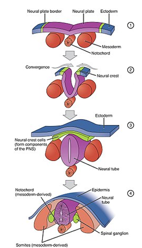
Responda
-
neural plate
-
dorsally
-
neural plate borders
-
neural crest
-
peripheral nervous system
-
notochord
-
somites
Questão 10
Questão
What happens to the notochord following the formation of the neural tube?
Responda
-
Degenerates completely
-
Forms the spinal cord
-
Forms the nucleus pulposus of the intervertebral discs
-
Forms the peripheral nervous system
Questão 11
Questão
What will result from an absence of joining of the neural folds at the cranial end of the neural plate?
Responda
-
Anencephaly - open brain, lack of skull
-
Craniorachischisis - completely open brain and spinal cord
-
Spina bifida - exposure of the spinal cord
-
Cyclopia - lack of division of the orbits of the eye into two cavities
Questão 12
Questão
Label this image showing the organisation of the embryo following neurulation.
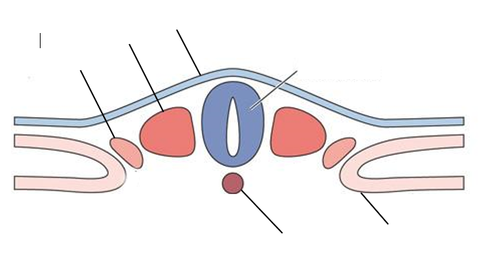
Responda
-
Epidermis
-
Paraxial mesoderm
-
Intermediate mesoderm
-
Neural tube
-
Notochord
-
Lateral plate mesoderm
Questão 13
Questão
Fill in the blanks to describe what each of the primitive tissues will develop into.
The [blank_start]lateral plate[blank_end] mesoderm will form the splanchnic and somatic nerves.
The [blank_start]intermediate[blank_end] mesoderm will form the kidneys and the gonads.
The [blank_start]paraxial[blank_end] mesoderm will form the head and the somites.
Responda
-
lateral plate
-
intermediate
-
paraxial
Questão 14
Questão
Why do the neural crest cells only occur in the anterior half of the vertebral column?
Responda
-
The posterior halves of the somites express inhibitory molecules
-
The anterior halves of the somites express attracting molecules
-
The posterior half of the vertebral column is already a solid structure - no other cells can migrate there
Questão 15
Questão
Fill in the blanks to describe the process of neural crest migration.
The first wave of migration forms the [blank_start]autonomic[blank_end] ganglia. The parasympathetic ganglia migrate to the walls of the [blank_start]organs[blank_end]. The sympathetic ganglia migrate in front of the [blank_start]aorta[blank_end].
The second wave of migration forms the [blank_start]posterior root[blank_end] ganglia either side of the spinal cord. It also forms the [blank_start]Schwann[blank_end] cells and satellite cells.
The final wave of migration forms the [blank_start]melanocytes[blank_end] throughout the epidermis. These give pigmentation to the hair, skin, iris etc.
The neural crest cells at the cranial end form the [blank_start]sensory ganglia[blank_end] of the cranial nerves. They also migrate to form the [blank_start]mesenchyme[blank_end] of the face and neck.
Responda
-
autonomic
-
organs
-
aorta
-
posterior root
-
Schwann
-
melanocytes
-
sensory ganglia
-
mesenchyme
Questão 16
Questão
Label this image to describe the body axes of the developing embryo.
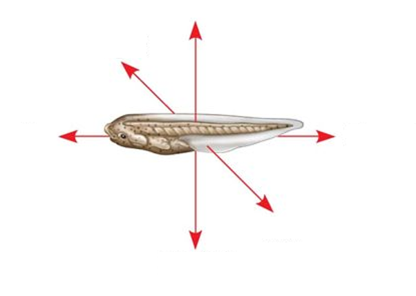
Responda
-
Anterior
-
Posterior
-
Dorsal
-
Ventral
-
Right
-
Left
Questão 17
Questão
How does a cell in a particular position in the developing embryo 'know' what tissue it will differentiate into?
Responda
-
It responds to a particular type of morphogen
-
It responds to a particular concentration of morphogen
-
It responds to a particular pH
-
It responds to different concentrations of oxygen
Questão 18
Questão
The notochord directs the development of the ventral spinal cord.
Responda
- True
- False
Questão 19
Questão
The Sonic Hedgehog gene and subsequent protein mediates the development of what?
Responda
-
Ventral spinal cord
-
Dorsal spinal cord
-
Limbs
-
Eyes
Questão 20
Questão
What embryonic structure secretes the SHH protein?
Responda
-
Neural tube
-
Notochord
-
Somites
-
Epidermis
Questão 21
Questão
Somites do not respond to the SHH protein.
Responda
- True
- False
Questão 22
Questão
A mutation in the [blank_start]SHH[blank_end] gene can result in holoprosencephaly. This results in a lack of development of the two hemispheres of the [blank_start]forebrain[blank_end]. It often presents with microcephaly and/or a cleft palate/lip. [blank_start]Cyclopia[blank_end] is the most extreme form of this condition whereby the orbits of the eye do not divide into two cavities, leaving a single central eye.
Responda
-
SHH
-
forebrain
-
Cyclopia
Questão 23
Questão
These diagrams show the blastocysts at different stages of their development. Label them.
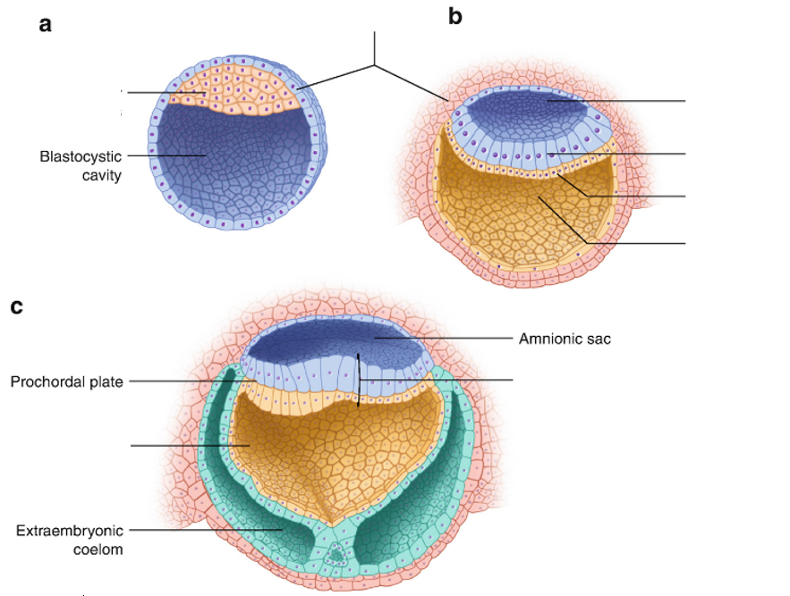
Responda
-
Inner cell mass
-
Trophoblast
-
Amnioic cavity
-
Epiblast
-
Hypoblast
-
Yolk sac
-
Blastodisc
-
Yolk sac
Questão 24
Questão
Failure of the neural tube to close at a particular point along the spine results in what birth defect?
Responda
-
Spina bifida
-
Craniorachischisis
-
Cyclopia
-
Anencephaly
Questão 25
Questão
Failure of the neural tube to close at any point results in what birth defect?
Responda
-
Craniorachischisis
-
Anencephaly
-
Spina bifida
-
Cyclopia
Questão 26
Questão
The paraxial mesoderm forms the head and the somites.
The somites will then differentiate into...
The [blank_start]sclerotome[blank_end] - this will form the vertebrae and the rib cartilage.
The [blank_start]myotome[blank_end] - this will form the muscles.
The [blank_start]dermatome[blank_end] - this will form the skin and connective tissue.
Responda
-
sclerotome
-
myotome
-
dermatome
Questão 27
Questão
What will cells in the dorsal portion of the neural tube develop into?
Responda
-
Sensory neurons
-
Motor neurons
Questão 28
Questão
What will cells in the ventral portion of the neural tube differentiate into?
Responda
-
Sensory neurons
-
Motor neurons
Questão 29
Questão
The different regions of the anterior neural tube receive different morphogens in different concentrations to determine what tissues they differentiate into based on location.
Responda
- True
- False
Questão 30
Questão
What will you see if you remove the notochord during development?
Responda
-
No motor neurons in the ventral spinal cord
-
No sensory neurons in the dorsal spinal cord
-
A single set of motor neurons in the ventral spinal cord
-
Two sets of motor neurons in the ventral spinal cord
Questão 31
Questão
What will you see if you transplant an additional notochord during development?
Responda
-
Two sets of motor neurons in the ventral spinal cord
-
A single net of motor neurons in the ventral spinal cord
-
No motor neurons in the ventral spinal cord
-
No sensory neurons in the dorsal spinal cord
Questão 32
Questão
In what direction do somites develop?
Responda
-
Posteriorly
-
Anteriorly
-
Superiorly
-
Inferiorly
-
Laterally
-
Medially
Questão 33
Questão
The somites organise the peripheral nerves.
Responda
- True
- False
Quer criar seus próprios Quizzes gratuitos com a GoConqr? Saiba mais.