11085605
Description
Flashcards by die zwei ??, updated more than 1 year ago
|
|
Created by die zwei ??
almost 7 years ago
|
|
| Question | Answer |
|
Image:
Image (binary/octet-stream)
|
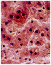
Pyknosis: nuclear shrinkage DNA condenses into shrunken basophilic mass |
|
Image:
Image (binary/octet-stream)
|
Karyiorrhexis: nuclear fragmentation |
|
Image:
Image (binary/octet-stream)
|
Karyolysis: nuclear fading |
|
Image:
Image (binary/octet-stream)
|
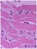
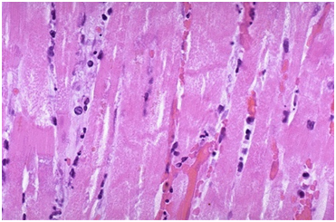
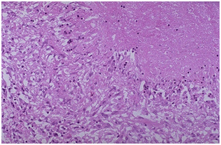
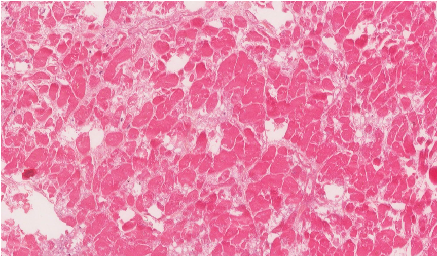
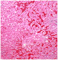
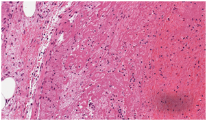
Coagulative necrosis recent MI |
|
Image:
Image (binary/octet-stream)
|
Coagulative necrosis of the heart |
|
Image:
Image (binary/octet-stream)
|
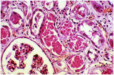
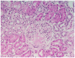
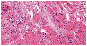
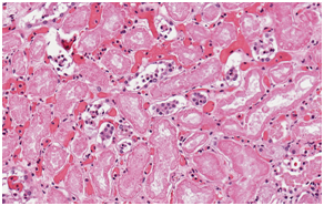
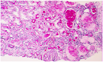
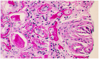
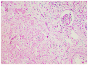
Coagulative necrosis of the kidney |
|
Image:
Image (binary/octet-stream)
|
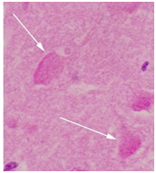
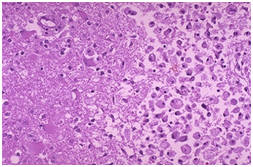
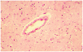
Liquefactive necrosis in brain |
|
Image:
Image (binary/octet-stream)
|
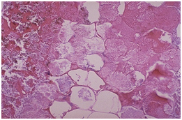
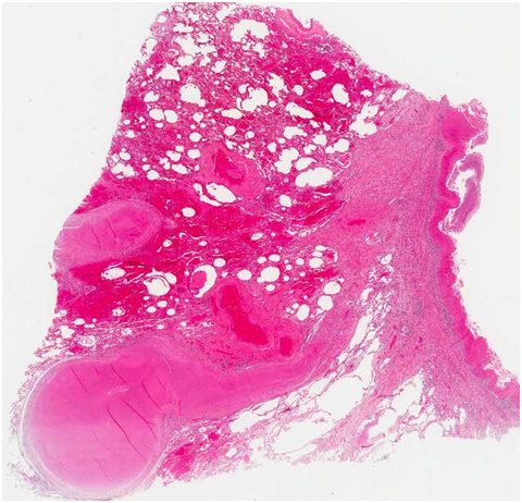
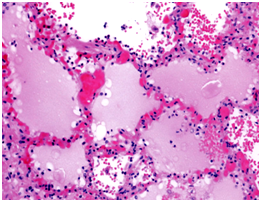
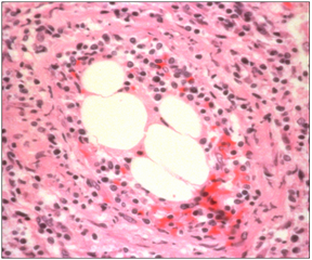
Fat necrosis (in pancreas) |
|
Image:
Image (binary/octet-stream)
|
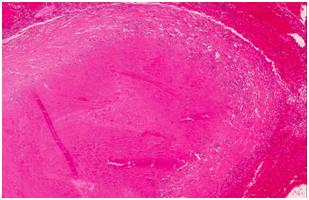
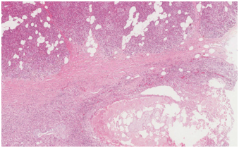
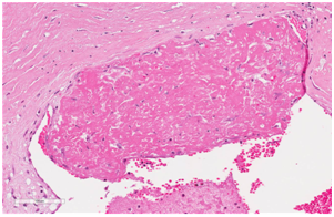
Caseous necrosis (due to TB) |
|
Image:
Image (binary/octet-stream)
|
Fibrinoid necrosis |
|
Image:
Image (binary/octet-stream)
|
Fibrinoid necrosis |
|
Image:
Image (binary/octet-stream)
|
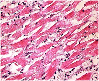
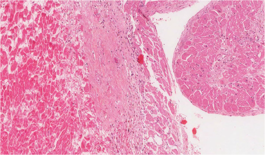
loss of nuclei coagulative necrosis as a consequence of tissue hypoxia (due to atherosclerosis and thrombosis) and cell death MI |
|
Image:
Image (binary/octet-stream)
|
loss of nuclei coagulative necrosis as a consequence of tissue hypoxia (due to atherosclerosis and thrombosis) and cell death MI |
|
Image:
Image (binary/octet-stream)
|
loss of nuclei coagulative necrosis as a consequence of tissue hypoxia (due to atherosclerosis and thrombosis) and cell death MI |
|
Image:
Image (binary/octet-stream)
|
loss of nuclei coagulative necrosis as a consequence of tissue hypoxia (due to atherosclerosis and thrombosis) and cell death MI |
|
Image:
Image (binary/octet-stream)
|
loss of nuclei coagulative necrosis as a consequence of tissue hypoxia (due to atherosclerosis and thrombosis) and cell death MI |
|
Image:
Image (binary/octet-stream)
|
loss of nuclei coagulative necrosis as a consequence of tissue hypoxia (due to atherosclerosis and thrombosis) and cell death MI |
|
Image:
Image (binary/octet-stream)
|
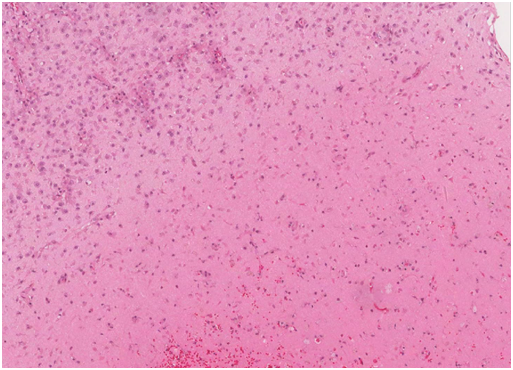
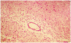
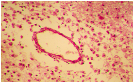
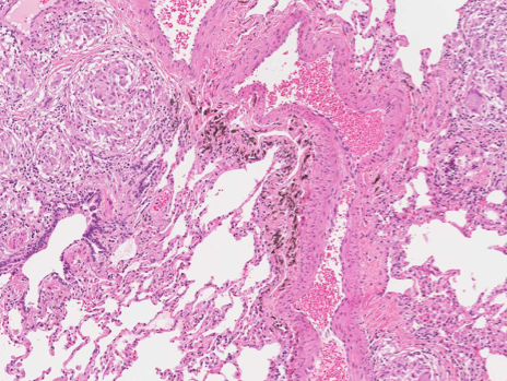
loss of brain parenchyma and macrophages which have phagocytosed the dead brain tissue brain infarct |
|
Image:
Image (binary/octet-stream)
|
loss of brain parenchyma and macrophages which have phagocytosed the dead brain tissue brain infarct |
|
Image:
Image (binary/octet-stream)
|
loss of brain parenchyma and macrophages which have phagocytosed the dead brain tissue brain infarct |
|
Image:
Image (binary/octet-stream)
|
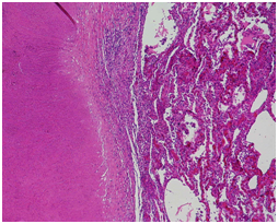
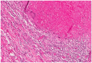
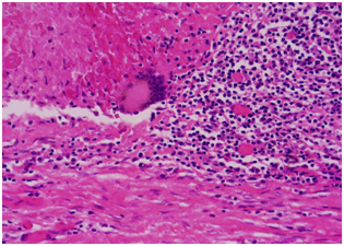
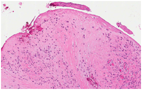
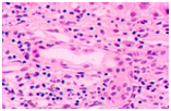
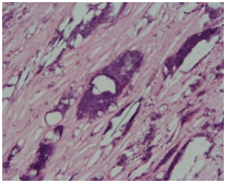
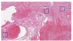
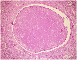
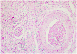
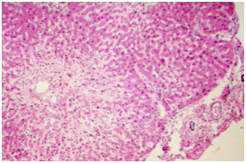
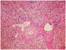
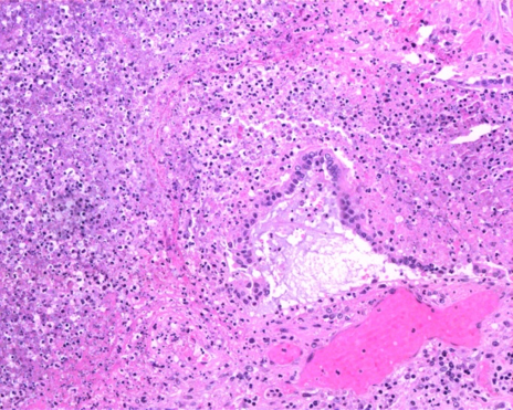
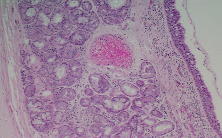
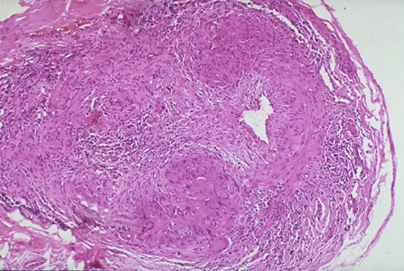
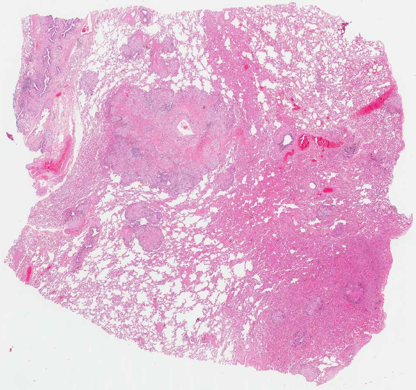
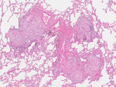
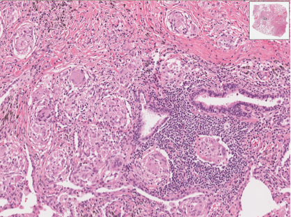
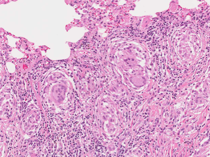
Granulomas with a necrotic center and a rim of epithelioid hystiocytes, and occasional multinucleated Langhans giant cells at the periphery Tuberculosis |
|
Image:
Image (binary/octet-stream)
|
Granulomas with a necrotic center and a rim of epithelioid hystiocytes, and occasional multinucleated Langhans giant cells at the periphery Tuberculosis |
|
Image:
Image (binary/octet-stream)
|
Granulomas with a necrotic center and a rim of epithelioid hystiocytes, and occasional multinucleated Langhans giant cells at the periphery Tuberculosis |
|
Image:
Image (binary/octet-stream)
|
Granulomas with a necrotic center and a rim of epithelioid hystiocytes, and occasional multinucleated Langhans giant cells at the periphery Tuberculosis |
|
Image:
Image (binary/octet-stream)
|
Granulomas with a necrotic center and a rim of epithelioid hystiocytes, and occasional multinucleated Langhans giant cells at the periphery Tuberculosis |
|
Image:
Image (binary/octet-stream)
|
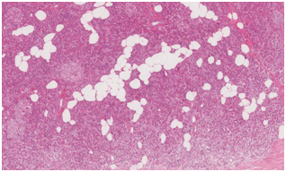
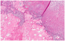
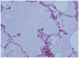
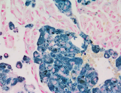
Fat necrosis (Steatonecrosis) |
|
Image:
Image (binary/octet-stream)
|
Fat necrosis (Steatonecrosis) |
|
Image:
Image (binary/octet-stream)
|
Fat necrosis (Steatonecrosis) |
|
Image:
Image (binary/octet-stream)
|
Fat necrosis (Steatonecrosis) |
|
Image:
Image (binary/octet-stream)
|
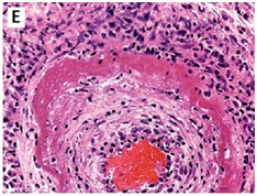
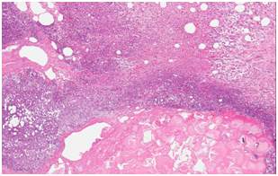
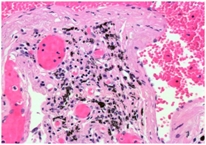
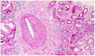
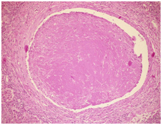
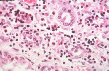
Fibrinoid necrosis is surrounded by rim of epithelioid histiocytes, lymphocytes, and plasma cells |
|
Image:
Image (binary/octet-stream)
|
Fibrinoid necrosis is surrounded by rim of epithelioid histiocytes, lymphocytes, and plasma cells |
|
Image:
Image (binary/octet-stream)
|
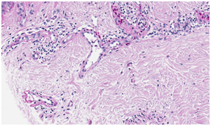
Synovia Rheumatoid arthritis (RA) – is a chronic inflammatory disorder of autoimmune origin |
|
Image:
Image (binary/octet-stream)
|
Synovia Rheumatoid arthritis (RA) – is a chronic inflammatory disorder of autoimmune origin |
|
Image:
Image (binary/octet-stream)
|
Synovia Rheumatoid arthritis (RA) – is a chronic inflammatory disorder of autoimmune origin |
|
Image:
Image (binary/octet-stream)
|
H&E: Hematoxylin-eosin stain: |
|
Image:
Image (binary/octet-stream)
|
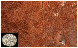
Histochemistry |
|
Image:
Image (binary/octet-stream)
|
Immunohistochemistry |
|
Image:
Image (binary/octet-stream)
|
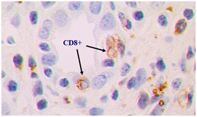
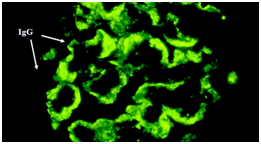
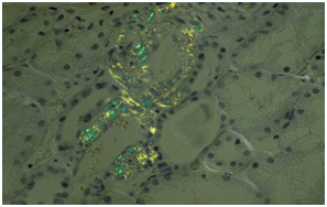
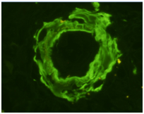
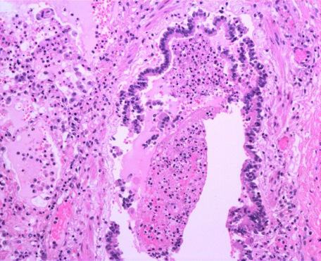
Immunofluorescence: IgG (antibodies) in green Shows human glomerulus in nephritis |
|
Image:
Image (binary/octet-stream)
|
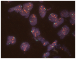
In situ hybridization: Her2 gene copy number in breast cancer tissue |
|
Image:
Image (binary/octet-stream)
|
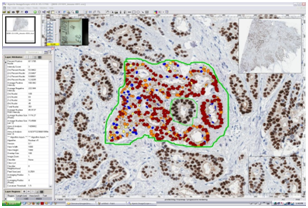
Digital image analysis |
|
Image:
Image (binary/octet-stream)
|
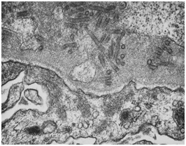
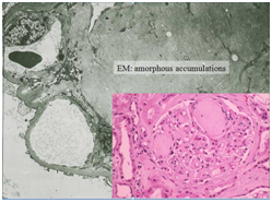
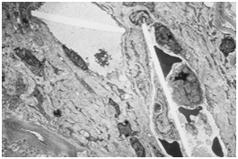
Electron microscopy: kidney glomerulus: |
|
Image:
Image (binary/octet-stream)
|
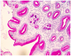
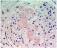
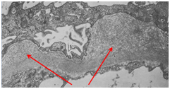
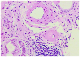
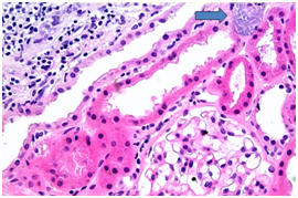
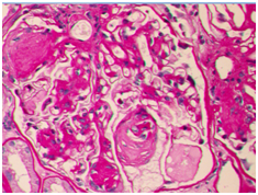
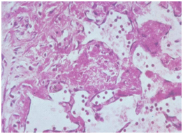
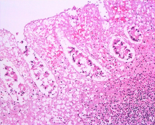
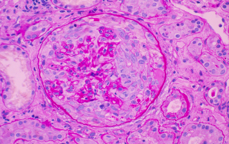
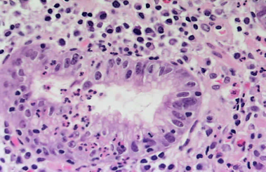
H&E stain of kidney: In glomerulus is an acellular part (pathologic accumulation) |
|
Image:
Image (binary/octet-stream)
|
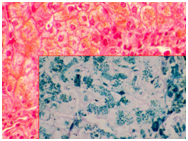
Congo Red staining: specific to amyloid process; brick red staining result |
|
Image:
Image (binary/octet-stream)
|
Congo Red staining: specific to amyloid process; brick red staining result |
|
Image:
Image (binary/octet-stream)
|
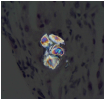
Congo Red – polarized light: |
|
Image:
Image (binary/octet-stream)
|
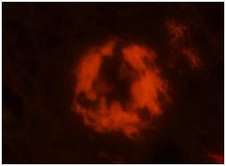
Amyloid, Congo Red, UV light |
|
Image:
Image (binary/octet-stream)
|
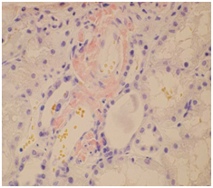
Amyloid under podocytes |
|
Image:
Image (binary/octet-stream)
|
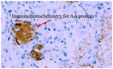
F component of AA amyloid |
|
Image:
Image (binary/octet-stream)
|
Image:
Image (binary/octet-stream)
|
|
Image:
Image (binary/octet-stream)
|
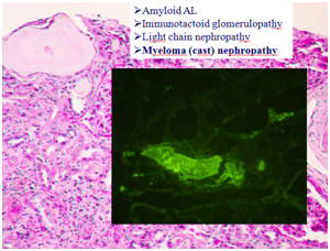
Amyloid AL Immunotactoid glomerulopathy --> Light chain nephropathy Myeloma (cast) nephropathy |
|
Image:
Image (binary/octet-stream)
|
Amyloid AL Immunotactoid glomerulopathy Light chain nephropathy --> Myeloma (cast) nephropathy |
|
Image:
Image (binary/octet-stream)
|
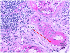
Hyalinosis |
|
Image:
Image (binary/octet-stream)
|
Hyalinosis |
|
Image:
Image (binary/octet-stream)
|
Hyalinosis |
|
Image:
Image (binary/octet-stream)
|
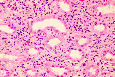
Hyalinosis: Kimmelstiehl-Wilson nodules in diabetes |
|
Image:
Image (binary/octet-stream)
|
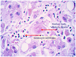
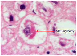
alcoholic hyalin |
|
Image:
Image (binary/octet-stream)
|
alcoholic hyalin |
|
Image:
Image (binary/octet-stream)
|
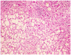
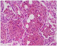
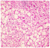
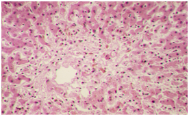
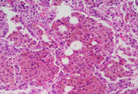
Steatosis: Lipocytes Normal hepatocytes |
|
Image:
Image (binary/octet-stream)
|
Atherosclerotic plaque |
|
Image:
Image (binary/octet-stream)
|
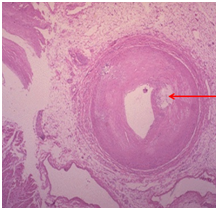
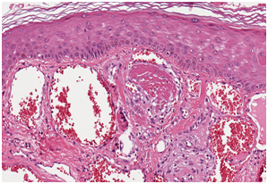
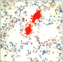
cholesterol crystals (due to atherosclerosis) |
|
Image:
Image (binary/octet-stream)
|
cholesterol crystals |
|
Image:
Image (binary/octet-stream)
|
Carbon (anthrax), anthracosis Black dots = pigments |
|
Image:
Image (binary/octet-stream)
|
Carbon (anthrax), anthracosis Black dots = pigments |
|
Image:
Image (binary/octet-stream)
|
Pigment accumulations |
|
Image:
Image (binary/octet-stream)
|
Pigment accumulations |
|
Image:
Image (binary/octet-stream)
|
Pigment accumulations |
|
Image:
Image (binary/octet-stream)
|
Pigment accumulations |
|
Image:
Image (binary/octet-stream)
|
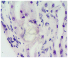
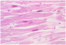
Calcium on H&E basophilic amorphous deposit |
|
Image:
Image (binary/octet-stream)
|
Calcium on H&E basophilic amorphous deposit |
|
Image:
Image (binary/octet-stream)
|
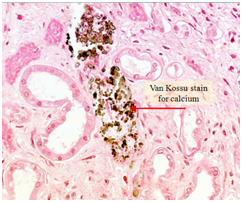
Van Kossu stain for calcium |
|
Image:
Image (binary/octet-stream)
|
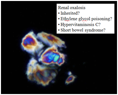
? |
|
Image:
Image (binary/octet-stream)
|
? |
|
Image:
Image (binary/octet-stream)
|
? |
|
Image:
Image (binary/octet-stream)
|
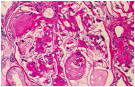
Hyalinosis, PAS stain |
|
Image:
Image (binary/octet-stream)
|
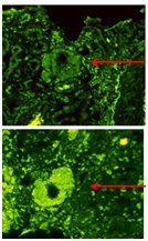
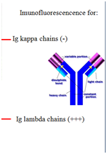
Immunofluoroscence Ig lambda in AL amyloid |
|
Image:
Image (binary/octet-stream)
|
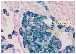
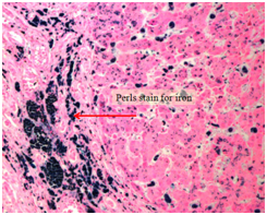
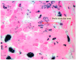
Pigments (H&E) Perl stain for iron |
|
Image:
Image (binary/octet-stream)
|
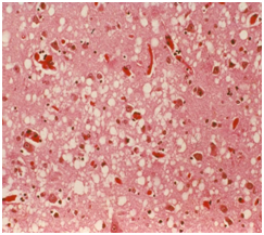
Steatosis, H&E |
|
Image:
Image (binary/octet-stream)
|
Edema (white spots) |
|
Image:
Image (binary/octet-stream)
|
Edema Inflammatory |
|
Image:
Image (binary/octet-stream)
|
Edema Inflammatory |
|
Image:
Image (binary/octet-stream)
|
Edema Inflammatory |
|
Image:
Image (binary/octet-stream)
|
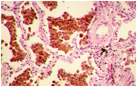
Edema Non-inflammatory lungs (alveoli) |
|
Image:
Image (binary/octet-stream)
|
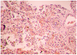
Pulmonary edema in CHF transudate accumulation in alveoli |
|
Image:
Image (binary/octet-stream)
|
Acute congestion: (can have) engorged capillaries edema focal hemorrhage |
|
Image:
Image (binary/octet-stream)
|
Chronic passive congestion: can show fibrosis & hemosiderin-laden macrophages |
|
Image:
Image (binary/octet-stream)
|
Chronic passive congestion: can show fibrosis & hemosiderin-laden macrophages |
|
Image:
Image (binary/octet-stream)
|
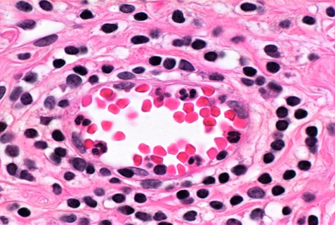
Petechia (hemorrhage) |
|
Image:
Image (binary/octet-stream)
|
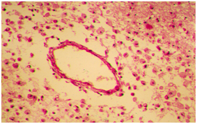
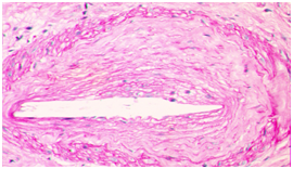
Venous thrombosis: composed of fibrin, platelets and blood cells |
|
Image:
Image (binary/octet-stream)
|
Venous thrombosis: composed of fibrin, platelets and blood cells |
|
Image:
Image (binary/octet-stream)
|
Venous thrombosis: composed of fibrin, platelets and blood cells |
|
Image:
Image (binary/octet-stream)
|
Venous thrombosis: composed of fibrin, platelets and blood cells |
|
Image:
Image (binary/octet-stream)
|
Acute congestion with edema |
|
Image:
Image (binary/octet-stream)
|
Venous thrombi |
|
Image:
Image (binary/octet-stream)
|
Venous thrombi (recanalized?!) |
|
Image:
Image (binary/octet-stream)
|
Venous thrombosis |
|
Image:
Image (binary/octet-stream)
|
Pulmonary "saddle" embolism |
|
Image:
Image (binary/octet-stream)
|
Arterial & venous thrombi Infective endocarditis |
|
Image:
Image (binary/octet-stream)
|
Fat embolism |
|
Image:
Image (binary/octet-stream)
|
Fat embolism? |
|
Image:
Image (binary/octet-stream)
|
Multiple Splenic... |
|
Image:
Image (binary/octet-stream)
|
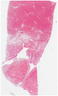
Hemorrhagic infarct of adrenal glands in septic shock |
|
Image:
Image (binary/octet-stream)
|
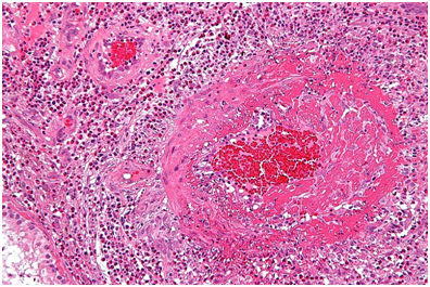
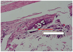
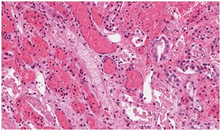
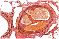
Atheroembolic renal disease |
|
Image:
Image (binary/octet-stream)
|
Atheroembolic renal disease |
|
Image:
Image (binary/octet-stream)
|
Atheroembolic renal disease |
|
Image:
Image (binary/octet-stream)
|
Atheroembolic renal disease |
|
Image:
Image (binary/octet-stream)
|
Atheroembolic renal disease |
|
Image:
Image (binary/octet-stream)
|
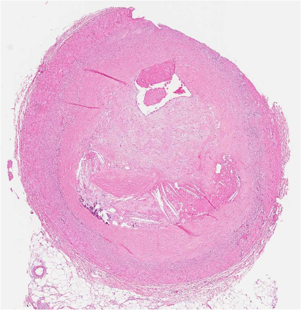
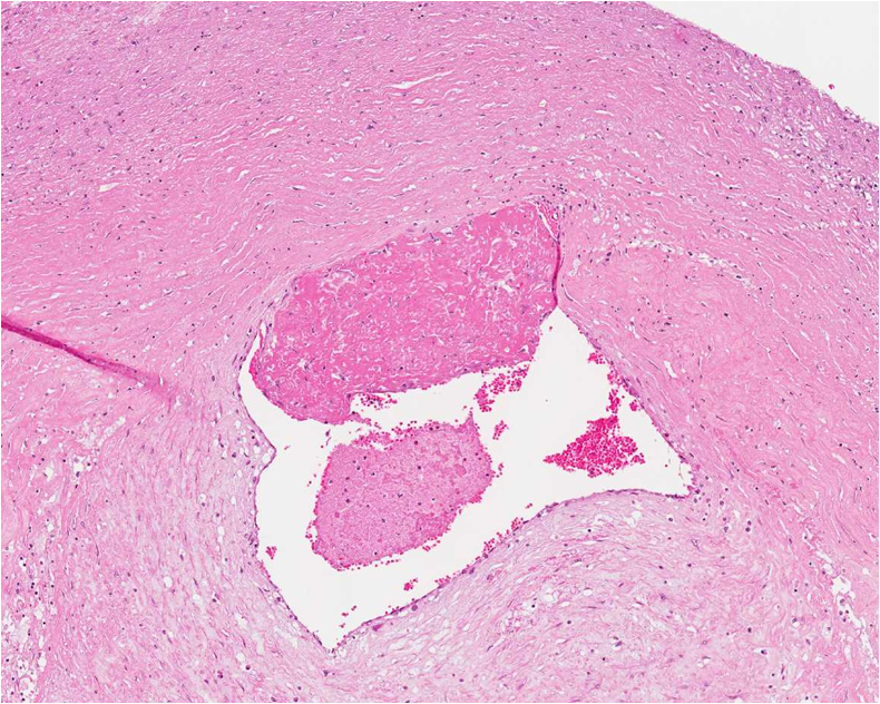
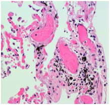
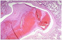
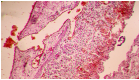
Rheumatic mitral valve disease |
|
Image:
Image (binary/octet-stream)
|
Rheumatic mitral valve disease |
|
Image:
Image (binary/octet-stream)
|
Rheumatic mitral valve disease |
|
Image:
Image (binary/octet-stream)
|
Rheumatic mitral valve disease |
|
Image:
Image (binary/octet-stream)
|
Rheumatic mitral valve disease |
|
Image:
Image (binary/octet-stream)
|
Rheumatic mitral valve disease |
|
Image:
Image (binary/octet-stream)
|
Rheumatic mitral valve disease |
|
Image:
Image (binary/octet-stream)
|
Venous thrombosis of the leg and pulmonary thromboembolism |
|
Image:
Image (binary/octet-stream)
|
Venous thrombosis of the leg and pulmonary thromboembolism |
|
Image:
Image (binary/octet-stream)
|
Venous thrombosis of the leg and pulmonary thromboembolism |
|
Image:
Image (binary/octet-stream)
|
Venous thrombosis of the leg and pulmonary thromboembolism |
|
Image:
Image (binary/octet-stream)
|
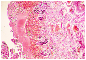
Infective endocarditis |
|
Image:
Image (binary/octet-stream)
|
Infective endocarditis |
|
Image:
Image (binary/octet-stream)
|
Infective endocarditis |
|
Image:
Image (binary/octet-stream)
|
Infective endocarditis |
|
Image:
Image (binary/octet-stream)
|
Infective endocarditis |
|
Image:
Image (binary/octet-stream)
|
Infective endocarditis |
|
Image:
Image (binary/octet-stream)
|
Infective endocarditis |
|
Image:
Image (binary/octet-stream)
|
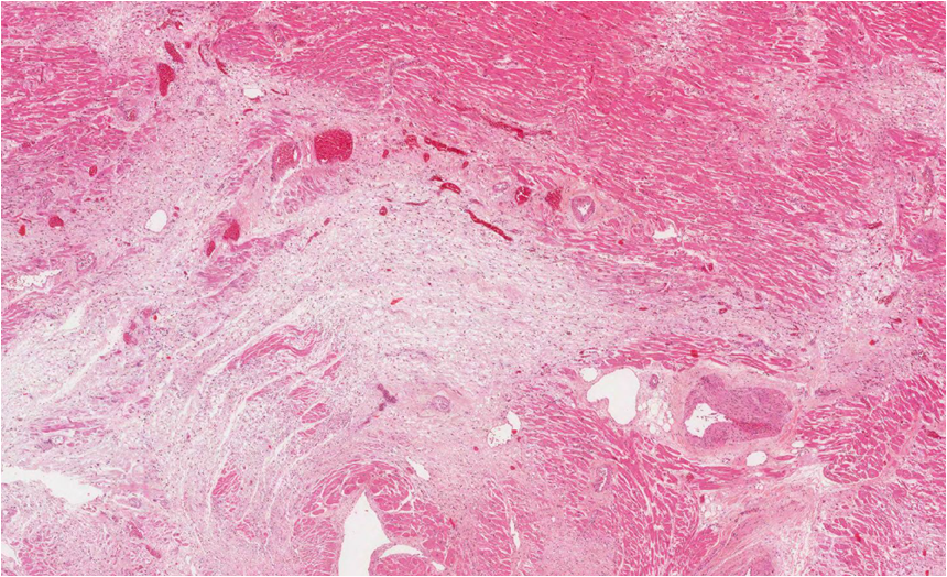
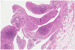
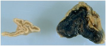
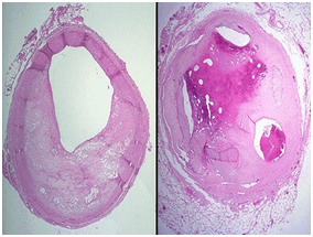
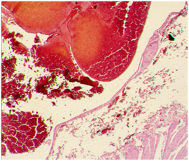
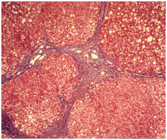
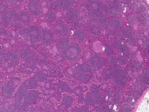
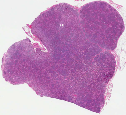
Liver Cirrhosis replacement of liver tissue by fibrosis (scar tissue) and regenerative nodules |
|
Image:
Image (binary/octet-stream)
|
Liver Cirrhosis replacement of liver tissue by fibrosis (scar tissue) and regenerative nodules |
|
Image:
Image (binary/octet-stream)
|
Liver Cirrhosis replacement of liver tissue by fibrosis (scar tissue) and regenerative nodules |
|
Image:
Image (binary/octet-stream)
|
Liver Cirrhosis replacement of liver tissue by fibrosis (scar tissue) and regenerative nodules |
| Appendicits dilated vein, leukocyte margination | |
| appendicitis exudate | |
| appendicitis exudate | |
| appendicits, irritation of capillaries-hyperemia, exudate | |
| serous inflammation viral inflammation of intestine, hyperemia, dilated veins, edema in glands, cellular infiltrate | |
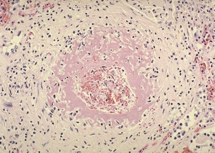
| fibrinous inflammation fibrinoid necrosis (pink circle), | |
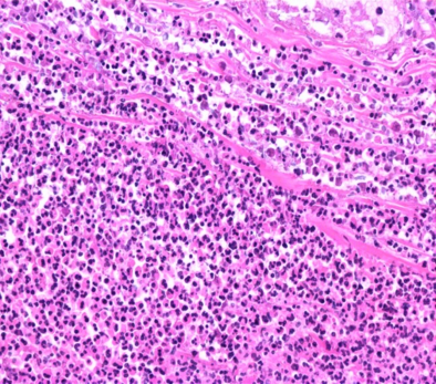
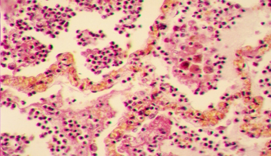
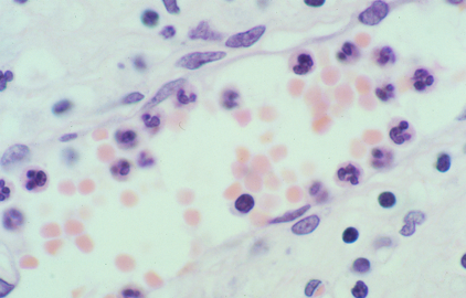
| active inflammation PMN (small dark points), macrophages, exudate | |
| acute bronchitis, exudate in lumen | |
| actue superlative inflammation, acute bronchitis, hyperemia, exudare | |
| hemorrhagic inflammation, crescentic and necrotizing glomerulonephritis | |
| hemorragic inflammation, hyperemia, macrophages, exudates | |
| hemorrhagic inflammation | |
| acute inflammation, swelling of cells, PMN&neutrophils and plasma cells, no fibrosis | |
| acute inflammation edema, not bacterial, no fibrosis, mononuclear cells (lymphocytes) | |
| chronic active inflammation, fibrosis, tubulitis, | |
| chronic active inflammation | |
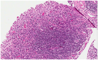
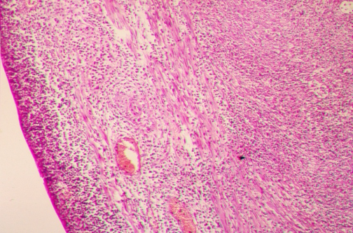
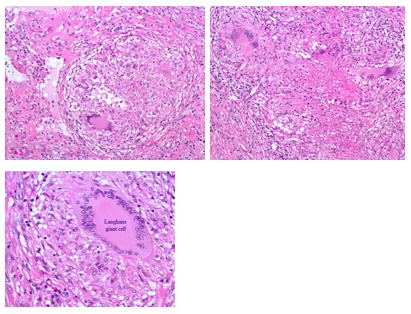
| granuloma capsule, giant cells | |
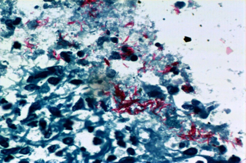
| granulomatous disease, TB, necrotizing granulomas | |
| margination, dilated vessel, RBC in center, show PMN | |
| temporal arteritis, giant cells, fibrosis of artery wall, lymphocytes (small dots) | |
| lung biopsy HE staining | |
| HE | |
| non-necrotizing granulomas, MMN | |
| foreign body giant cell, langerhans giant cell | |
| anthracosis | |
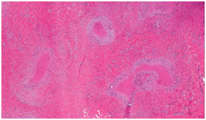
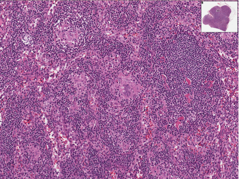
| lymph node biopsy | |
| lymph node biopsy | |
| lymph node biopsy | |
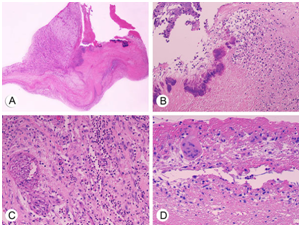
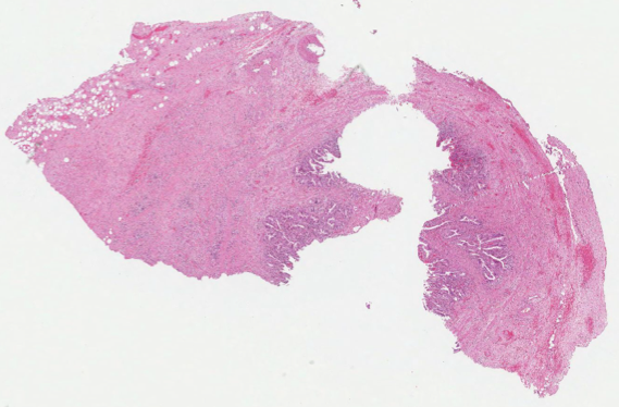
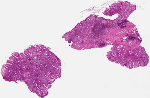
| chronic cholecystitis | |
| cirrhosis | |
| chronic cholecystitis | |
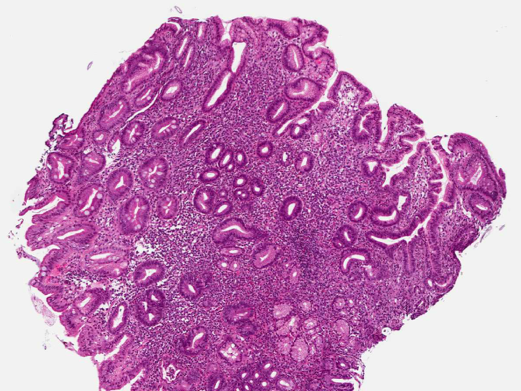
| lymphoid follicule | |
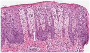
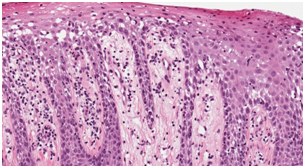
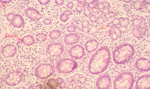
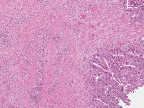
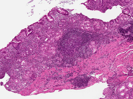
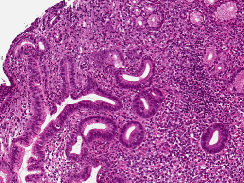
| stomach biopsy | |
| PMN, MMN, inflammation | |
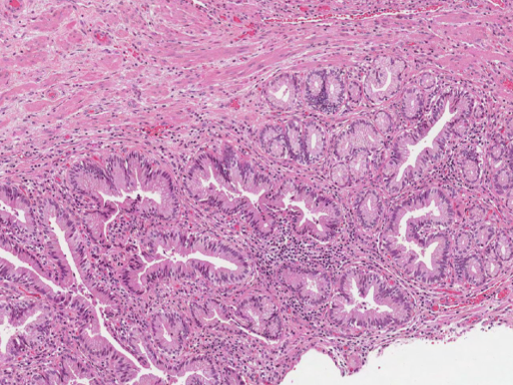
| intestinal metaplasia |
Want to create your own Flashcards for free with GoConqr? Learn more.