11155286
Beschreibung
Mindmap von Samantha Jarlette, aktualisiert more than 1 year ago
|
|
Erstellt von Samantha Jarlette
vor fast 7 Jahre
|
|
Diabetic Foot Ulcers
- Nursing Implementation
- Health
promotion
- Patient education
- Foot care (Montada-Atin, 2014)
- Correctly fitting and
supportive foot wear. Do
not go bare foot, and
inspect shoes before
putting them on in order
to reduce chance of
injury.
- Wash feet daily
with warm water,
pat dry; use
lanoline to
prevent drying
and cracking of
skin, do not apply
between toes
- Wear clean, cotton or
wool socks/stockings;
do not wear tight or
constrictive clothing
- Guard against
frost bite or
extreme heat
(e.g. heading
pads or hot
water bottles)
- Avoid crossing of legs
and prolong
sitting/standing that
may reduce
circulation
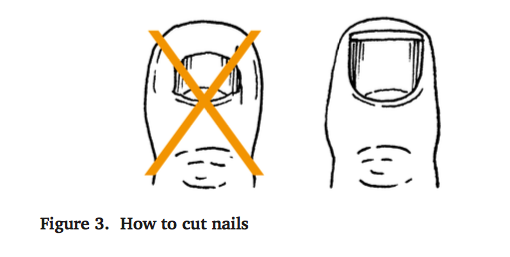
- Use nail file and
appropriate nail
cutting technique
(straight across); do
not use sharp
objects on feet
- Correctly fitting and
supportive foot wear. Do
not go bare foot, and
inspect shoes before
putting them on in order
to reduce chance of
injury.
- Smoking cessation to
reduce circulation
impairment (RNAO, 2013)
- Recognizing and
reporting the signs and
symptoms of potential
ulcer or increasing
infection (Bakker,
Apelqvist, Schaper, 2012)
- Foot care (Montada-Atin, 2014)
- Prevention, detection and monitoring
- Visual examination of foot
to be preformed daily
(Montada-Atin, 2014).
- Comprehensive foot
examination: includes
assessment of structural
abnormalities, neuropathy,
vascular disease (peripheral
pulses), ulcerations, and
evidence of infection
(Montada-Atin, 2014
- Type 1 diabetic: to be
preformed annually starting
5 years after onset of
diabetes (Montada-Atin,
2014)
- Type 2 diabetic: to be
preformed at diagnosis and
annually (Montada-Atin,
2014)
- Patients with high risk factors
should be examined every 1-6
months (Bakker, Apelqvist,
Schaper, 2012)
- Patients with high risk factors
should be examined every 1-6
months (Bakker, Apelqvist,
Schaper, 2012)
- Type 2 diabetic: to be
preformed at diagnosis and
annually (Montada-Atin,
2014)
- Type 1 diabetic: to be
preformed annually starting
5 years after onset of
diabetes (Montada-Atin,
2014)
- Semmes-Weinstein
monofilament screening
(Bakker, Apelqvist, Schaper,
2012).
- Visual examination of foot
to be preformed daily
(Montada-Atin, 2014).
- Patient education
- Treatment
- Wound care
- Debridement: If healable, remove
necrotic tissue and eschar through
surgical, mechanical, enzymatic or
autolytic methods (RNAO, 2013)
- Infection and inflammation
control: treat localized or
systemic spread of infection
(RNAO, 2013)
- Moisture balance: Provide moist wound
environment to encourage wound healing
and formulation of new tissue (RNAO, 2013).
Wound irrigation with noncytotoxic
solutions using a gentle pressure of 4-15 psi
to reduce trauma and injury (Montada-Atin,
2014)
- Debridement: If healable, remove
necrotic tissue and eschar through
surgical, mechanical, enzymatic or
autolytic methods (RNAO, 2013)
- Pressure relief
- Redistribute pressure
on feet through
custom orthotics, foam
or pressure
mattresses, foam or air
boots, sheet lifts, and
frequent positioning
(Aalaa et al., 2012)
- Redistribute pressure
on feet through
custom orthotics, foam
or pressure
mattresses, foam or air
boots, sheet lifts, and
frequent positioning
(Aalaa et al., 2012)
- Protecting
extremity from
injury
- Sheepskin under heels
and lower legs , footboard
at the end of patient bed,
and correct fitting shoes
(Aalaa et al., 2012)
- Sheepskin under heels
and lower legs , footboard
at the end of patient bed,
and correct fitting shoes
(Aalaa et al., 2012)
- Glucose control
- Nutritional therapy
- Eating three meals a day at regular, uniform
times. Eating at intervals no greater than 6
hours apart. Limiting simple sugar intake
(candies, pop, jam, etc.) and high-fat foods
(chips, fried foods, etc.). Consuming more
high-fibre foods such as vegetables, brown
rice, whole-grain bread, etc. Type 1 diabetics
may need to increase caloric intake, and
follow consistent timing of meals for glucose
control. Type 2 diabetics may need to
reduce caloric intake for weight control,
with emphasis on achieving glucose, lipid,
and blood pressure goals (Montada-Atin,
2014).
- Target glucose levels
- To reduce risk of vascular complications,
Canadian Diabetes Association
recommends: fasting plasma glucose level of
4.0 to 7.0 mm/L and A1c less than or equal to
7.0% (CDA, 2008)
- To reduce risk of vascular complications,
Canadian Diabetes Association
recommends: fasting plasma glucose level of
4.0 to 7.0 mm/L and A1c less than or equal to
7.0% (CDA, 2008)
- Exercise
- Exercise as tolerated: increases insulin activity,
lowers blood glucose levels, contributes to
weight loss, and increase peripheral
circulation (Montada-Atin, 2014).
- Exercise as tolerated: increases insulin activity,
lowers blood glucose levels, contributes to
weight loss, and increase peripheral
circulation (Montada-Atin, 2014).
- Nutritional therapy
- Wound care
- Health
promotion
- Pathophysiology
- Sensory neuropathy is a form of
diabetic neuropathy that damages
the nerves because of metabolic
derangements associated with
diabetes mellitus. Sensory
neuropathy can affect the hands
and feet, which cause paresthesias,
abnormal sensations, pain and loss
of sensation (Michel, 2014).
- Due to this development the skin can
become so sensitive that even light
pressure, like bed sheets, can’t be
tolerated and can break the skin (Michel,
2014). There is usually also a complete or
partial loss of sensitivity to touch,
temperature, and pain, which worsens
the patient’s susceptibility to foot ulcers
and their development.
- Sensory neuropathy is a form of
diabetic neuropathy that damages
the nerves because of metabolic
derangements associated with
diabetes mellitus. Sensory
neuropathy can affect the hands
and feet, which cause paresthesias,
abnormal sensations, pain and loss
of sensation (Michel, 2014).
- Clinical Manifestations
- Diabetic foot ulcers are most
commonly found on the soles of
the feet due to the constant
pressure, and typically cannot be
felt due to the neuropathy and
therefore worsen (Weledji &
Fokam, 2014). Clinical
manifestations include:
- Fever and/or chills in
advanced stages
- Swelling and warmth
around the wound
- Discolouration: red,
black, or blue
- Foul-smelling
discharge draining
from wound
- Thickened or
callused skin
around the ulcer
- Pain when the wound is
touched
- Fever and/or chills in
advanced stages
- Diabetic foot ulcers are most
commonly found on the soles of
the feet due to the constant
pressure, and typically cannot be
felt due to the neuropathy and
therefore worsen (Weledji &
Fokam, 2014). Clinical
manifestations include:
- Complications
- Amputation of lower extremities such as toes,
feet, and lower leg is an unfortunate but
common complication of diabetic foot ulcers
caused by diabetes mellitus. Due to the loss of
sensitivity a patient can have a foot ulcer
without even realizing it, the ulcer will then fail
to heal which can lead to a serious infection
(Weledji & Fokam, 2014).
- A patient suffering from diabetes
mellitus is more susceptibly
infection due to a defect in the
mobilization of inflammatory
cells and an impairment of
phagocytosis by neutrophils and
monocytes (Michel, 2014).
- The ulcer can go right
down to the bone and
allow for the infection to
spread into the whole foot,
which will then result in
amputation (Weledji &
Fokam, 2014).
- The ulcer can go right
down to the bone and
allow for the infection to
spread into the whole foot,
which will then result in
amputation (Weledji &
Fokam, 2014).
- A patient suffering from diabetes
mellitus is more susceptibly
infection due to a defect in the
mobilization of inflammatory
cells and an impairment of
phagocytosis by neutrophils and
monocytes (Michel, 2014).
- Amputation of lower extremities such as toes,
feet, and lower leg is an unfortunate but
common complication of diabetic foot ulcers
caused by diabetes mellitus. Due to the loss of
sensitivity a patient can have a foot ulcer
without even realizing it, the ulcer will then fail
to heal which can lead to a serious infection
(Weledji & Fokam, 2014).
- Assessment
- Assessment of foot ulcers is
extremely important because it
allows for healthcare
professionals to determine
appropriate treatment and
follow up care
- The Wagner-Meggitt
classification is what’s
universally used to
determine wound depth and
once wound depth is
clarified treatment can then
begin (Doupis & Veves,
2008).
- The Wagner-Meggitt
classification consists of 6
wound grades:
- Grade 0: skin intact
- Grade 1:
superficial
ulcer
- Grade 2: deep
ulcer to
tendon, bones,
or joint
- Grade 3: deep ulcer with
abscess or osteomyelitis
- Grade 4:
forefoot
gangrene
- Grade 5: whole foot
gangrene
- Grade 0: skin intact
- The Wagner-Meggitt
classification consists of 6
wound grades:
- The Wagner-Meggitt
classification is what’s
universally used to
determine wound depth and
once wound depth is
clarified treatment can then
begin (Doupis & Veves,
2008).
- Assessment of foot ulcers is
extremely important because it
allows for healthcare
professionals to determine
appropriate treatment and
follow up care
Medienanhänge
Möchten Sie kostenlos Ihre eigenen Mindmaps mit GoConqr erstellen? Mehr erfahren.