15863314
Brain Tumors
- Differential
Diagnosis of
Double Vision
- Lens
Problems
- Cataract
- Cataract
- Cornea Problems
- Astigmatism\Dry eyes
syndrome\Infections
- Astigmatism\Dry eyes
syndrome\Infections
- Eye Muscle Problems
- Strabismus\
Graves
disease
- Strabismus\
Graves
disease
- Nerve problem
- Diabetes\Myasthenia
gravis \MS\ Guillain Barre
syndrome
- Diabetes\Myasthenia
gravis \MS\ Guillain Barre
syndrome
- Brain problems
- Brain
tumors\Migraine\Increased
ICP
- Brain
tumors\Migraine\Increased
ICP
- Lens
Problems
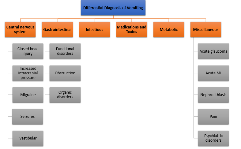
- Differential Diagnosis of Vomiting
- Differential Diagnosis of
Limb Weakness
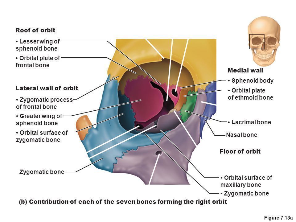
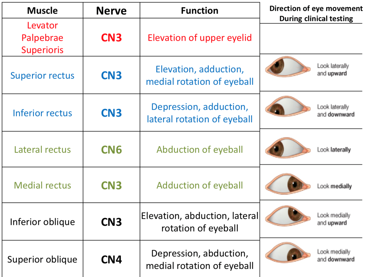
- Anatomy of orbit
- Anatomy of
extraocular
muscles
- Anatomy of
extraocular
muscles
- The Visual Pathway
- 1st order neuron
- its cell bodies lie in
the retina
(ganglion cells)
- their axons form the
optic nerve, optic
chiasma & optic tract.
- their axons form the
optic nerve, optic
chiasma & optic tract.
- their axons form the
optic nerve, optic
chiasma & optic tract.
- their axons form the
optic nerve, optic
chiasma & optic tract.
- its cell bodies lie in
the retina
(ganglion cells)
- 2nd order neuron
- its cell bodies lie in the L.G.B
- their axons form the optic
radiation that terminate in the
visual cortex of the occipital
lobe of the brain
- their axons form the optic
radiation that terminate in the
visual cortex of the occipital
lobe of the brain
- its cell bodies lie in the L.G.B
- 1st order neuron
- Types of speech
- Apraxia of speech
- and involves inconsistent production of speech sounds and rearranging of sounds in a word
- and involves inconsistent production of speech sounds and rearranging of sounds in a word
- Cluttering
- a rapid rate of speech, which makes speech difficult to understand.
- a rapid rate of speech, which makes speech difficult to understand.
- Dysarthria
- is a weakness or paralysis of speech muscles
- is a weakness or paralysis of speech muscles
- Dysprosody
- It is characterized by alterations in intensity, in the timing of utterance segments, and in rhythm,
cadence, and intonation of words.
- It is characterized by alterations in intensity, in the timing of utterance segments, and in rhythm,
cadence, and intonation of words.
- Muteness
- is complete inability to speak
- is complete inability to speak
- Speech Sound Disorders
- involve difficulty in producing specific speech sounds
- involve difficulty in producing specific speech sounds
- Stuttering
- Voice disorders
- are impairments, often physical, that involve the function of the larynx or vocal resonance
- are impairments, often physical, that involve the function of the larynx or vocal resonance
- Apraxia of speech
- Clinical
presentation
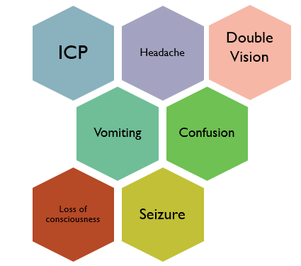
- Risk Factors of Brain Tumors
- Age\Gender\Home & Work Exposures\Family
History\Exposure to infections, viruses and
allergens\Electromagnetic fields \Race and
ethnicity\Ionizing Radiation\Head Injury & Seizures
- Age\Gender\Home & Work Exposures\Family
History\Exposure to infections, viruses and
allergens\Electromagnetic fields \Race and
ethnicity\Ionizing Radiation\Head Injury & Seizures
- Types of brain tumors
- Adults tumors
- primary
tumors are
usually
supratentorial.
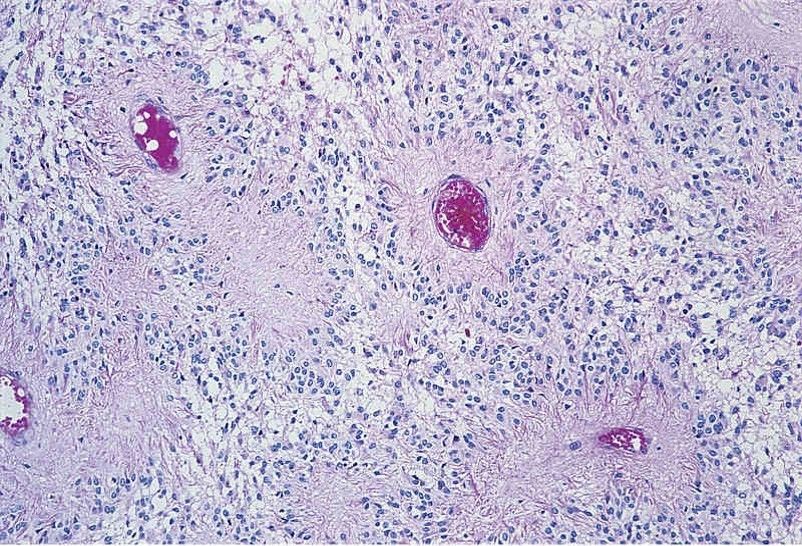
- glioblastoma multiforme
- Malignant, high-grade tumor of astrocytes
- Most common primary malignant CNS tumor in adults
- Usually arises in the cerebral hemisphere;
characteristically crosses the corpus callosum
- Butterfly lesion
- Histology
- Characterized by regions of necrosis surrounded by tumor
cells (pseudopalisading) and endothelial cell proliferation;
tumor cells are GFAP positive.
- Characterized by regions of necrosis surrounded by tumor
cells (pseudopalisading) and endothelial cell proliferation;
tumor cells are GFAP positive.
- Poor prognosis
- Malignant, high-grade tumor of astrocytes
- schwannoma
- Benign tumor of Schwann cells
- involves cranial or spinal nerves; within the cranium, most
frequently involves cranial nerve VIII at the cerebellopontine
angle (presents as loss of hearing and tinnitus)
- Tumor cells are S-100 positive.
- Histology
- prognosis
- Good prognosis after resection
- Good prognosis after resection
- Benign tumor of Schwann cells
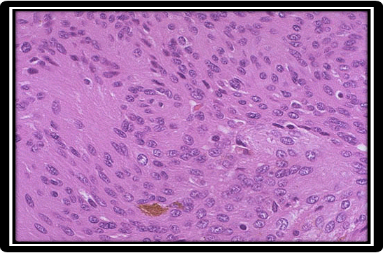
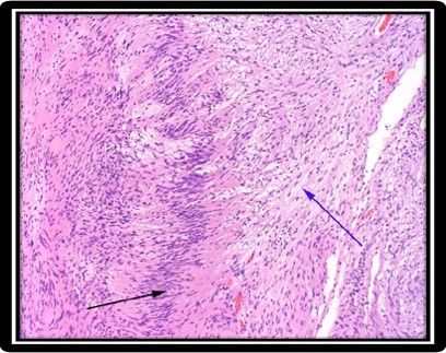
- meningioma
- Benign tumor of arachnoid cells
- Most common benign CNS tumor in adults
- More commonly
seen in women; rare
in children
- More commonly
seen in women; rare
in children
- May present as seizures; tumor compresses, but does not invade, the cortex.
- Imaging reveals a round mass attached to the dura.
- Histology
- shows a whorled pattern; psammoma bodies may
be present.
- shows a whorled pattern; psammoma bodies may
be present.
- Prognosis
- Good prognosis
- Grade 1
- 93%
- 93%
- Grade 2
- 5%
- 5%
- Grade 3
- 2%
- 2%
- Grade 1
- Good prognosis
- Benign tumor of arachnoid cells
- primary
tumors are
usually
supratentorial.
- Children tumors
- primary tumors are
usually infra tentorial
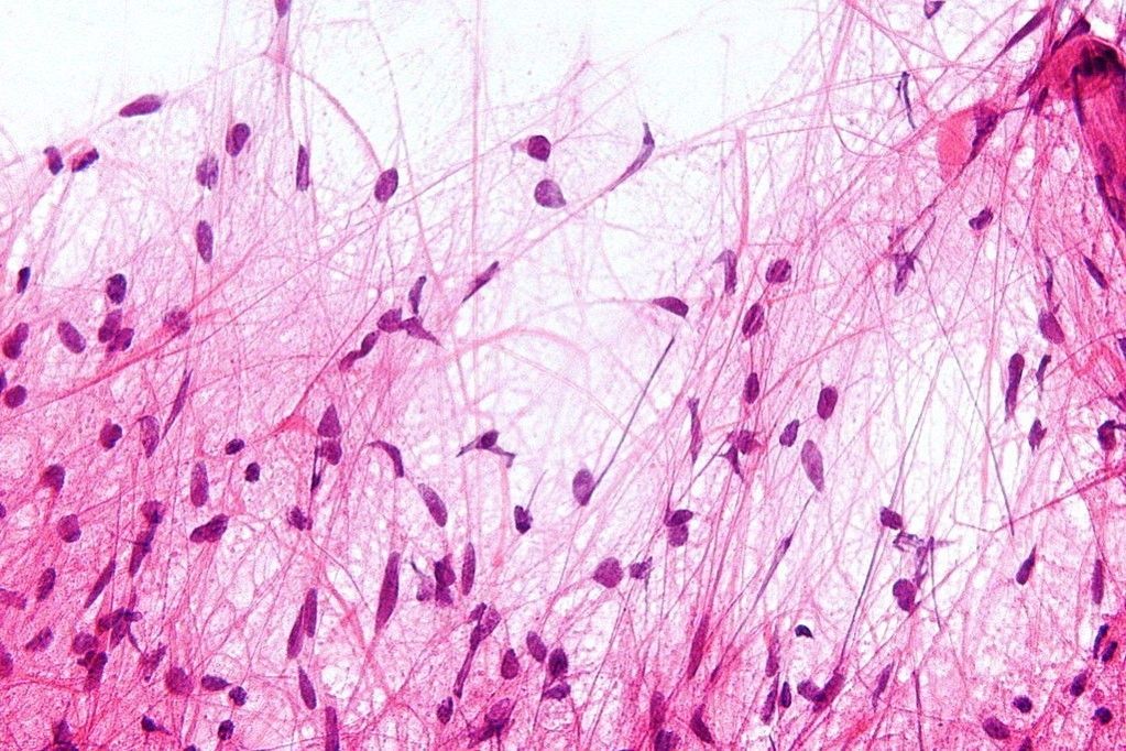
- pilocytic astrocytoma
- Benign tumor
of astrocytes
- Most common CNS tumor in
children; usually arises in the
cerebellum
- Imaging reveals a
cystic lesion with a
mural nodule
- Histology
- Biopsy shows Rosenthal fibers (thick
eosinophilic processes of astrocytes)
and eosinophilic granular bodies;
tumor cells are GFAP positive.
- Biopsy shows Rosenthal fibers (thick
eosinophilic processes of astrocytes)
and eosinophilic granular bodies;
tumor cells are GFAP positive.
- Grading
- Benign tumor
of astrocytes
- ependymoma
- Malignant tumor of
ependymal cells
- Most commonly arises in
the 4th ventricle; may
present with
hydrocephalus
- Histology
- Perivascular pseudorosettes are a
characteristic finding on biopsy
- Perivascular pseudorosettes are a
characteristic finding on biopsy
- Prognosis
- Recur after
surgery,
acquire more
aggressiveness
- Prognosis depends
on the location
and its histologic
grade.
- Prognosis depends
on the location
and its histologic
grade.
- Recur after
surgery,
acquire more
aggressiveness
- Malignant tumor of
ependymal cells
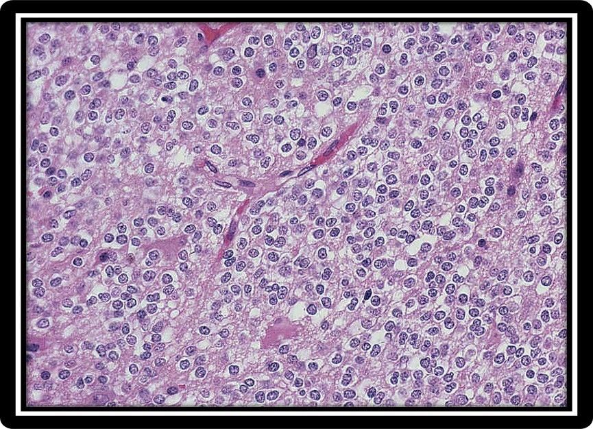
- medulloblastoma
- Malignant tumor derived
from the granular cells of the
cerebellum (neuroectoderm)
- Histology
- Histology reveals small, round
blue cells; Homer-Wright
rosettes may be present.
- Histology reveals small, round
blue cells; Homer-Wright
rosettes may be present.
- Poor prognosis; tumor
grows rapidly and spreads
via CSF.
- Metastasis to the cauda equina is
termed 'drop metastasis
- Metastasis to the cauda equina is
termed 'drop metastasis
- Prognosis
- Resection & radiation
allow 5-year survival
of 75%.
- Resection & radiation
allow 5-year survival
of 75%.
- Malignant tumor derived
from the granular cells of the
cerebellum (neuroectoderm)
- primary tumors are
usually infra tentorial
- Metastatic
Brain
tumors
- Breast\Lung\Skin\Kidney\GIT
- Breast\Lung\Skin\Kidney\GIT
- Other tumors
- OLIGODENDROGLIOMA
- Malignant tumor of oligodendrocytes
- Imaging reveals a calcified tumor in the
white matter, usually involving the frontal
lobe; may present with seizures
- Histology
- 'Fried-egg' appearance of cells on biopsy
- 'Fried-egg' appearance of cells on biopsy
- prognosis
- Slow-growing, long survival (5-10 yrs)
- Grade 2 (with treatment): 10-20 years
- Grade 3 (with treatment): 5-10 years
- 5-year survival rate is 75%,
10-year survival rate is 50%.
- Slow-growing, long survival (5-10 yrs)
- Malignant tumor of oligodendrocytes
- OLIGODENDROGLIOMA
- Adults tumors
- Investigations of space
occupying lesions of the brain
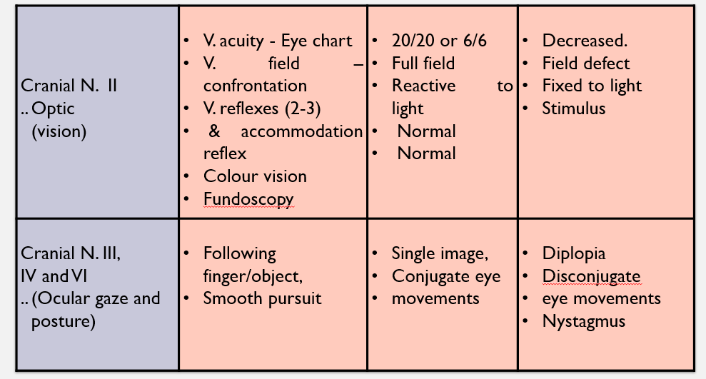
- Eye Examination
- Risks of Brain Surgery
- Allergic reaction to anesthesia\Bleeding in the brain\A blood clot\Brain swelling\Coma\Impaired
speech, vision, coordination, or balance\Infection in the brain or at the wound site\Memory
problems\Seizures\Stroke
- Allergic reaction to anesthesia\Bleeding in the brain\A blood clot\Brain swelling\Coma\Impaired
speech, vision, coordination, or balance\Infection in the brain or at the wound site\Memory
problems\Seizures\Stroke
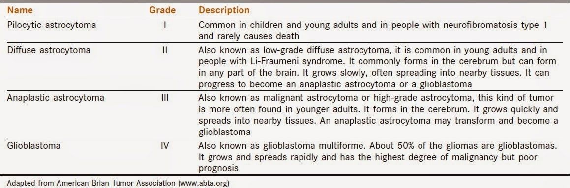
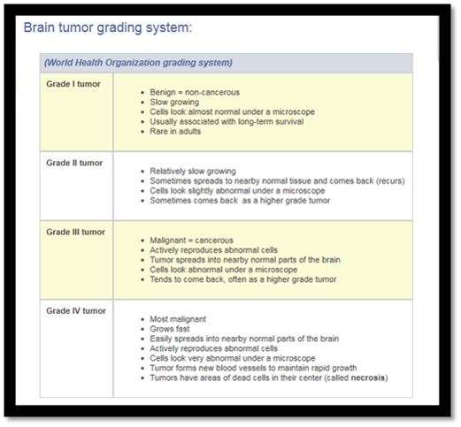
- Brain tumor grading system
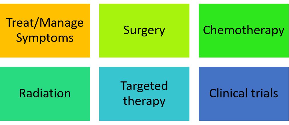
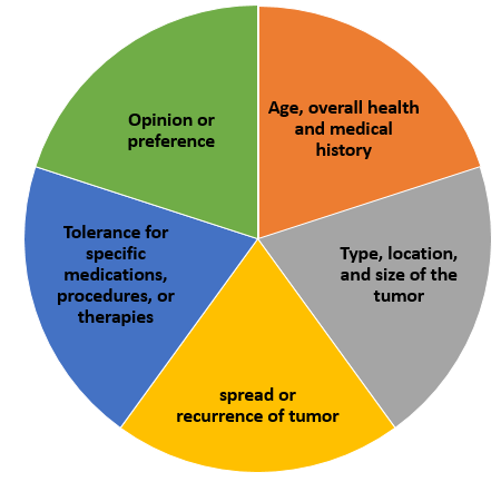
- Management plan
- Consider
Medienanhänge
Möchten Sie kostenlos Ihre eigenen Mindmaps mit GoConqr erstellen? Mehr erfahren.