3614477
Beschreibung
Mindmap von izyan fartini, aktualisiert more than 1 year ago
|
|
Erstellt von izyan fartini
vor mehr als 9 Jahre
|
|
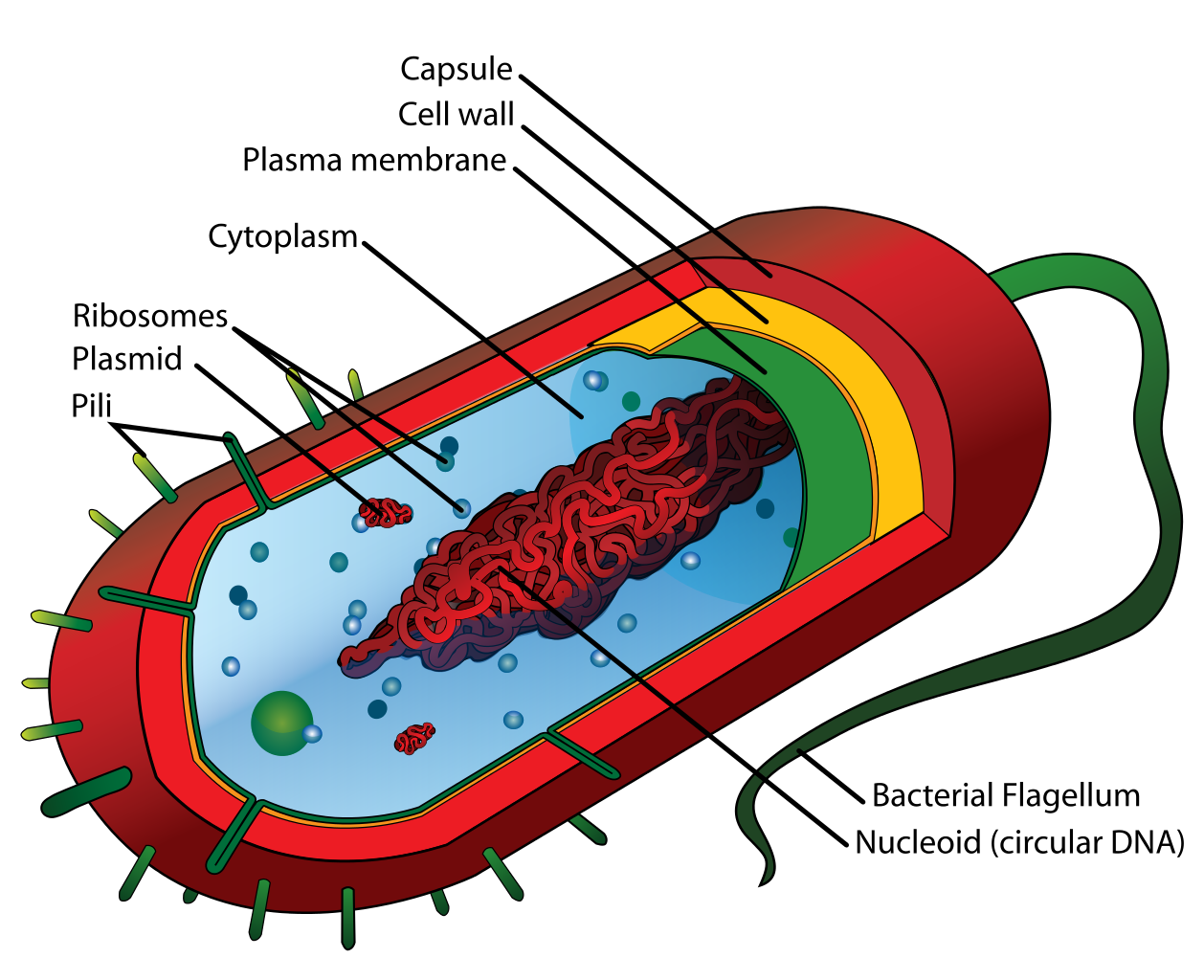
INTERNAL STRUCTURE OF
PROKARYOTES
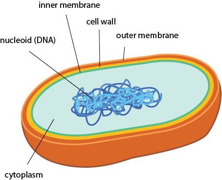
- STRUCTURE OF
PROKARYOTES
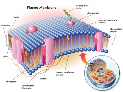
- PLASMA MEMBRANE
Anmerkungen:
- The prokaryotic plasma membranes are composed of phospholipids bilayer with embedded proteins. In the middle of the bilayer, the fatty acids of the phospholipids are found, which is called as hydrophobic region. Prokaryotic cells can have multiple plasma membranes. In prokaryotic organisms, plasma membranes are responsible for controlling the entry and exit of the cell
- Functions
- 1. Separate the cell from
its outside environment
- 2. Regulates what enter
and out of the cell.
- 3. Protecting the integrity
of the interial of the cell
- - allowing only
selected substance
into the cell and
keeping other
substances out.
- - allowing only
selected substance
into the cell and
keeping other
substances out.
- 4. serves as a base of attachment for
the cytoskeleton in some organisms
and the cell wall in others
- 5. The lipid bilayer is semi-permeable, which
allows only selected molecules to diffuse
across the membrane
Anmerkungen:
- https://www.youtube.com/watch?v=UgN76naeA1Q
- 1. Separate the cell from
its outside environment
- Functions
- CYTOPLASM
- gel-like, yet fluid, substance in which all of the
other cellular components are suspended.
- gel-like, yet fluid, substance in which all of the
other cellular components are suspended.
- RiBOSOMES
- protein builders or the protein
synthesizers of the cell.
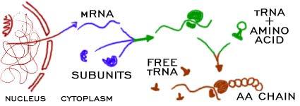
- when are ribosomes used in the process
of protein synthesis?
- when the cell needs to make a protein,
mRNA is created in the nucleus
- The mRNA is then sent out of the
nucleus and to the ribosomes.
- when it is time to make protein,
the 2 sub units come together
and combine with the mRNA.
- the 2 sub units lock onto
the mRNA and start the
protein synthesis.
- the 2 sub units lock onto
the mRNA and start the
protein synthesis.
- when it is time to make protein,
the 2 sub units come together
and combine with the mRNA.
- The mRNA is then sent out of the
nucleus and to the ribosomes.
- when the cell needs to make a protein,
mRNA is created in the nucleus
- when are ribosomes used in the process
of protein synthesis?
- protein builders or the protein
synthesizers of the cell.
- INCLUSIONS
- - nuclear or cytoplasmic aggregates of
stainable substances, usually protein.
- TYPES : 1.
metachromatic
granules 2.
Polysaccharide
granules 3. Lipid
inclusion 4.
Sulphur granules
5. Carboxysomes
6. Magnetosomes
7. Gas vacuoles
- - nuclear or cytoplasmic aggregates of
stainable substances, usually protein.
- ENDOSPORE
Anmerkungen:
- https://www.youtube.com/watch?v=7zCQLITFEb0
- a dormant, tough and
non-reproductive
structure produced by
certain bacteria from the
Firmicute phylum.
- Special stain technique
- Moeller stain : Allow the endospore
to show up as red while the rest of
the cell stains blue.
- Schaeffer-Fulton stain : Stains
endospore green and bacterial
bodies red.
- Moeller stain : Allow the endospore
to show up as red while the rest of
the cell stains blue.
- Special stain technique
- FORMATION OF ENDOSPORE
- FORMATION OF ENDOSPORE
- PLASMA MEMBRANE
Medienanhänge
Möchten Sie kostenlos Ihre eigenen Mindmaps mit GoConqr erstellen? Mehr erfahren.