17210432
Description

Flashcards by Med Student , updated more than 1 year ago
|
|
Created by Med Student
almost 6 years ago
|
|
| Question | Answer |
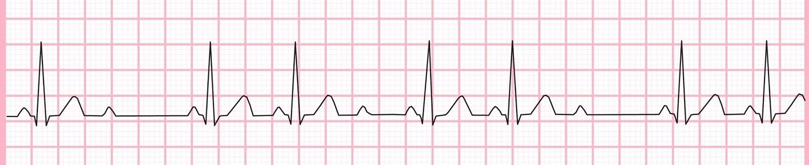
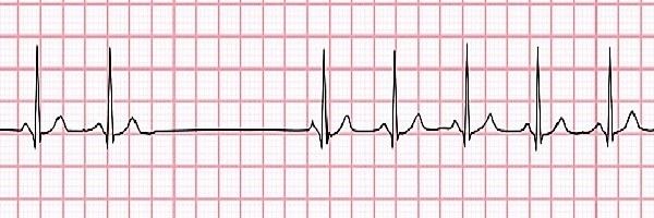
| Premature Atrial Contraction (Supra-ventricular Premature Contraction) ECG 1. Premature beat appears early 2. After this there is a compensatory pause before the next sinus p wave 3. This means that there is a really short RR interval and the very next RR interval is longer but is u combine these 2 their length will be shorter than 2 normal RR intervals due to the incomplete compensatory pause | |
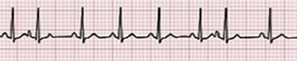
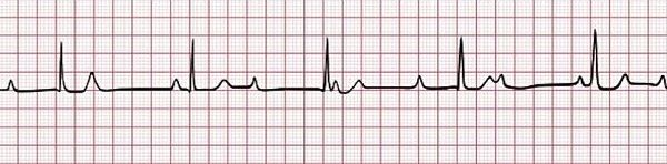
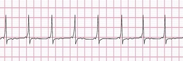
| Supraventricular Tachycardia ECG No p wave Very short RR interval Normal QRS | |
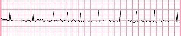
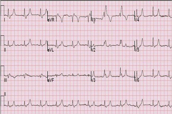
| Atrial Fibrillation ECG No P waves Arrhythmia F waves No isoelectric line | |
| Atrial Flutter ECG F waves- sawtooth No p waves No isoelectric line | |
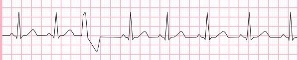
| Premature Ventricular Contractions (Ventricular Extrasystole) ECG No P wave preceeding PVC Wide & Bizzare QRS. QRS will be at least 0.12s-0.14s | |
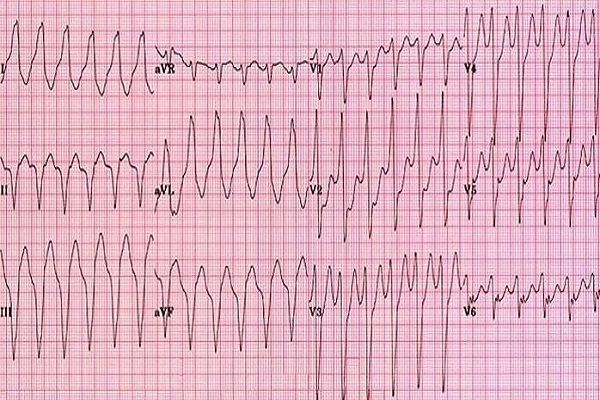
| Ventricular Tachycardia ECG Monomorphic QRS complex- all look the same- from re-entrant loop Polymorphic QRS Increased heart rate Regular rhythm No p-wave or disassociated p-wave No PR interval Wide QRS width | |
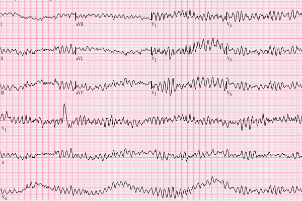
| Ventricular Fibrillation ECG Chaotic rhythm No p wave No PR interval No QRS complex is identifiable Looks like a bunch of squiggly line | |
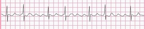
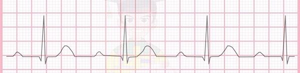
| AV Block Type 1 ECG The signal to AV node is delayed Increased PR Interval | |
| AV Block Type 2a (Mobitz 1) ECG The signal (PR interval) gets progressively delayed until a ‘dropped beat’ | |
| AV Block Type 2b (Mobitz 2) ECG There are intermittent random dropped beats No PR lengthening | |
| AV Block Type 3 (Complete) ECG P waves have no relation to QRS intervals | |
| Right Bundle Branch Block ECG 1. Shows RR waves due to slow activation of contraction left/right ventricles • ‘Bunny ears’ 2. Wide QRS Complex 3. RR wave in right leads- V1 and V2 4. S wave in Lead 1 is wider >0.04 and this S wave is also in V5 and V6 | |
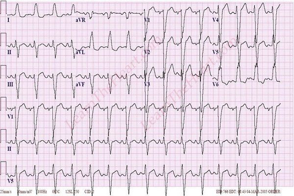
| Left Bundle Branch Block ECG 1. Shows RR waves due to slow activation of contraction left/right ventricles • ‘Bunny ears’ 2. Wide QRS complex 3. RR wave in left leads- V5 and V6 4. Also RR in first and aVL 5. R wave don’t increase in V1 to V4- Pathological progression | |
| SA Node Block Heart Rate- Varies Rhythm- Irregular P waves- Present and normal Pr Interval- Normal Dropped Beats- present | |
|
Image:
Wpw (binary/octet-stream)
|
Wolf Parkinson White Syndrome Short PR interval “Delta wave“ - A delta wave is slurring of the upstroke of the QRS complex. Short PR interval Wider QRS complexes |
Want to create your own Flashcards for free with GoConqr? Learn more.