
Notes
Dirofilaria - Dog & Cat
Content of the notes (1)
Page 1
Dirofilaria Dog
1.
Epidemiology/microbiology
·
Dirofilaria immitus most common cause of
heartworm and endemic in US.
·
Canids are definitive host, but found in a
number of other species.
·
Endemic in North and South America, coastal
Africa, southern Europe & Asia, cases increasing every year.
·
Transmission
o
Reservoir of disease in domestic and feral dogs.
o
Mosquito vector (more than 70 species have been
shown to transmit).
o
Environmental temperature above 57 0F
(13 0C) for development of infective L3 larvae.
·
Life Cycle
o
Life cycle of 7-9 months
o
Female mosquitoes ingest microfilariae from
host.
o
Microfilariae transform into L1 larvae after
entering mosquito.
o
Two moults over 2-4 weeks (depending on
temperature) into L3.
o
L3 deposited onto dog’s skin in a droplet of
hemolymph when feeding.
o
Enter through bite would and moult to L4 within
a couple of days.
o
L4 migrate through body and from 50-70 days
after infection undergo moult to juvenile worms.
o
Worms penetrate circulation, mature over several
months in pulmonary vessels. Produce microfilariae around 180-210 days.
o
Mature adults have a life expectancy of 5-7
years and vary in length from 15-18cm (males) to 25-30cm (females).
·
Pathogenesis
o
Damage to pulmonary arteries and lungs –
eosinophilic pneumonitis and endothelial damage.
o
Wolbachia also implicated in disease process but
mechanism unknown.
o
Severity of disease related to interaction with
host, exercise seems to make clinical signs appear more quickly.
o
As worms die lodge in small distal pulmonary
arteries – PTE. Leads to haemorrhage and fibrosis – Pulmonary hypertension.
o
Leads to cor pulmonale and right-sided heart
failure.
o
Caval syndrome occasionally reported in large
worm burdens due to obstruction of right atrium.
o
Glomerulonephropathy is also reported.
2.
Clinical
findings
·
Frequently asymptomatic, especially if inactive.
·
Mild cough often noted first, followed by
exercise intolerance and unthrifty appearance.
·
Advanced disease – PTE, ascites, lethargy,
haemoglobinaemia.
3.
Diagnostic
investigations
·
Radiography – reversed D cardiac silhouette,
caudal lobar pulmonary arteries enlarged and tortuous. May be accompanied by
pulmonary parenchymal disease.
·
Microfilarial detection – direct examination
using ‘Knott’s procedure’. Negative in up to 80% of cases due to single sex
infections, immune response, heartworm preventative. Positive from 6 months
after infection.
·
Serology - Antigen detection – sensitivity
95-100%, specificity 100%, as long as 3 or more female worms. False negatives
in all male infections and with low number of females. Detected by 5-7 months
after infection.
4.
Treatment/prognosis
·
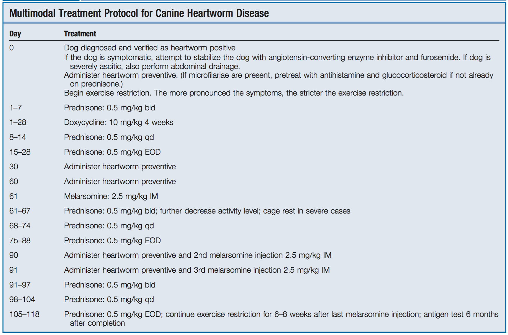
Medical management:
o
Melarsomine – arsenical-based drug, effective
against adults and filarial stages from 4 months. Two dose protocol only kills
90% of adults, 3 dose protocol 98%
o
Macrocyclic lactones – ivermectin, milbemycin,
moxidectin, selamectin. Preventatives. Administered for 2 months before an
adulticide to eliminate juvenile stages and allow maturation of adults for
melarsomine to be effective. Pre-treat with antihistamines/glucocorticoids.
o
Doxycycline – for Wolbachia.
·
Prevention – macrocyclic lactone heartworm
preventatives.
·
Prognosis – varies with severity of worm burden
and clinical signs from excellent to grave.
·
Public health – rare reports of human
infections.
Feline Heartworm Disease
·
Same pathogen can infect cats, distribution the
same as canine disease.
·
Life cycle the same as in dogs up to the
immature adult stage, at this point in most cats the juvenile worms die 3-4
months after infection resulting in an abbreviated infection. In a very small
number of cats the worms mature to adults and live 2-4 years.
·
Pathogenesis:
o
Acute vascular and parenchymal inflammatory
response to the worms (whether or not disease progresses).
o
Can lead to severe lung injury and sudden death
in some cats (10-20% of cats with adult infections).
o
Can develop chronic respiratory disease (whether
abbreviated or adult infection).
·
Clinical findings – coughing or dyspnoea (64%),
intermittent vomiting (38%), 28% no clinical signs. Respiratory distress,
ataxia, collapse, seizures, sudden death rarely reported.
·
Diagnosis – difficult.
o
Radiology – Vascular enlargement (64%),
hyperinflation.
o
Echocardiography – worms visible in 64%
o
Antigen testing – positive in 55% usually don’t
get adult worms so will be negative.
o
Antibody testing – positive in 82%
o
Microfilarial detection – negative in 90%
·
Therapy – no adulticide proven to be of benefit,
therefore use long-term macrocyclic lactones and steroids.
·
10-20% mortality within 4 years.
·
Prevention – as dogs.
Literature:
·
Case report of an adult Dirofilaria worm being
found in the spinal canal of an adult cat leading to hind limb paraparesis
(JFMS 2013).