20499244
Description
Quiz by Charlotte Jakes, updated more than 1 year ago
|
|
Created by Charlotte Jakes
about 5 years ago
|
|
Question 1
Question
What is the central nervous system?
Answer
-
The brain and spinal cord from which axons extend to supply motor fibres to muscles and collect sensory information
-
Axons gathered into bundles (nerves) as well as their ganglia and supporting cells
-
The neurons within the brain
-
The spinal cord
Question 2
Question
What is the peripheral nervous system?
Answer
-
Axons gathered into bundles (nerves), their ganglia and supporting cells
-
The brain and spinal cord
-
The branch of the nervous system that is not under our voluntary control
-
The branch of the nervous system that innervates the GI tract only
Question 3
Question
Select all the functions of the support cells of the peripheral nervous system
Answer
-
Support the neuron structure
-
Regulate interstitial fluid
-
Supply cells with nutrients
-
Receive synaptic connections from neighbouring neurons
-
Regulate the Na+/K+ gradient across the axonal membrane
-
Form the dendrites of the neuron
Question 4
Question
Neurons are highly differentiated and have almost no capacity for division.
Answer
- True
- False
Question 5
Question
Which neuroglial cell type stabilises structures following injury to produce scar tissue, directs neural growth during development, controls the interstitial environment and maintains the blood-brain barrier?
Answer
-
Astrocytes
-
Microglia
-
Ependymal cells
-
Oligodendrocytes
Question 6
Question
Which type of neuroglial cell makes up only 5% of glial cells but numbers increase in the event of infection due to their behaviour as macrophages?
Answer
-
Microglia
-
Asrocytes
-
Ependymal cells
-
Oligodendrocytes
Question 7
Question
Which neuroglial cell lines the chambers and passageways of CNS that are filled with CSF that monitor the composition of as well as secrete this CSF?
Answer
-
Microglia
-
Astrocytes
-
Ependymal cells
-
Oligodendrocytes
Question 8
Question
Which neuroglial cell type forms myelin sheaths around axons by their processes wrapping around them in a layered structure?
Answer
-
Oligodendrocyes
-
Ependymal cells
-
Microglia
-
Astrocytes
Question 9
Question
The cerebrospinal fluid protects the CNS from shock. Where is it found?
Answer
-
Surrounds the brain and spinal cord in the subarachnoid space and the ventricles
-
Occurs in the brain in the ventricles only
-
Occurs around the spinal cord in the vertebral column only
-
Surrounds the brain in the subarachnoid space only
Question 10
Question
Neuroglial cells are found throughout the entire nervous system.
Answer
- True
- False
Question 11
Question
Which is the only type of neuroglial cell that forms branches to connect with other neuroglial cells?
Answer
-
Ependymal cells
-
Oligodendrocytes
-
Astrocytes
-
Microglia
Question 12
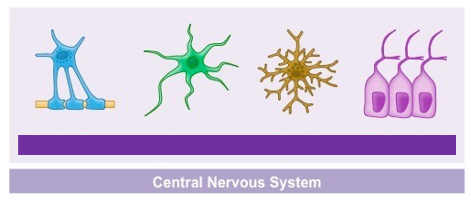
Question
Label this image to name each neuroglial cell according to its structure.
Answer
-
Oligodendrocyte
-
Astrocyte
-
Microglia
-
Ependymal cell
Question 13
Question
Which type of support cell in the peripheral nervous system surrounds cell bodies in the ganglia to regulate the levels of oxygen, carbon dioxide, nutrients etc around them?
Answer
-
Satellite cells
-
Schwann cells
-
Oligodendrocytes
-
Microglia
Question 14
Question
Which type of support cell in the peripheral nervous system surrounds axons forming the myelin sheath?
Answer
-
Schwann cells
-
Satellite cells
-
Oligodendrocytes
-
Ependymal cells
Question 15
Question
Schwann cells form nodes of Ranvier by breaking down their myelin at regular intervals.
Answer
- True
- False
Question 16
Question
What name is given to collections of cell bodies lying outside the CNS?
Answer
-
Ganglia
-
Satellite cells
-
Synapses
-
Soma cords
Question 17
Question
Which ganglia do not contain synapses?
Answer
-
Sensory ganglia
-
Motor ganglia
Question 18
Question
Which ganglia contain synapses?
Answer
-
Motor ganglia
-
Sensory ganglia
Question 19
Question
For the most part, pre-ganglionic fibres are myelinated and post-ganglionic fibres are unmyelinated.
Answer
- True
- False
Question 20
Question
Fill in the blanks below to describe the different types of neuron in terms of their poles.
[blank_start]Unipolar[blank_end] neurons are found almost exclusively in the cerebellum and have one one axon extending from their cell body.
[blank_start]Bipolar[blank_end] neurons have two axons extending from one cell body.
[blank_start]Pseudounipolar[blank_end] neurons have a single branch from their cell body splitting into two axons - one that goes to the spinal cord and the other that goes to the peripheral tissues.
[blank_start]Multipolar[blank_end] neurons have multiple dendrites extending from their cell body with a single axon extending in the opposite direction.
Answer
-
Bipolar
-
Unipolar
-
Pseudounipolar
-
Multipolar
Question 21
Question
What name is given to the layers of connective tissue covering the brain and spinal cord to protect them?
Answer
-
Meninges
-
Camper's fascia
-
Cerebrospinal membranes
-
Ependymal cells
Question 22
Question
What is the correct layering of the meninges from outermost to innermost?
Answer
-
Dura mater, arachnoid mater, pia mater
-
Dura mater, pia mater, arachnoid mater
-
Aranchoid mater, dura mater, pia mater
-
Arachnoid mater, pia mater, dura mater
-
Pia mater, dura mater, arachnoid mater
Question 23
Question
Fill in the blanks to describe the structure of the dura mater.
The dura mater is the outermost layer of the meninges that has a [blank_start]two[blank_end]-layered structure. The outer layer fuses with the [blank_start]periosteum[blank_end] to line the cranial cavity - this is the [blank_start]periosteal[blank_end] layer. The inner layer is known as the [blank_start]meningeal[blank_end] layer which has specialised [blank_start]folds[blank_end] known as [blank_start]dural[blank_end] reflections that support the brain and [blank_start]partition[blank_end] it. Venous [blank_start]sinuses[blank_end] run in gaps between the two layers.
Answer
-
two
-
periosteum
-
periosteal
-
meningeal
-
folds
-
dural
-
partition
-
sinuses
Question 24
Question
How does the arachnoid mater link to the pia mater?
Answer
-
Meshwork of collagen and elastic fibres in the subarachnoid space
-
Desmosomes linking each individual cell of the layer
-
Venous sinuses run between them
-
Mucus layer secreted by the pia mater
Question 25
Question
What is found in the subarachnoid space which allows the layer to act as a 'suspension' system to protect the brain and spinal cord from sudden impact?
Answer
-
Cerebrospinal fluid
-
Venous drainage
-
Lymph
-
Mucus
Question 26
Question
Which cells tightly attach the pia mater to the brain, allowing it to follow the brain's contours?
Answer
-
Astrocytes
-
Microglia
-
Oligodendrocytes
-
Cell bodies of neurons
Question 27
Question
The pia mater is highly vascularised.
Answer
- True
- False
Question 28
Answer
-
Periosteal dura mater
-
Meningeal dura mater
-
Arachnoid mater
-
Subarachnoid space
-
Pia mater
Question 29
Question
The cerebrospinal fluid is an ultrafiltrate of blood that will eventually return back into circulation.
Answer
- True
- False
Question 30
Question
Fill in the blanks below to describe the flow of cerebrospinal fluid around the brain.
1. CSF is secreted by the [blank_start]choroid[blank_end] plexus in each [blank_start]lateral[blank_end] ventricle. These are plexuses of cells including modified [blank_start]ependymal[blank_end] cells.
2. CSF flows through the [blank_start]interventricular foramina[blank_end] into the [blank_start]third[blank_end] ventricle.
3. Choroid [blank_start]plexus[blank_end] in the third ventricle secretes more CSF.
4. CSF flows down [blank_start]cerebral aqueduct[blank_end] to the fourth ventricle.
5. Choroid plexus in the [blank_start]fourth[blank_end] ventricle secretes more CSF.
6. CSF flows out via two [blank_start]lateral[blank_end] apertures and one [blank_start]median[blank_end] aperture.
7. CSF fills the [blank_start]subarachnoid[blank_end] space to bath the external surfaces of the brain and spinal cord.
8. CSF is reabsorbed into venous drainage of the [blank_start]dural[blank_end] venous sinuses/
Answer
-
choroid
-
lateral
-
ependymal
-
interventricular foramina
-
third
-
plexus
-
cerebral aqueduct
-
fourth
-
lateral
-
median
-
subarachnoid
-
dural
Question 31
Question
Fill in the blanks below to describe the subdivisions of the peripheral nervous system.
The [blank_start]somatic[blank_end] nerves supply the body wall, skeletal muscle and skin. They contain both motor and sensory fibres that facilitate voluntary movement.
Nerves of [blank_start]special sensation[blank_end] facilitate the experience of sight, smell, taste, hearing and balance.
[blank_start]Autonomic[blank_end] nerves supply the internal organs with motor fibres to smooth/cardiac muscle and sensory fibres to control involuntary functions of the body.
Answer
-
somatic
-
special sensation
-
Autonomic
Question 32
Question
All sensory axons - somatic or autonomic - have their cell bodies outside the CNS in ganglia with no synapses.
Answer
- True
- False
Question 33
Question
All motor ganglia belong to the autonomic nervous system and always contain synapses.
Answer
- True
- False
Question 34
Question
What do the dorsal roots of the spinal cord carry?
Answer
-
Afferent sensory axons
-
Efferent motor axons
-
Efferent sensory axons
-
Afferent motor axons
Question 35
Question
What do the ventral roots of the spinal cord carry?
Answer
-
Afferent motor fibres
-
Efferent motor fibres
-
Afferent sensory fibres
-
Efferent sensory fibres
Question 36
Question
Where are the cell bodies of the motor efferent neurons found?
Answer
-
Grey matter of the spinal cord
-
White matter of the spinal cord
-
Ganglia outside the CNS
-
In the subarachnoid space
Question 37
Question
Outside of the spinal cord, the spinal nerves divide into branches known as rami.
The [blank_start]dorsal[blank_end] ramus carries nerves that carry visceral motor, somatic motor and sensory information to and from the skin and muscles of the back.
The [blank_start]ventral[blank_end] ramus contains nerves that carry visceral motor, somatic motor and sensory information to and from the ventrolateral surface, the body wall and limbs.
Answer
-
dorsal
-
ventral
Question 38
Question
What is the rami communicantes?
Answer
-
Branches of the spinal nerve connecting it to the sympathetic trunk carrying autonomic fibres
-
Branches of the spinal nerve connecting it to the anterior body trunk (muscles, skin, organs)
-
Branches of the spinal nerve connecting it to the posterior body trunk (muscles, skin, organs)
-
Collections of afferent sensory cell bodies outside the CNS
Question 39
Question
Fill in the blanks to label this cross section of the spinal cord.
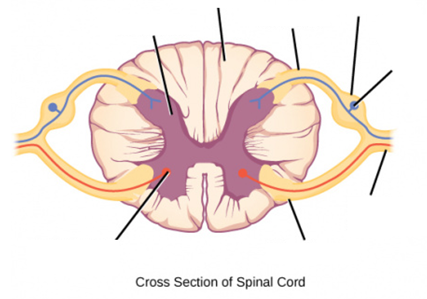
Answer
-
Grey matter
-
White matter
-
Dorsal root
-
Ventral root
-
Sensory soma
-
Dorsal root ganglion
-
Spinal nerve
-
Motor soma
Question 40
Question
Fill in the blanks to label this diagram showing the rami communicantes of the spinal nerves.
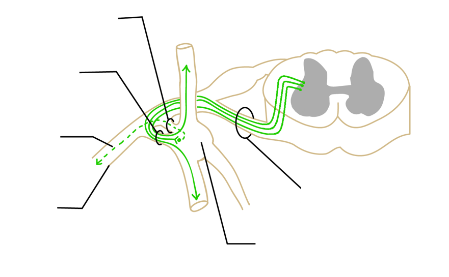
Answer
-
Sympathetic chain ganglion
-
Spinal nerve
-
Postganglionic axon
-
White ramus communicans
-
Grey ramus communicans
-
Preganglionic axons
Question 41
Question
Anything referred to as 'white' in regards to the nervous system contains myelinated axons.
Answer
- True
- False
Question 42
Question
There are [blank_start]31[blank_end] left-right pairs of spinal nerves in humans.
There are [blank_start]8[blank_end] cervical spinal nerve pairs.
There are [blank_start]12[blank_end] thoracic spinal nerve pairs.
There are [blank_start]5[blank_end] lumbar spinal nerve pairs.
There are [blank_start]5[blank_end] sacral spinal nerve pairs.
There is 1 [blank_start]coccygeal[blank_end] pair of spinal nerves.
Answer
-
31
-
8
-
12
-
5
-
5
-
coccygeal
Question 43
Question
What is a myotome?
Answer
-
Set of muscles innervated by single specific spinal nerve
-
All the spinal nerves that innervate a single muscle
-
A bundle of motor fibres
-
The ganglion in which afferent motor cell bodies gather
Question 44
Question
What is a dermatome?
Answer
-
Area of skin supplied by afferent nerve fibres from a single dorsal root of a spinal nerve
-
All the afferent nerve fibres that supply a particular area of skin
-
Bundles of sensory cell bodies outside the CNS
-
A set of muscles innervated by a single specific spinal nerve
Question 45
Question
What type of neurons are sensory neurons?
Answer
-
Pseudounipolar
-
Unipolar
-
Bipolar
-
Multipolar
Want to create your own Quizzes for free with GoConqr? Learn more.