5818188
Description
Quiz by Rachel Nall, updated more than 1 year ago
|
|
Created by Rachel Nall
about 8 years ago
|
|
Question 1
Question
A [blank_start]Standard of Care[blank_end] is the highest mandate for clinical behavior.
Answer
-
Standard of Care
Question 2
Question
Select the standard of nursing practice that most applies to CRNAs.
Answer
-
Standard III
-
Standard IV
-
Standard V
-
Standard VI
Question 3
Question
Fill in the blanks for the AANA Standards for Nurse Anesthesia Practice:
Standard V
A. Monitor [blank_start]ventilation[blank_end] continuously.
B. Monitor [blank_start]oxygenation[blank_end] continuously.
C. Monitor [blank_start]cardiovascular[blank_end] status continuously.
D. Monitor [blank_start]body temperature[blank_end] continuously.
E. Monitor [blank_start]neuromuscular function[blank_end] and status.
F. Monitor and assess the [blank_start]patient positioning[blank_end].
Answer
-
ventilation
-
oxygenation
-
cardiovascular
-
body temperature
-
neuromuscular function
-
patient positioning
Question 4
Question
One of the key components to patient safety is to: [blank_start]Avoid turning your back on the patient[blank_end] and surgical procedure for long periods of time.
Answer
-
Avoid turning your back on the patient
Question 5
Question
[blank_start]Monitors[blank_end] are placed first and removed last.
Answer
-
Monitors
Question 6
Question
Name two ways (in alpha order) that CRNAs monitor the respiratory system:
1. [blank_start]Capnography[blank_end]
2. [blank_start]Pulse oximetry[blank_end]
Answer
-
Capnography
-
Pulse oximetry
Question 7
Question
What is the standard of care for monitoring of oxygenation?
Answer
-
Arterial blood gas
-
Capnography
-
Pulse oximetry
Question 8
Question
The Beer-Lambert Law is a method for measuring what?
Answer
-
Capnography
-
Arterial blood gas
-
Pulse oximetry
Question 9
Question
Drag and drop the spectrum of light to the appropriate type.
Red: [blank_start]660[blank_end] nm
Infrared: [blank_start]940[blank_end] nm
Answer
-
660
-
940
Question 10
Question
The Beer-Lambert Law and pulse oximetry measures the difference between absorbed light by [blank_start]oxyhemoglobin[blank_end] relative to [blank_start]deoxyhemoglobin[blank_end] in a pulsatile ([blank_start]arterial[blank_end]) bed.
Answer
-
oxyhemoglobin
-
deoxyhemoglobin
-
arterial
Question 11
Question
[blank_start]Oxyhemoglobin Dissociation Curve[blank_end]: The percentage of hemoglobin saturation with oxygen at different partial pressures of oxygen in blood is described by this S-shaped curve.
Answer
-
Oxyhemoglobin Dissociation Curve
Question 12
Question
Insert the appropriate corresponding numbers for the oxygen-hemoglobin dissociation curve.
At an 02 reading of 97 percent, the Pa02 is likely [blank_start]100[blank_end].
At an 02 reading of [blank_start]90[blank_end] percent, the Pa02 is usually 60.
At an 02 reading of 83 percent, the Pa02 is usually [blank_start]50[blank_end].
Answer
-
100
-
50
-
90
Question 13
Question
Your patient's pulse oximeter isn't reading well. What are some potential causes?
Low flow conditions
• [blank_start]Hypotension[blank_end]→vasoconstrictionoftheperiphery
– Motion artifact
– Nail polish
– Ambient light interference – [blank_start]Dysfunctional hemoglobin[blank_end]
• Fetal hemoglobin
• Hemoglobin S
– Carboxyhemoglobinemia
• 240timestheaffinityforhgbvs.O2. – Methemoglobinemia
– Methylene blue, indigo carmine
Answer
-
Hypotension
-
Dysfunctional hemoglobin
Question 14
Question
Name the three ways we verify intubation:
1. [blank_start]Auscultation[blank_end]
2. [blank_start]Chest excursion[blank_end]
3. Confirmation of [blank_start]CO2 in expired gases[blank_end].
Answer
-
Auscultation
-
Chest excursion
-
CO2 in expired gases
Question 15
Question
What is an early indicator of esophageal intubation and airway disconnect?
Answer
-
Arterial blood gas
-
Capnography
-
Pulse oximetry
Question 16
Question
[blank_start]Capnography[blank_end] continuously monitors all of the following:
– Alveolar ventilation
– Pulmonary perfusion
– Respiratory patterns
– Correct placement of endotracheal tube
Answer
-
Capnography
Question 17
Question
Match the type of capnography with its description:
[blank_start]Main-Stream Capnographs[blank_end] (non-diverting or flow-through):
CO2 sensor located between endotracheal tube and breathing circuit
[blank_start]Side-Stream Capnographs[blank_end]: (Diverting or aspiration):
Sensor is located in the main unit and CO2 is aspirated via a sampling tube connected to a T-piece adapter located between endotracheal tube and breathing circuit.
Answer
-
Main-Stream Capnographs
-
Side-Stream Capnographs
Question 18
Question
Name two potentially fatal conditions that may first be indicated by capnographic changes:
[blank_start]Pulmonary embolism[blank_end]
[blank_start]Malignant hyperthermia[blank_end]
Answer
-
Pulmonary embolism
-
Malignant hyperthermia
Question 19
Question
Use of [blank_start]capnography[blank_end] has markedly decreased incidence of unrecognized esophageal intubation and their associated brain injuries and deaths
Answer
-
capnography
Question 20
Question
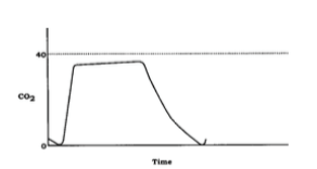
Label the following image with the appropriate portions of the expiratory segment.
Answer
-
Beginning of exhalation
-
Gas exhalation alveolar capillary bed
-
Alveolar plateau, C02-rich gas
-
Inspiration
Question 21
Question
Label the appropriate angle segments of the expiration of capnography.
Answer
-
Alpha angle
-
Beta angle
Question 22
Question
Which of the following options describes Phase I of the expiratory segment?
Answer
-
Exhalation of gas from the alveolar capillary bed mixing with dead space gas
-
Positive slope due to continuous excretion of CO2 into alveoli
-
Gas exchange that is free of CO2
-
Used to assess extent of breathing
Question 23
Question
In capnography, what is part of the expiratory segment?
Answer
-
Phase 0
-
Phase I
-
Phase II
-
Phase III
Question 24
Question
In capnography, the [blank_start]alpha[blank_end] angle is between Phases II and III.
The [blank_start]alpha[blank_end] angle is an indirect indication of the V/Q status of the lung.
The [blank_start]beta[blank_end] angle is between Phase III and descending limb of inspiratory segment.
The [blank_start]beta[blank_end] angle is used to assess the extent of rebreathing.
Answer
-
alpha
-
beta
-
alpha
-
beta
-
alpha
-
beta
-
alpha
-
beta
Question 25
Question
Fill in the blanks for the five characteristics of capnogram that should be evaluated:
-Frequency
-Rhythm
-[blank_start]Height[blank_end]
-Baseline
-[blank_start]Shape[blank_end]
Answer
-
Height
-
Shape
Question 26
Question
Your patient has low or no ETCO2 -- what are two of the major causes?
[blank_start]Decreased CO2 production/delivery[blank_end].
Causes: Hypothermia
[blank_start]Decreased pulmonary perfusion[blank_end].
Causes: Hypovolemia
Hypotension
Pulmonary embolism
Decreased cardiac output (arrest)
Answer
-
Decreased CO2 production/delivery
-
Decreased pulmonary perfusion
Question 27
Question
Select some causes of low ETCO2.
Answer
-
Altered alveolar ventilation
-
Increase CO2 production/delivery
-
Increased pulmonary perfusion
-
Technical errors/machine faults
Question 28
Question
Match the causes of low ETCO2 to the examples (in alpha order)
Altered Alveolar Ventilation
[blank_start]Apnea (accidental extubation)[blank_end]
[blank_start]Hyperventilation[blank_end]
[blank_start]Increase in dead space ventilation[blank_end]
[blank_start]Partial/total airways obstruction[blank_end]
Technical Errors/Machine Faults
[blank_start]Esophageal intubation[blank_end][blank_start]Sampling line leak[blank_end]
[blank_start]Ventilator malfunction[blank_end]
Answer
-
Apnea (accidental extubation)
-
Hyperventilation
-
Increase in dead space ventilation
-
Partial/total airways obstruction
-
Esophageal intubation
-
Sampling line leak
-
Ventilator malfunction
Question 29
Question
Name the two chief causes of elevated CO2.
[blank_start]Increased CO2 production/delivery[blank_end].
[blank_start]Increased pulmonary perfusion[blank_end]
Answer
-
Increased CO2 production/delivery
-
Increased pulmonary perfusion
Question 30
Question
Drag and drop the cause of the elevated ETCO2 with its cause:
Increased CO2 production/delivery.
[blank_start]Fever[blank_end]
[blank_start]Hyperthyroid storm[blank_end]
[blank_start]Laparoscopic surgeries[blank_end]
[blank_start]Malignant hyperthermia[blank_end]
[blank_start]Right-to-Left Shunts[blank_end]
[blank_start]Sepsis[blank_end]
[blank_start]Shivering[blank_end]
[blank_start]Sodium bicarbonate[blank_end]
[blank_start]Tourniquet[blank_end]
Increased pulmonary perfusion
[blank_start]Increased blood pressure[blank_end]
[blank_start]Increased cardiac output[blank_end]
Answer
-
Fever
-
Hyperthyroid storm
-
Laparoscopic surgeries
-
Malignant hyperthermia
-
Right-to-Left Shunts
-
Sepsis
-
Shivering
-
Sodium bicarbonate
-
Tourniquet
-
Increased blood pressure
-
Increased cardiac output
Question 31
Question
Select some examples of elevated CO2 causes.
Answer
-
Hyperventilation
-
Hypoventilation
-
Bronchial intubation
-
Exhausted CO2 absorber
-
Apnea/circuit disconnect
-
Malignant hyperthermia
-
Sepsis
Question 32
Question
You notice your patient is experiencing small breaths at various points in the ETCO2 waveform. What is NOT a potential cause of this?
Answer
-
Inadequate muscle paralysis
-
Improper ventilator settings
-
Excessive oxygenation
-
Pressure on the patient's chest
Question 33
Question
Cardiac oscillations are most prominent in what patient population?
Answer
-
Adult
-
Elderly
-
Pediatric
-
Pregnant
Question 34
Question
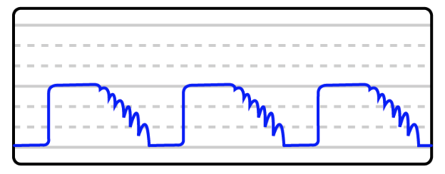
[blank_start]Cardiac oscillations[blank_end] are small gas movements produced by pulsations of the aorta and heart.
Answer
-
Cardiac oscillations
Question 35
Question
This image is an example of what on a capnograph?
Answer
-
Cardiac oscillations
Question 36
Question
Other considerations/things that may mimic [blank_start]cardiac oscillations[blank_end]
– negative intrathoracic pressure
– low respiratory rate
– low I:E ratio
– Waning muscle relaxation
Answer
-
cardiac oscillations
Question 37
Question
Phase II and III are prolonged or slanted when a patient experiences a [blank_start]prolonged expiratory upstroke[blank_end].
Answer
-
prolonged expiratory upstroke
Question 38
Question
You are administering anesthesia when you suddenly notice your patient's baseline is elevated. You immediately:
Answer
-
Check for disconnections
-
Look at your CO2 absorbent
-
Change the I:E ratio
-
Increase your patient's sedative
Question 39
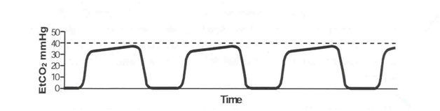
Question
You see the capnograph below during your case. You know this is a sign of:
Answer
-
Exhausted CO2 absorbent
-
Airway obstruction
-
Cardiac oscillations
-
Inspiratory valve malfunction
Question 40
Question
An [blank_start]expiratory valve[blank_end] defect allows exhaled CO2-rich gases to be rebreathed with each inspiration.
Answer
-
expiratory valve
Question 41
Question
ETC02 is indicative of [blank_start]alveolar[blank_end] CO2.
Answer
-
alveolar
-
arterial
Question 42
Question
You have two CO2 readings: 38 and 43. Which one is the arterial concentration or PACO2?
Answer
-
38
-
43
Question 43
Question
The [blank_start]facial[blank_end] nerve is the better site to monitor for onset of block because the larynx also mimics the response of the diaphragm.
Answer
-
facial
-
ulnar
Question 44
Question
The [blank_start]ulnar[blank_end] nerve is the best nerve to monitor for recovery.
Answer
-
facial
-
ulnar
Question 45
Question
Match the pattern of stimulation to its stimulus.
[blank_start]Single stimulus:[blank_end] The simplest mode of stimulation; consists of a single supramaximal electrical stimulus that is delivered from every 1 to 10 seconds (1‐0.1 Hz)
[blank_start]Train of Four:[blank_end] Four successive 200 μs stimuli at 2Hz delivered every 0.5 seconds for 2 seconds
[blank_start]Double Burst Stimulation:[blank_end] delivers two bursts (at 50Hz) of three electrical stimulations separated by 750 sec followed later by two such impulses
[blank_start]Tetanus:[blank_end] Repetitive, high-frequency stimulation at frequencies of 50 Hz or greater for five seconds
Answer
-
Single stimulus:
-
Train of Four:
-
Double Burst Stimulation:
-
Tetanus:
Question 46
Question
Which form of peripheral nerve monitoring is based on the concept that acetylcholine is depleted by successive stimulation?
Answer
-
Single stimulus
-
Train of four
-
Double-burst stimulation
-
Tetanus
Question 47
Question
In Train of Four stimulation, no twitches equals what kind of blockage?
Answer
-
75
-
80
-
90
-
100
Question 48
Question
Clinical relaxation usually require what percentage of neuromuscular blockage?
Answer
-
50 to 60 percent
-
60 to 70 percent
-
75 to 95 percent
-
100 percent
Question 49
Question
What PNM improves accuracy over the T4/T1 ratio by delivering a stronger stimulation and eliminating the second and third twitches?
Answer
-
Double burst stimulation
-
Single burst stimulation
-
Tetanus
Question 50
Question
Which PNM option may provide an indication of the time until return of the first response in the TOF and subsequent readiness for reversal of neuromuscular blockage?
Answer
-
Tetanus
-
Post-Tetanic Potentiation
-
Train of Four
-
Double-Burst Stimulation
Question 51
Question
Which of the following muscle groups are most sensitive to neuromuscular blocking agents?
Answer
-
Diaphragm and Masseter
-
Extra ocular and adductor pollicis
-
Diaphragm and Orbicularis oculi
-
Adductor pollicis and Orbicularis oculi
Want to create your own Quizzes for free with GoConqr? Learn more.