9556983
Descripción
Fichas por Elizabeth Babcock, actualizado hace más de 1 año
|
|
Creado por Elizabeth Babcock
hace casi 7 años
|
|
| Pregunta | Respuesta |
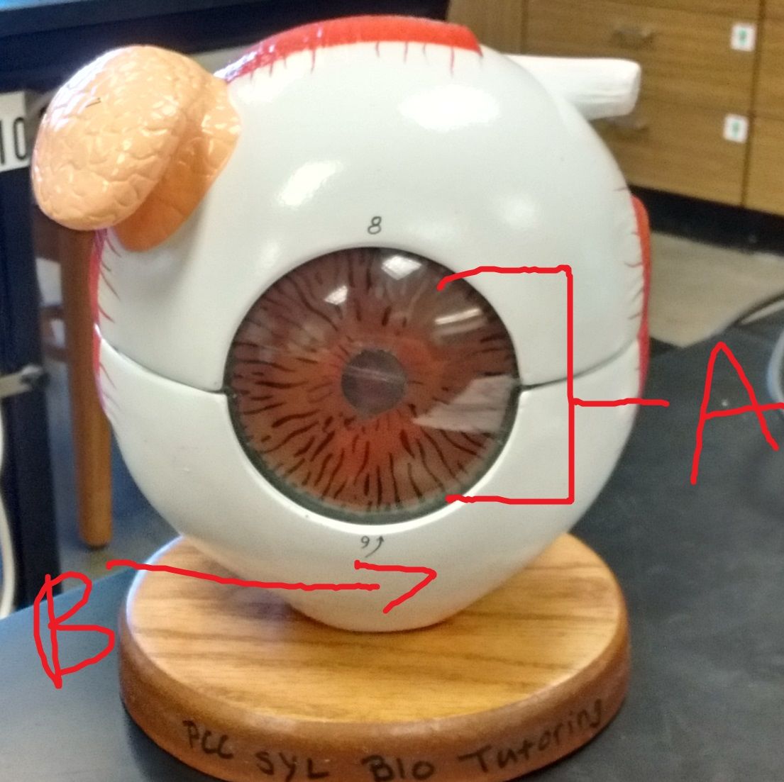
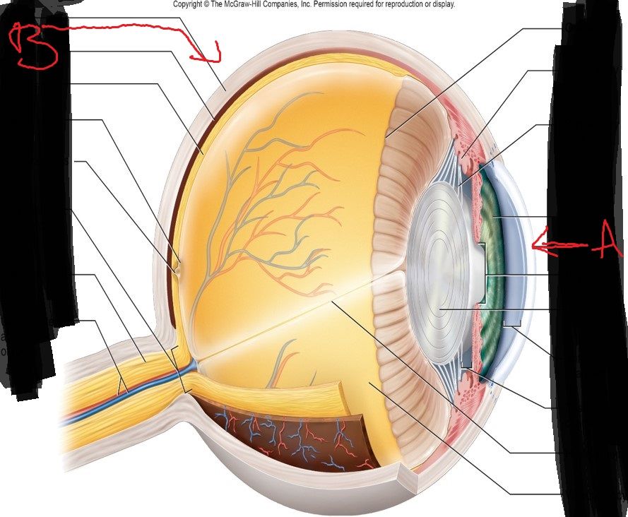
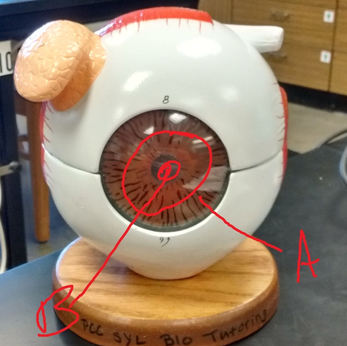
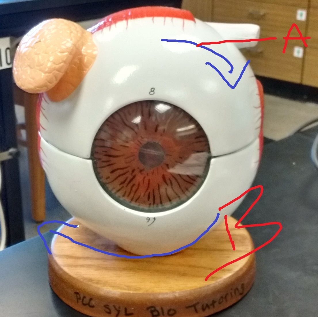
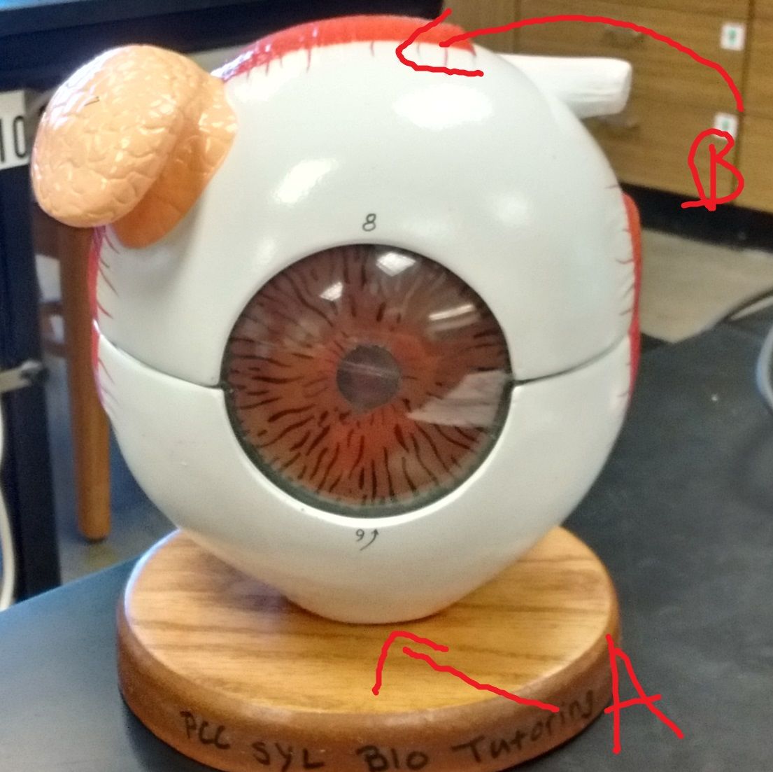
| Name the structures | A. Cornea. B. Sclera |
| What sysyems? Explain | A. Scotopic vision: Many rods converge on each bipolar cell and many bipolar cells converge on each ganglion cell. Allows rods to combine their effects thru spatial summation and stimulate the ganglion cell generating in dim light. - Ganglion cell represents large area of retina to produce grainy image. B. Photopic vision: Little neural convergence. Fovea each cone has a private line to the brain so eahc optic nerve fiber represents tiny area of retina. Vision is pretty sharp but lack of convergence means photopic vision can't function well in dim light because weakly stimulated cones cannot collaborate to stimulate ganglion cell. |
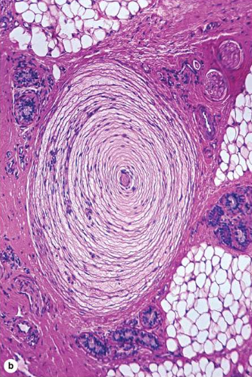
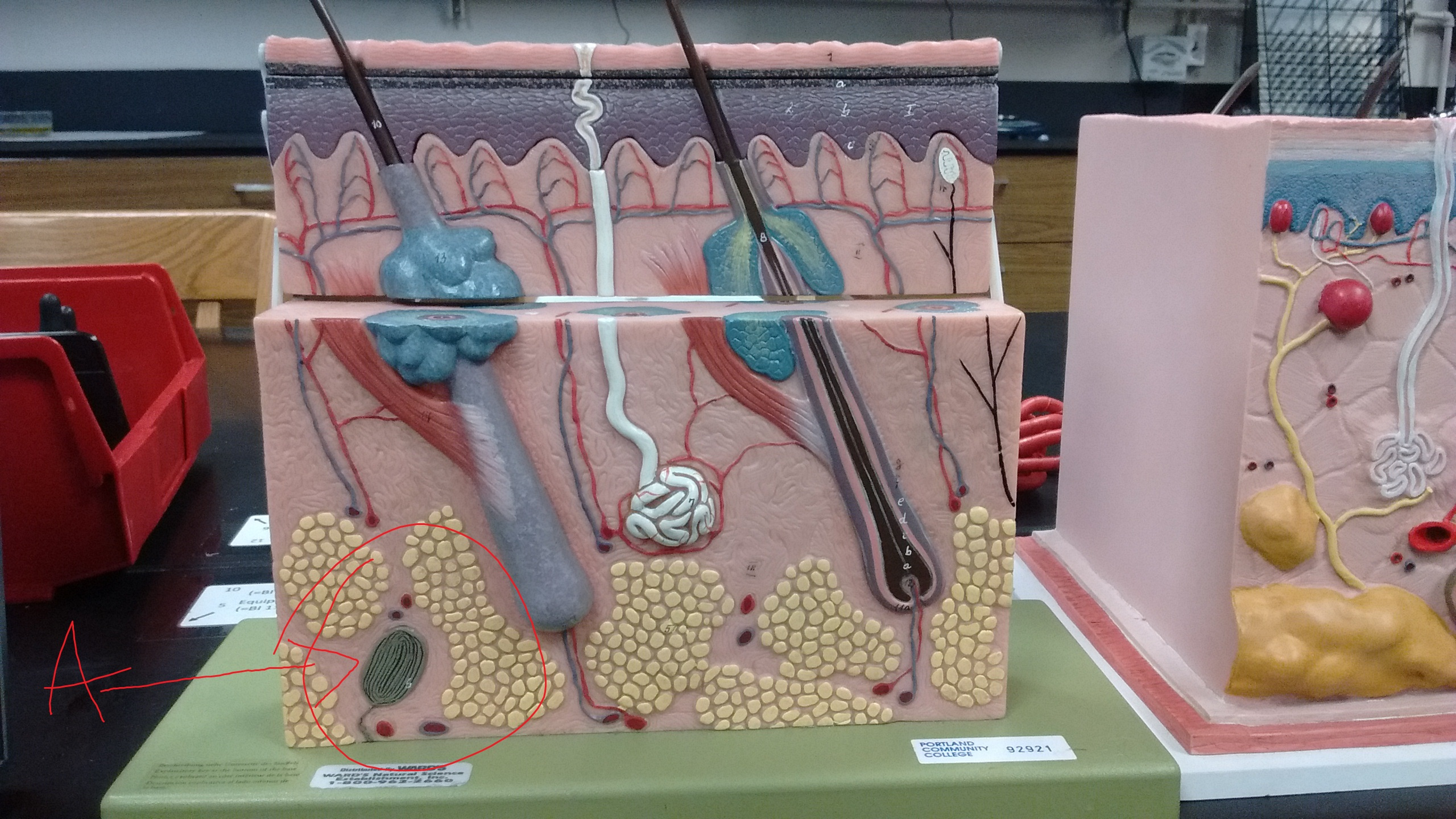
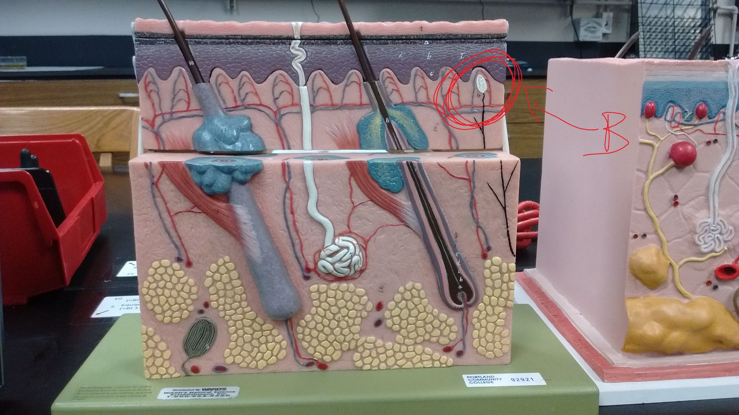
| What is this receptor? Type, stimulus detection? Where is it found? | A. Pacinian corpuscle. B. Deep Pressure. C. Lies Deep to Dermis |
| What is this receptor? Type, stimulus detection? Where is it found? | A. Pacinian corpuscle. B. Deep Pressure. C. Lies Deep to Dermis |
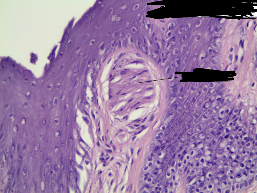
| What is this receptor? Type, stimulus detection? Where is it found? | A.Meissner's Corpuscle. B. Light touch. C. Dermal Papillae of skin |
| What is this receptor? Type, stimulus detection? Where is it found? | A.Meissner's Corpuscle. B.Light touch. C. Dermal Papillae of skin |
| How does the density of heat receptors correspond to that of touch receptors? What about cold receptors? | A. More Touch receptors because of slow adaptation. B. More Cold receptors because we are warm blooded and detect with nociceptors. Depends on where in the body |
| What do calipers test for? | Differentiate two distinct sensations when skin is touched simultaneously at two points |
| Which areas have the smallest distance from 2pt discrimination? Which has the greatest? | 1. Fingertips/tongue. 2. Back |
| Put your hand in 45 degree C water. What sensation do you feel? | Warm |
| What external anatomy is a transparent mucous membrane. Lines eyelids and covers anterior side of eye except the cornea. Prevents eye ball from drying | What is Conjunctiva |
| What is the site of tear production? Where are they drained? Name 1 function of tears What enzyme prevents the infection? | A. Lacrimal gland, secrete via lacrimal duct. B. Drained via Lacrimal canals, sac, or nasolacrimal duct. C. Help with diffusion of 02 and CO2 D. Lysozyme |
| Fibrous Tunic (2) | A. Cornea. B. Sclera. Which Layer? |
| Type of Tissue. Function. Innervation | Fibrous Tunic -- A.Cornea. -Astigmatism, 3 lyr nonK-Strat.Squamous.Epith. simple squamous. Collagen and fibroblasts. Nourished by tears and aqueous humor. B. Sclera- white of eye. Provides support and shape. Dense irregular CT. - Posteriorly pierced by Optic Nerve II. |
| Vascular Tunic (5) | A. Iris. B. Pupil. C. Lens. D.Ciliary Body. E. Choroid |
| Which tunic? What does it contain? | Vascular Tunic. A. Iris. B. Pupil. C. Lens |
| What is A? Function/Structure? Innervation? Attachment? | A. Iris. Colored portion of eye. Between Cornea and lens. Attaches to ciliary body. . Regulates amt of light entering eye. Autonomic reflexes. -- Circular muscle fibers contract in bright light to shrink pupil. ---> Radial muscle fibers contract to dim light to enlarge pupil |
| What is B? Function/Structure? Innervation? | B. Pupil. Hole in center is pupil |
| What is C? Description. What is it held by? Function? | C. Lens. Avascular, crystalline proteins arranged like layers in an onion. Transparent. --Held in place by suspensory ligaments. Focuses light on Fovea |
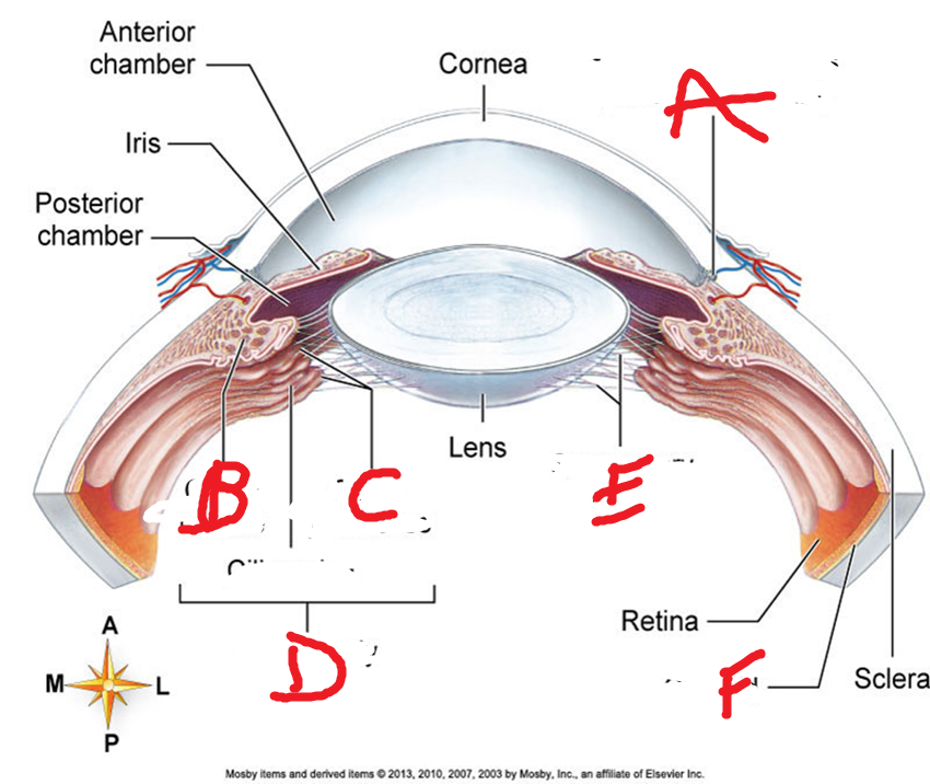
| What layer is this? Name what each is | Vascular tunic. A. Canal Schlemm, B. Ciliary muscle. C. Ciliary Processes. D. Ciliary Body. E. Suspensory Ligaments. F. Choroid. |
| What are B,C,D, E? Functions? | B. Ciliary Muscle: Controls tension on ligaments and lens. C: Ciliary Processes- Folds on ciliary body, secrete aqueous humor. D. Ciliary Body. |
| Which structure secretes aqueous humor? What tunic? | 1. Ciliary processes. 2. Vascular tunic |
| What are the autonomic reflexes of the pupil? | Autonomic reflexes.. 1. Circular muscle fibers contract in bright light to shrink pupil. 2. Radial muscle fibers contract to dim light to enlarge pupil |
| What is F? Structures? Function | F. Choroid. Pigmented epithelia cells (melanocytes and blood vessels). Melanocytes absorb scattered light. Provides nutrients to retina. |
| Which structure provides nutrients to retina and contains melanocytes and blood vessels of the eye? Which layer? | 1. Choroid. 2. Vascular tunic |
| What layer is the Retina? What its layers? Where does it attach? What happens if it detaches? | 1. Neural Tunic. 2. Outerlayer: Pigmented retina. 3. Inner layer: Sensory retina. 4. Attached only at optic disk where optic nerve begins and ora serrata. 5. Trauma( fluid btw layers, distortion or blindness |
| What are the 3 layers of the sensory retina? | 1.Photoreceptor: Sensitive to light rays. A. Rods- shades of gray in dim light. Cones (sharp color vision). Fovea Centralis- densely packed region. Sharpest resolution. 2. Bipolar cells. 3. Ganglionic cells |
| What is the Fovea Centralis? | Densely packed region of cones. Sharpest resolution or acuity |
| Optic Nerve (II), Function, and location | Extends from eyeball to brain |
| Optic Disc: Location and what it creates. | Optic nerve exits back of eyeball. Creates blindspot (no presence of photoreceptors) |
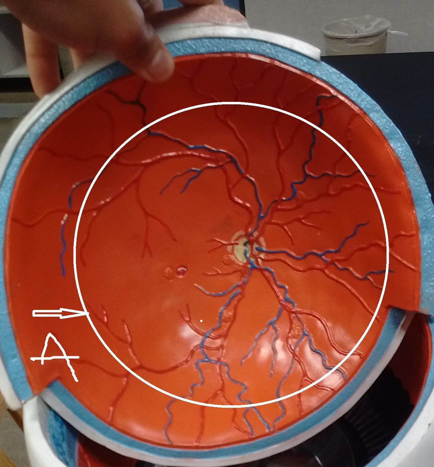
| Central retinal artery and vein. Function | Fan out to supply nourishment to retina. Visible for inspection |
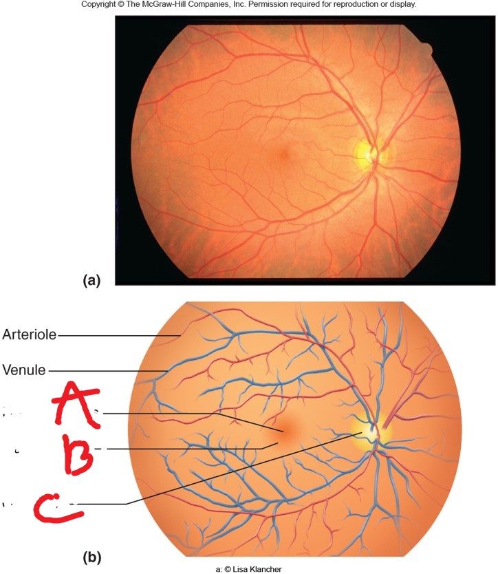
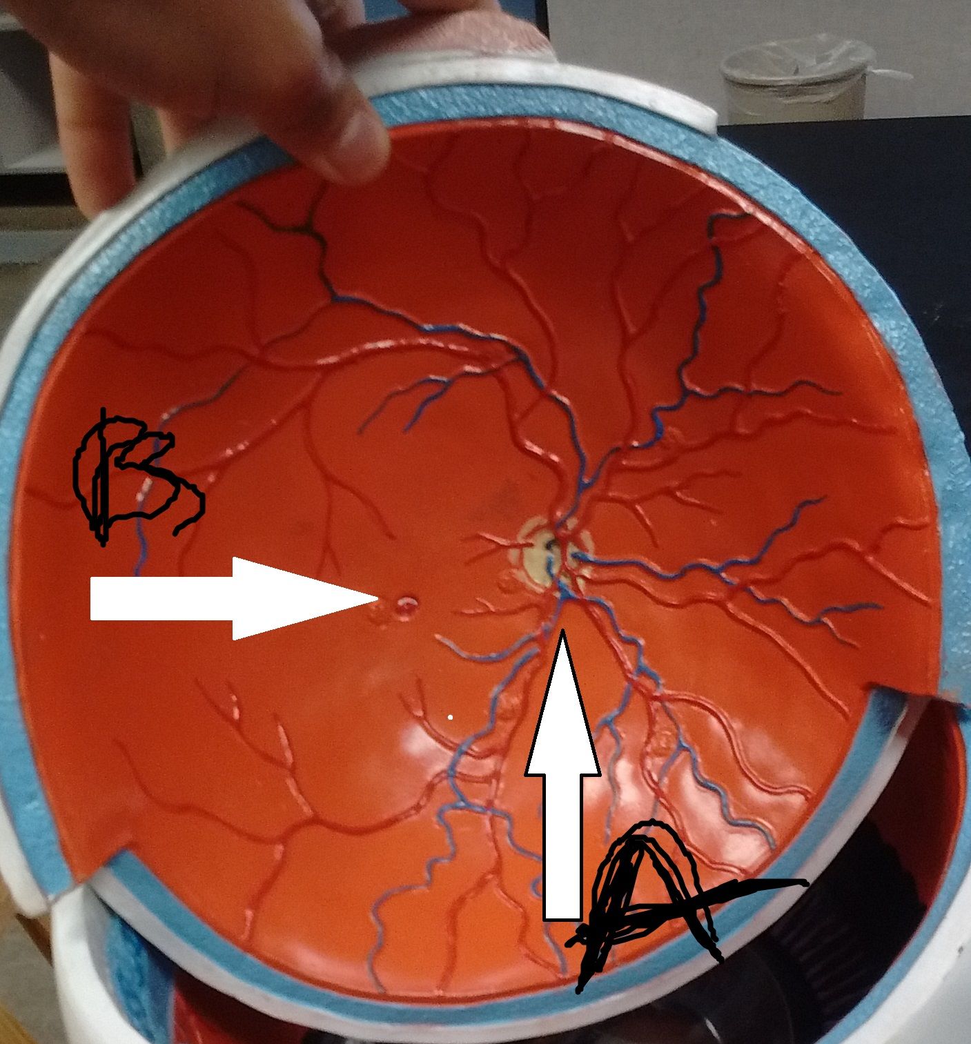
| What is A,B,C. Fu | A. Fovea centralis: located in center of macula where visual acuity is highest, high concentration of retinal cones. B. Macula lutea (yellow spot) Just lateral to center of retina, which constitutes region of maximum visual acuity. C. Optic Disk. Blind spot, Optic Nerve |
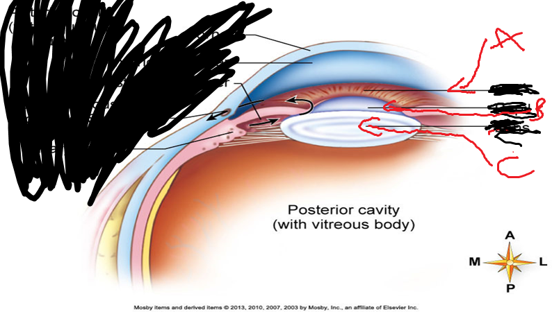
| Which cavity is filled with a jellylike substance? | Posterior cavity posterior to lens. Filled with vitreous body/humor. |
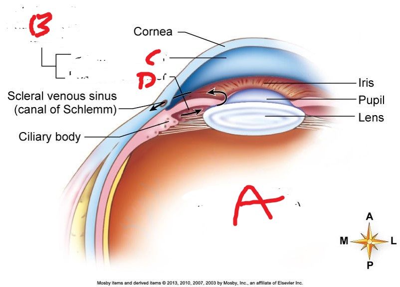
| What is A,B,C,D? Location? | Interior cavities: A'. Posterior cavity- posterior to lens. Filled with Vitreous body. Floaters are debris in vitreous of older ppl. B. Anterior Cavity- Anterior to lens. Filled with Aqueous humor. C.Anterior chamber. D. Posterior Chamber. |
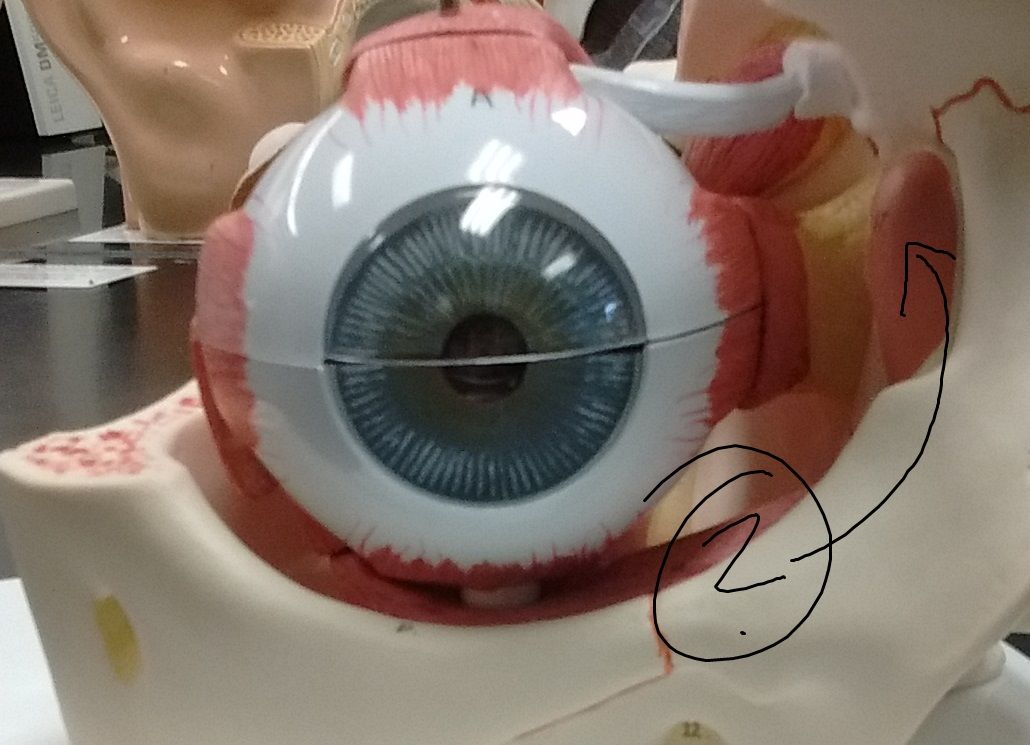
| Name the extrinsic eye muscles | 1.Superior oblique. 2. Inferior Oblique. 3. Superior rectus. 4. Inferior rectus. 5. Lateral rectus. 5. Medial rectus. |
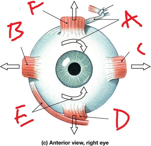
| Name what A,B,Care Describe the eye movements. and CN that enervate each | A.Superior Oblique- Inferior medial. Trochlea ligament sling, CNIV. B. Lateral Rectus. Moves eye laterally from nose. CNVI. C. Medial rectus. Moves eye medially toward nose. CN III. |
| What are the 2 intrinsic muscles of the eye? What are the functions? What tissue are they made of? | 1. Iris., Regulates size of pupil 2. Ciliary muscle. Controls shape of lens. |
| What are the muscles of the iris? What fibers innervate them? In what situations would they involved in? | 1. Constrictor pupillae- Innervated by parasympathetic fibers (Close vision and bright light) 2. Dilator pupillae- Innervated by sympathetic fibers. (Distant vision and dim light) |
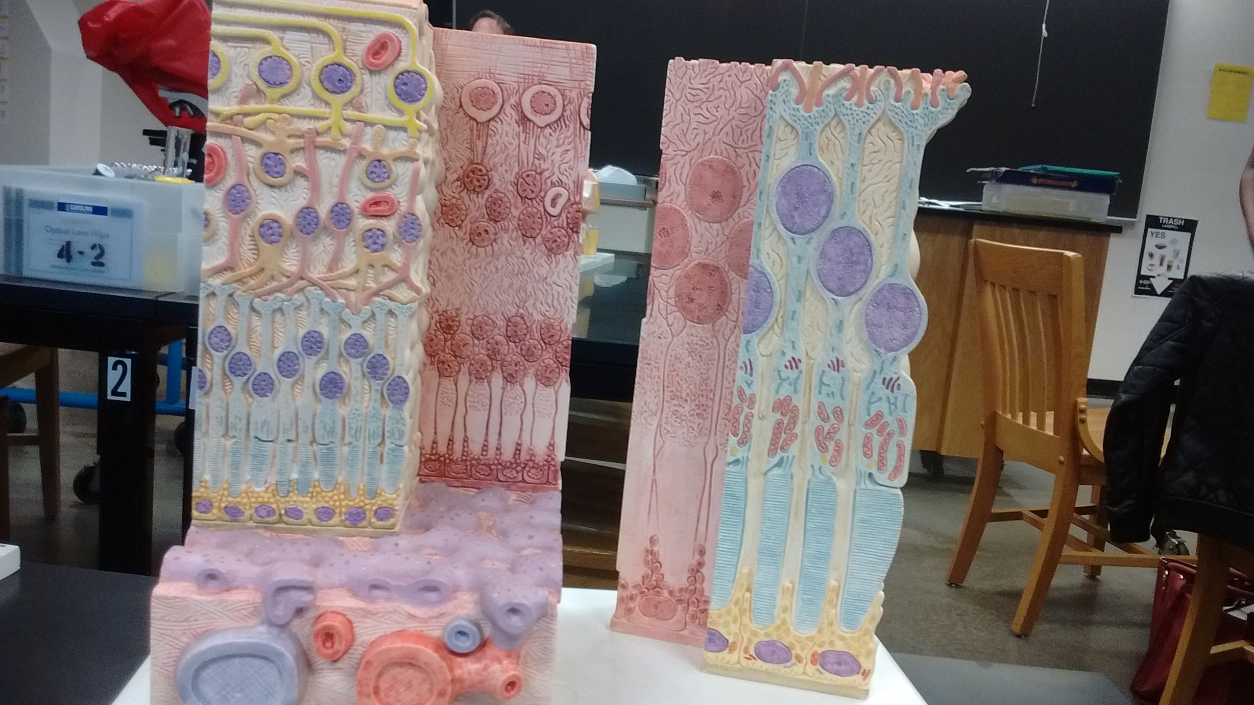
| Histology of the Retina. Name the layers. | 1. Choroid. 2. Pigmented Epithelial layer of retina. 3. Rods and cones. 4. Bipolar cells. 5. Ganglion cells |
| What is the blind spot? Why does the image disappear? | Created by optic disk. No photoreceptors |
| What are the 2 afterimage definitions? | 1. Positive afterimage: Bright image seen because of the continued firing of rods (source: sun). 2. Negative afterimage. Altered imaged that is an indication of affected photoreceptor cells that have been bleached. Specific cones that have been affected will change the image's color. (Source bright colored) |
| What is the photosensitive pigment of rods? Why bleached? | Rhodopsin Photosensitive pigment of the rods. Composed of a light-sensitive chemical, retinal, and protein, opsin. When light strikes retina, rhodopsin splits into its two component parts and becomes pale (bleached). . |
| What are the photoreceptors that detect color? What about responsible for vision in low light? | A. Photopsins, blue, green, red. Rhodopsin |
| What is color blindness? | Inability to distinguish between certain colors. Absence of certain cone photopsins |
| Night Blindness, difficulty seeing in low light | Nyctalopia, inability to make normal amount of rhodopsin |
| What is the bending of light rays? | Refraction |
| What refracts more light anterior to the eyeball? | Cornea refracts light more. Lens fine-tunes images as shift focus between near and distant objects |
| What does light pass thru? What is the end point of which it hits? | 1. Cornea. 2. Aqueous humor. 3. Lens. 4. Vitreous humor. Endpoint: Retina |
| Specific point of intersection on the retina | Focal point |
| Distance between the center of the lens and its focal point | Focal distance. |
| Focal distance is determined by what 2 factors? What makes for shorter and greater focal distances? | 1. Distance from the object to the lens.- Closer the object the greater focal distance. 2. Shape of lens- Rounder the lens, the more refraction occurs, resulting in shorter focal distance. |
| Define Accommodation | Alteration in curvature of lens of the eye to focus an image on the retina |
| Lens accommodation for near objects and distance objects. What happens to the ciliary muscle? | 1. Near objects= round lens as the muscle contracts. 2. Distance objects= flatter lens as the muscle relaxes. |
| Normal vision | Emmetropia |
| Eyeball is too deep or curvature is too great. Focal point is in front of retina. What visual dysfunction is this? How is it corrected? | Myopia- nearsighted. Diverging lens, concave lens |
| Eyeball is too shallow or the curvature of lens is too flat. Focal point is behind retina. What visual dysfunction is this? How is it corrected? | Hyperopia- farsighted. Converging lens- convex lens |
| Corneal surface is wavy, parts of image out of focus | Astigmatism |
| What tests for visual acuity? What is normal? What is better? 20/15 or 20/30? | Snellen eye chart, 20/15. You can see at 20ft what ppl can see at 15ft. |
| What is sensation? | Interpretation of sensory nerve impulses by the brain as awareness of internal and external environment. Punctate |
| What are the somatosensory vs special sense receptors? | Somatosensory: temp, pain, touch, pressure, vibration, proprioception. Special: Vison, Taste, Smell, Auditory |
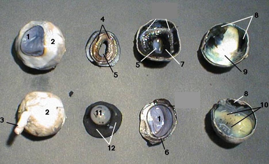
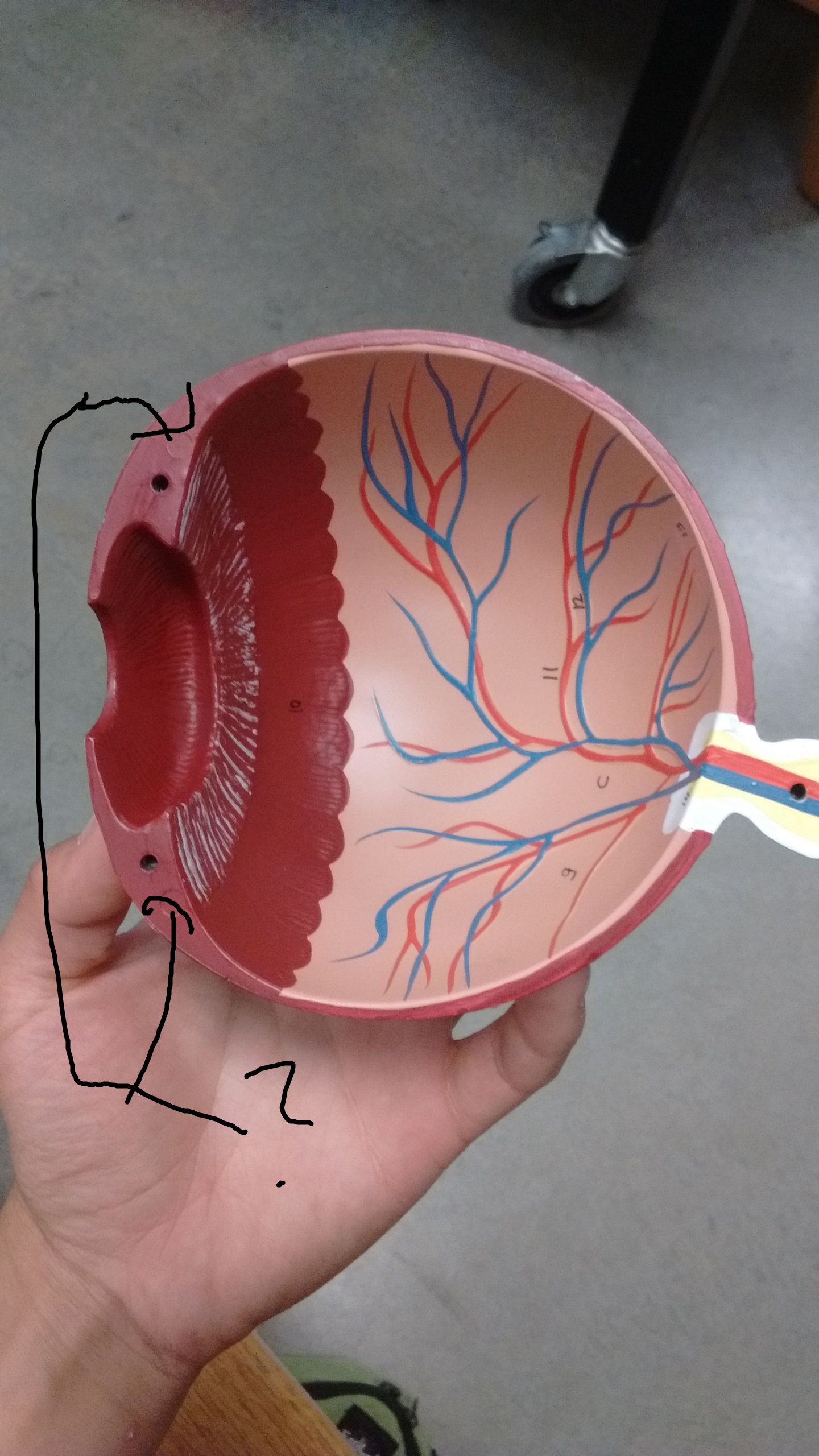
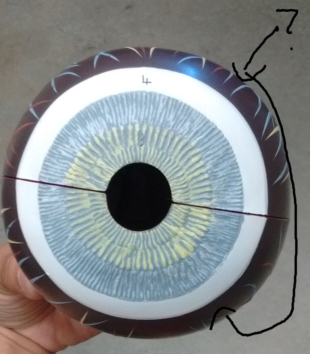
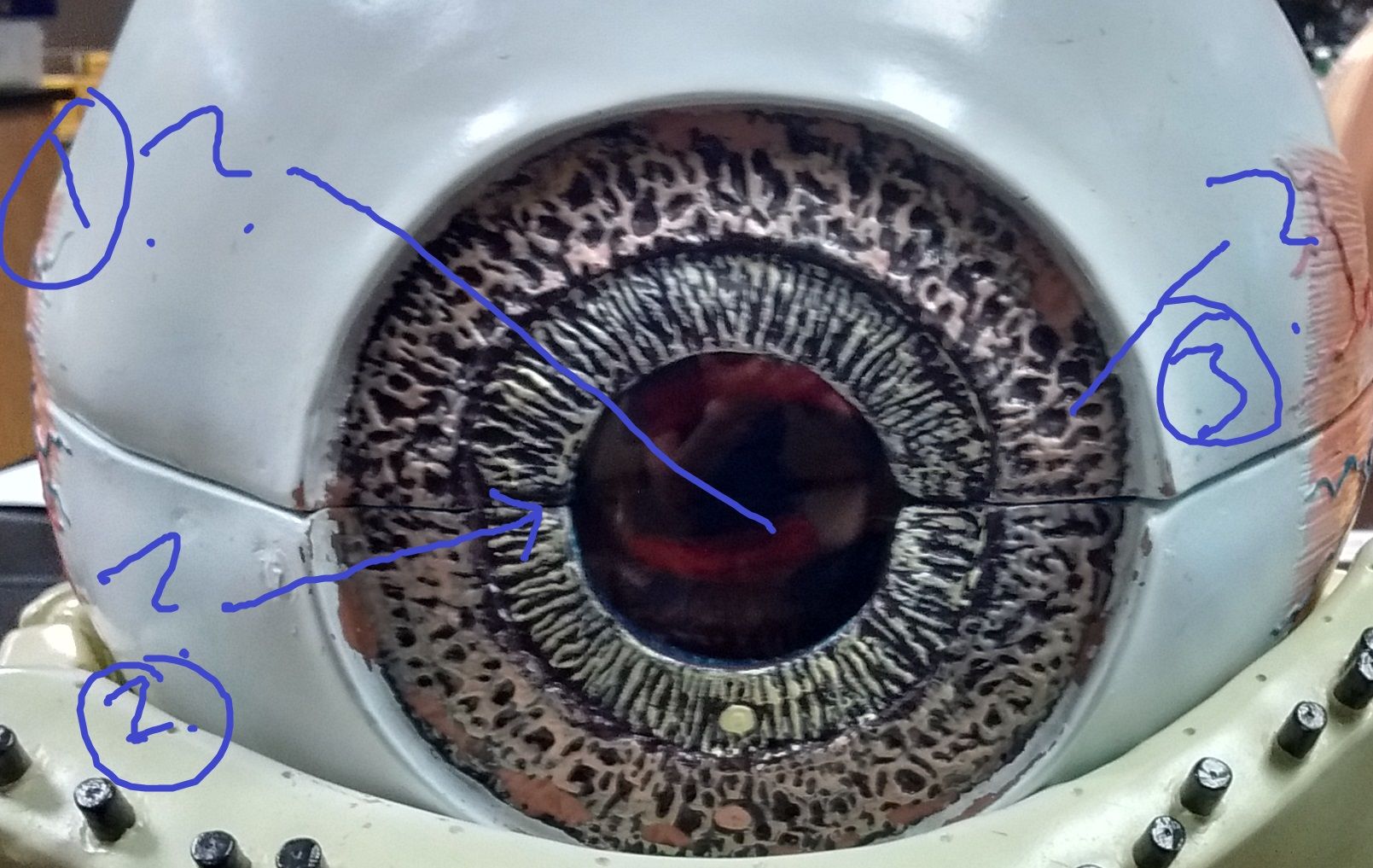
| Identify each number | 1.Cornea. 2. Sclera. 3. Optic nerve. 4. Iris. 5. Pupil. 6. Ora serrata. 7. Ciliary body. 8. Choroid. 9. Tapetum lucidum. 10. Retina. 11. Lens. 12. Vitreous humor |
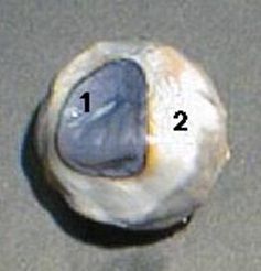
| Identify | 1. Cornea. 2. Sclera |
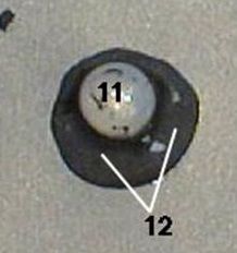
| Identify | 11. Lens. 12. Vitreous humor |
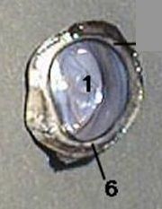
| Identify | 1. Cornea. 6. Ora serrata (non photo sensitive to photosensitive) Between ciliary body and retina |
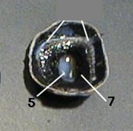
| Identify | 5. Pupil. 7. Ciliary body |
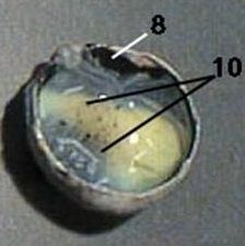
| Identify | 8. Choroid (melanocytes +blood vessels) . 10. Retina (2 layers) |
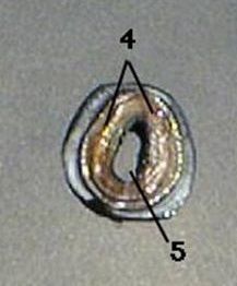
| Identify | 4. Iris. 5. Pupil |
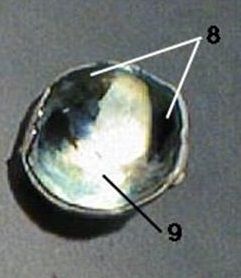
| Identify | 9. Choroid. 9. Tapetum Lucidum (Lying immediately behind the retina, it reflects visible light back through the retina, increasing the light available to the photoreceptors, though blurring the initial image of the light on focus.) |
| Disease: Blood Shot eyes. Inflammed | Conjunctiva |
| What fills the cavity of the posterior chamber posterior to the lens of the eye? | Vitreous humor |
| What fills the cavity anterior of the lens in the anterior chamber of the eye? | Aqueous humor |
| Chylamidia conjunctivitis | redness and swelling of the clear membrane that lines the inside of your eyelids and covers the white of your eye. Chlamydia can be spread when you get bacteria in your eyes |
| sympathetic ophthalmitis | This is an inflammatory condition affecting both eyes that occurs after a penetrating injury (accidental or surgical) to one of the eyes. |
| Neonatorum | Conjunctivitus in babdies |
| Strabismus ---> Amblyopia (Kids) and Diplopia (Adults) | General term: Squint, Strabismus, or squint, is any misalignment of the eyes. Name the form kids and adults might have. |
| Hyperopia | Farsighted |
| Disorders of the retina | 1. Macular degeneration. 2. Diabetic Retinopathy |
| Myopia | Nearsighted |
| Nerves for Vision | Optic II, Oculomotor nerve III, Trochlea IV, Opthalmamic branch of trigeminal V, Abducens VI |
| What nerve carries visual impulses from rods and cones to the brain? | Optic nerve II |
| What does the Opthamamic branch of trigeminal nerve V do for the eye? | Carries impulses of temperature, pain, touch |
| What nerve supplies lateral rectus, intrinsic eye muscle? | What does Abducens VI nerve supply? |
| What does Trochlea IV supply? | What nerve supplies superior oblique, extrinsic eye muscles |
| What nerve supplies voluntary and involantary motor impulses? | What does the Oculomotor nerves III supply? |
| Name these structures. Function/insertion | A. Iris, responsible for controlling the diameter and size of the pupil and thus the amount of light reaching the retina. B. Pupil: Light enters thru |
| Name these structures. Function/insertion/ direction | A. Superior Oblique. Medial- inferior direction B. Inferior Oblique: Lateral- Superior direction |
| Name these structures. Function/insertion | A: Inferior rectus. Downward B. Superior Rectus: Upward |
| Name these structures. Function/insertion | A. Optic disk, Optic nerve exits eye posterior eyeball. Creates blindspot B. Fovea Centralis, located in center of macula lutea, visual acuity is highest. (Retinal cones) |
|
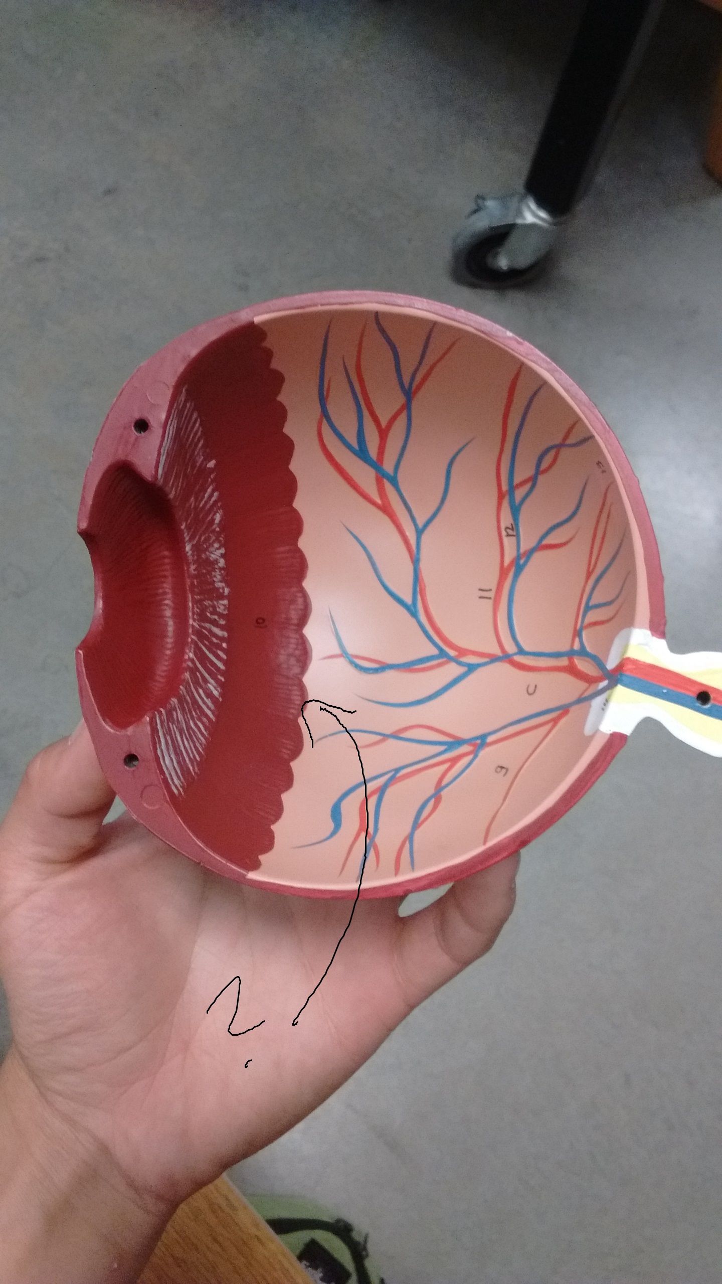
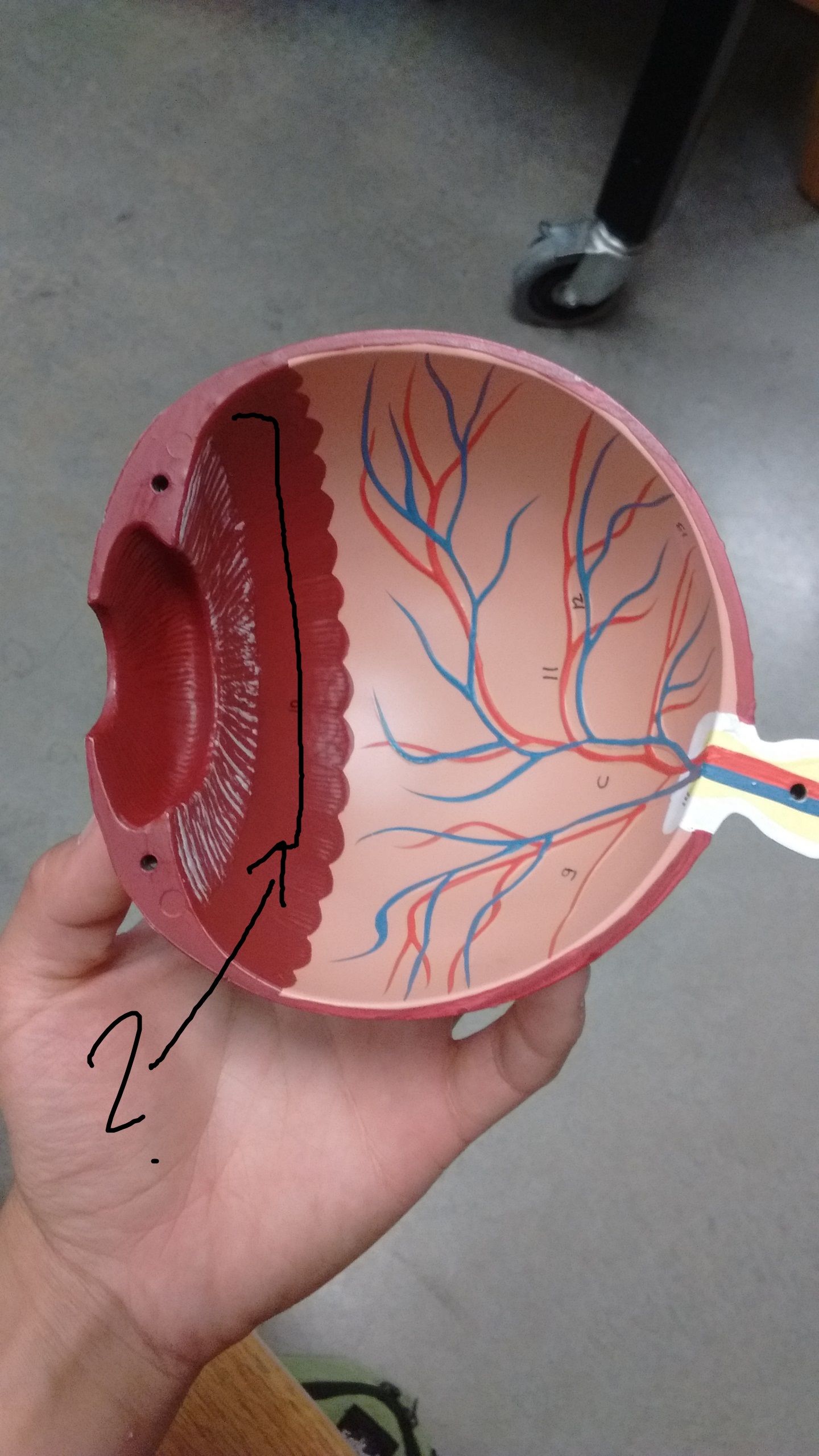
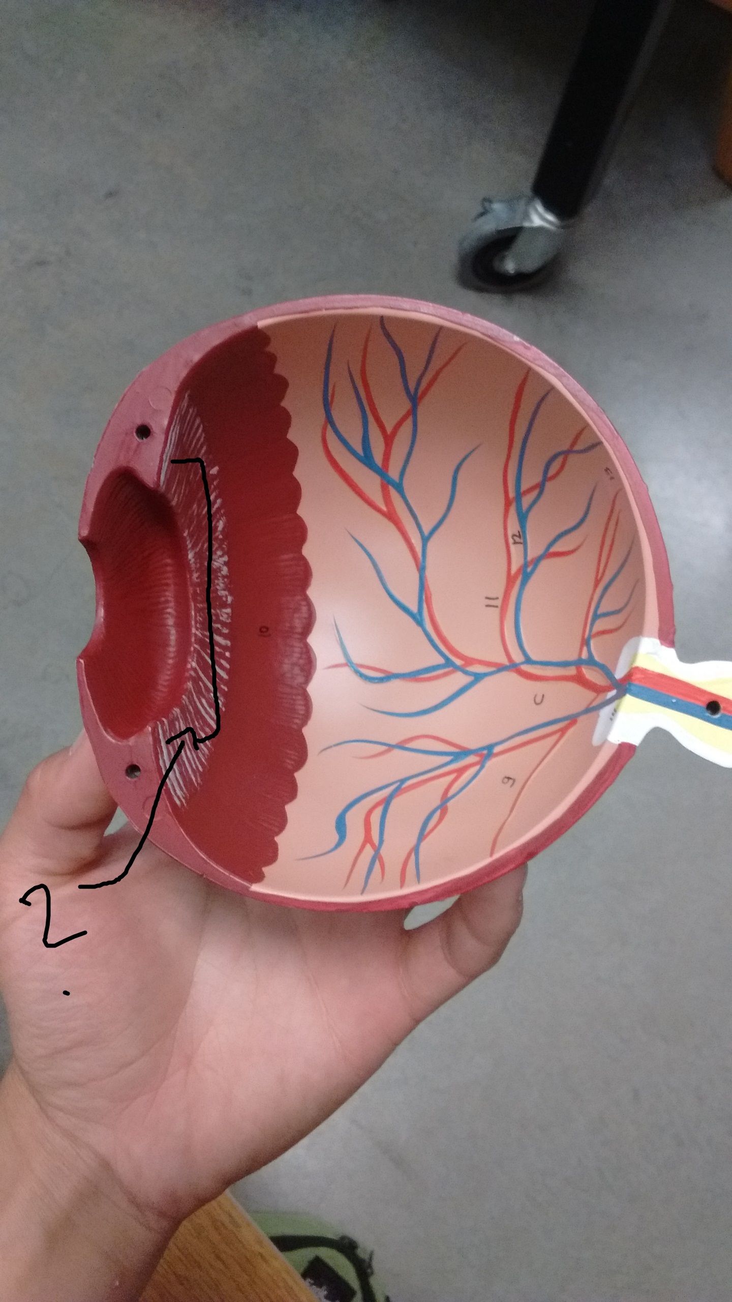
Name this structure Function/insertion
Image:
Retina (binary/octet-stream)
|
A. Retina, Outer layer: pigmented epithelium, inner- sensory retina. Attached only at optic disk and ora serrata |
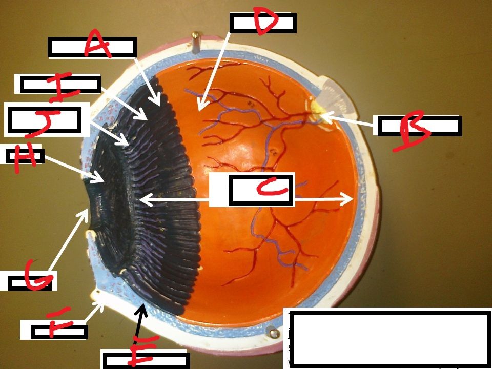
| Name these structures | A. Ora serrata B. Optic nerve C. Posterior cavity E. Choriod F. Sclera G: Pupil H:Iris I: Ciliary Muscle J: Ciliary Processes |
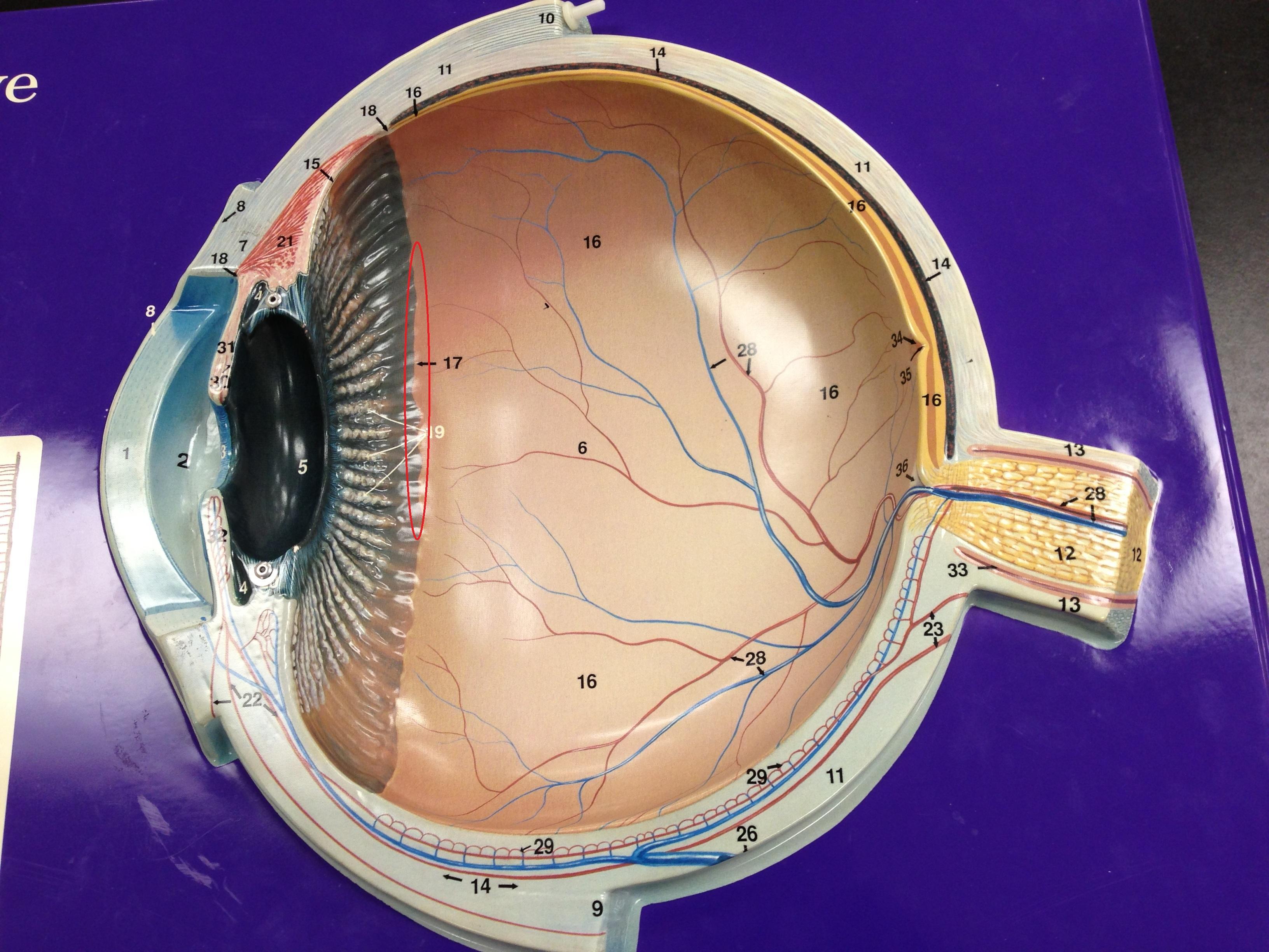
| 1. Lens. 2. Anterior chamber of the anterior cavity. 3. Pupil. 5. Iris. 21. Ciliary Muscle 19. Ciliary Processes 17. Ora Serrata 35. Fovea Centralis 36. Optic disk | |
| What can you see with an ophthalmoscope? (4) including retina | What instrament can see? Fovea centralis, optic disk (blind spot), central retinal artery. (Retina) |
| What is this a representative of? Explain it | This is the histology of the Retina Pigment epithelium. - Capture stray light. Rods and cones aren't neurons, but part of the ependymal cells. Bipolar cells: First order neurons. Ganglion cells: largest neurons of the retina. 2nd order neurons. Their axons form optic nerve II. (Detect light intensity) |
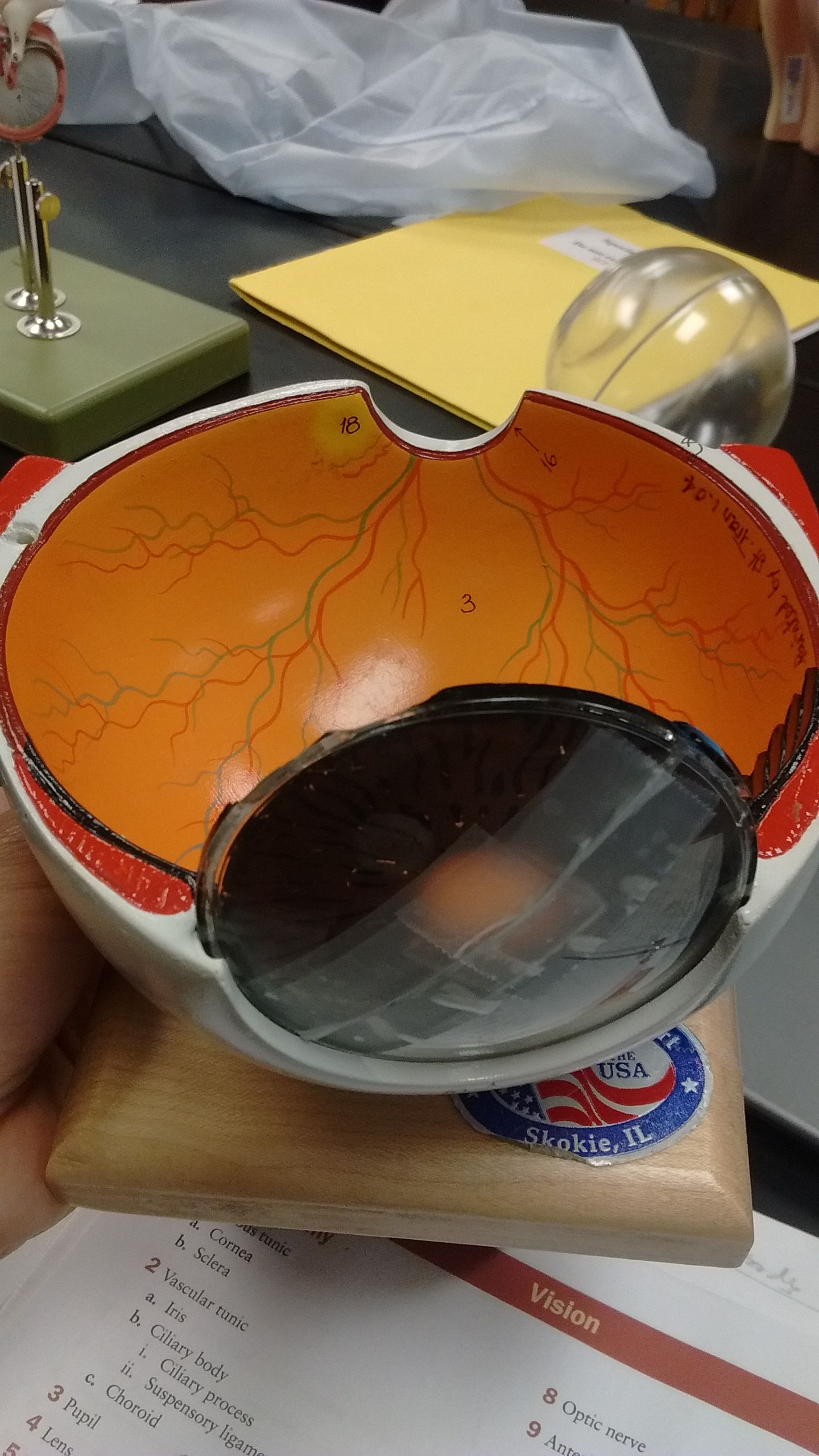
| Identify the numbers | 3. Retina 18. Fovea Centralis 16. Optic disk 4. Sclera |
| Circular muscle fibers contract in bright light to shrink pupil. Postganglionic parasympathetic fiber Radial muscle fibers contract to dim light to enlarge pupil. Postganglionic sympathetic fibers | Name the 2 muscles of the iris Functions and innervated postganglionic fibers |
| What collects aqueous humor from the anterior chamber and delivers it into the episcleral blood vessels via aqueous veins | What does the Canal Schlemm do? |
| D,E,F. Describe the eye movements. and CN that enervate each | D. Inferior Rectus. Moves eye inferiorly. CN III. Oculomotor E. Inferior Oblique. Superior ->Lateral. CNIII. Oculomotor F. Superior Rectus. Moves eye superior. CN III. Oculomotor |
| Scotopic vision | Night vision, what is responsible? |
| Photopic vision | Day vision |
| What are the parts of Rhodopsin? The moieties? | 1. Protein opsin and vit. A- Retinal. |
| What are the parts of Cones? | Pigment: Photopsin. 3 kinds of cones, same moeity, but diff amino acids which can detect different wavelengths of light. |
| How do you generate an optic nerve signal? Night and day scenerio | The retina adapts to change in light through the use of the rods. In the dark, the chromophore retinal has a bent shape called cis-retinal (referring to a cis conformation in one of the double bonds). When light interacts with the retinal, it changes conformation to a straight form called trans-retinal and breaks away from the opsin. This is called bleaching because the purified rhodopsin changes from violet to colorless in the light. At baseline in the dark, the rhodopsin absorbs no light and releases glutamate which inhibits the bipolar cell. This inhibits the release of neurotransmitters from the bipolar cells to the ganglion cell. -- BC bipolar cells are excited when there is no glutamate to inhibit it. They are excited by rising light intensities. When there is light present, glutamate secretion ceases thus no longer inhibiting the bipolar cell from releasing neurotransmitters to the ganglion cell and therefore an image can be detected. The Ganglion cells are the only retinal cells that produce AP. |
| Identify | 1. Ora Serrata |
| Identify and what does it contain? | 1. Ciliary body. 2. Contains Ciliary process, ciliary muscle and suspensory ligaments |
| Identify and function | 1. Ciliary Processes 2. They are folds on ciliary body. 3. It secretes aqueous humor |
| Identify and function | 1. Ciliary muscle 2. Controls tension on ligaments and lens |
| Identify and function | 1. Choroid. 2. Site of melanocytes and blood vessles. 3. Melanocytes's black pigments help absorb scattered light 4. Provides nutrients to retina |
| Identify and function | 1. Pupil 2. Iris. 2 muscles to change amt of light thru to the pupil 3. Ciliary body |
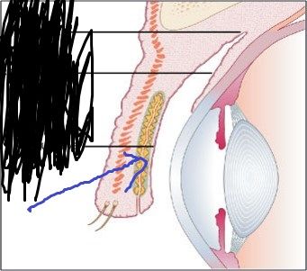
| Identify and function | 1. Lacrimal sac 2. Drainage of tears towards the nasal cavity |
| Eyelids - what it is separated by and where they meet | Palpabrae 2. Palpebral fissure 3. Medial and lateral commissures |
| What gets swollen when you have a cold? What duct? | Nasolacrimal duct. Get's what when you have a cold? |
| What tendon inserts on the superolateral area of the eye? | Trochlea inserts where? |
| What gives the iris it's black, brown, hazel color? What about blue, green, gray? | Chromatophors does what for the eye? What if the melanin is scanty? |
| What is the pupillary constriction in response to light? To admit less light in | Photopupillary reflex |
| Explain the 3 processes for near response? | 1. Convergence of eyes to have eyes orient. 2. Constriction of pupil: Screen out peripheral light rays. 3. Acommodation: change the curvature of lens to focus. Ciliary muscle |
| Description of Rod vs Cone and it's moeities. Bleaching and regeneration | RodsEach disk studded with Rhodopsin.- contains retinal and opsin. Produce shades of gray. - Rhodopsin regenerates faster than it bleaches in the dark/dim. Cones: Day and color vision. Have photopsin- Retinal and opsin. |
| Retinal circuitry and veisual sensitivity | ejxjnz |
| The visual projection pathway | Optic nerve leave each orbit thru optic canal and converge to form an X, optic chiasm on hemidecussation Right cerebral lhemisphere sees objects in left visual field because images fall on the right half of each retina. |
¿Quieres crear tus propias Fichas gratiscon GoConqr? Más información.