11839555
Description
Flashcards by Laura Gennaro , updated more than 1 year ago
|
|
Created by Laura Gennaro
almost 7 years ago
|
|
| Question | Answer |
| The Orbit | anatomic compartment enclosed medially, laterally, & posteriorly by bone |
| Proptosis | -anterior displacement of the eye within the orbit |
| Causes of Proptosis | -Graves disease (accumulation of extracellular matrix proteins & fibrosis in rectus muscles) -Glioma, meningioma of the optic nerve -Lacrimal gland enlargement results in inferior/medial displacement of the eye (sarcoidosis; lymphoma, pleomorphic adenoma, adenoid cystic carcinoma) |
| Other names for the eyelid | -palpebra -blepharon |
| The eyelid is composed of... Functions to... | -skin externally and palpebral conjunctiva (mucosa) internally -Protect the eye; elements of tear film generated by Meibomian glands |
| Blepharitis | chronic inflammation at the eyelid margin -affects tear production -result of skin disease, staph infections -periorbital skin can be red |
|
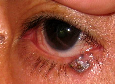
Basal cell carcinoma
Image:
Image (binary/octet-stream)
|
-most common malignancy of the eyelid -predilection for lower eyelid, medial canthus -may distort tissue and prevent eyelid closure -locally aggressive behavior (can be difficult to remove) |
| Sebaceous carcinoma | -rare, aggressive malignancy with predilection for eyelid -high rate of metastasis (spreads first to the parotid and submandibular nodes) |
| The conjunctiva is comprised of... | -transparent, non-keratinized squamous epithelium containing goblet cells |
| When the conjunctiva is inflamed... | the eye appears red due to presence of many fine blood vessels |
|
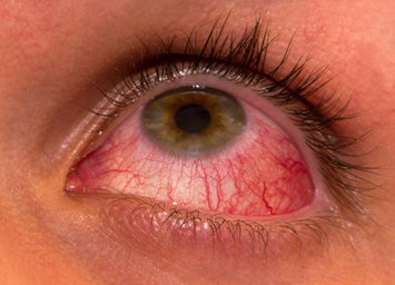
Acute Viral Conjunctivitis
Image:
Image (binary/octet-stream)
|
-most common cause of acute conjunctivitis; usually caused by adenovirus -Eyes have watery discharge & gritty/burning feeling -Highly contagious -Self-limiting (1-3 weeks) |
|
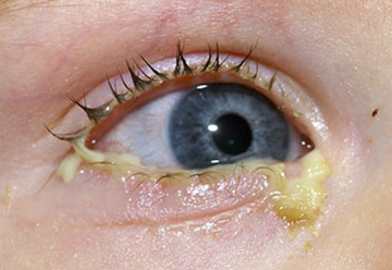
Acute Bacterial Conjunctivitis
Image:
Image (binary/octet-stream)
|
-usually caused by Staph. aureus -redness & thick, purulent discharge in one eye (sometimes bilateral) -Highly contagious -Typically self-limiting -N. gonorrhea can cause severe/sight-threatening conjunctivitis -Chlamydia trachomatis can cause severe conjunctival scarring |
| Acute Allergic Conjunctivitis | -caused by airborne allergens contacting the eye -typically presents as bilateral redness, watery discharge, itching |
|
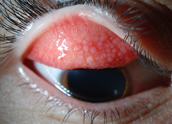
Secondary chronic conjunctivitis
Image:
Image (binary/octet-stream)
|
- may occur in contact lens wearers -should be seen by ophthalmologist if symptoms do not improve 12-24hrs following discontinuation of contact lens use |
| Conjunctival neoplasms | -tend to develop at limbus (border of cornea and sclera; site of stem cells of ocular surface) -Conjunctival nevi, squamous cell carcinoma, melanoma are the most common |
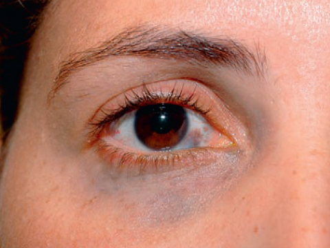
| The sclera may appear blue in a variety of conditions... | -Osteogenesis imperfecta -Heavily pigmented congenital nevus in underlying uvea (congenital melanosis oculi) |
| Nevus of Ota |
congenital melanosis oculi accompanied by periocular cutaneous pigmentation
Image:
Image (binary/octet-stream)
|
| What makes up the major refractive surface of the eye? | Cornea and overlying tear film |
| Myopia | Nearsightedness -inability to refract light onto retina -occurs because the eyes grow too long anterio-posteriorly -distant objects appear blurry |
| Hyperopia | Farsightedness -near objects appear blurry -occurs because the eyes grow too short |
| Presbyopia | age-related farsightedness due to declining converging power of the lens -due to progressive weakening of the ciliary muscles |
| Astigmatism | -irregularity of cornea or lens resulting in inability to properly focus light on the retina -Vision is blurry at any distance, people may have eye discomfort or headaches |
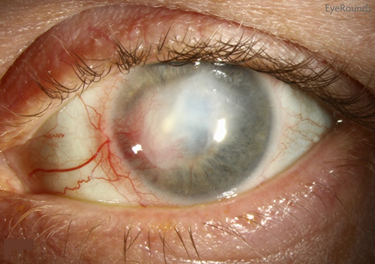
| Keratitis |
-inflammation of the cornea caused by infection (herpes simplex, herpes zoster), injury, wearing contact lenses too long
-eye redness, pain, blurred vision
-loss of vision may ensue & prompt medical attention necessary (inflamed corneal fibroblasts haphazardly deposit collagen)
Image:
Image (binary/octet-stream)
|
| Corneal collagen | precisely aligned to facilitate optic transparency |
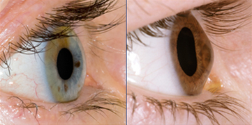
| Keratoconus |
-common corneal disease
-progressive thinning and ectasia of cornea results in conical rather than spherical shape
-generates irregular astigmatism
-rigid contact lenses may generate appropriate surface shape to cornea
Image:
Image (binary/octet-stream)
|
| Anterior chamber of the lens | -bounded anteriorly by cornea -bounded posteriorly by iris |
| Posterior chamber | -bounded anteriorly by iris -bounded posteriorly by lens |
| The aqueous humor enters the ...chamber, bathes the ..., and enters the ...chamber via the ... | -posterior -lens -anterior -pupil |
| Cataract | -opacities of lens resulting from physical changes to lens -results in blurry, foggy vision |
| Cataracts are associated with... | -diabetes mellitus -corticosteroids -radiation -trauma -age-related |
| Glaucoma | -group of eye diseases characterized by optic nerve damage and progressive visual field loss -strongly but not invariably associated with elevated intraocular pressure (IOP) -elevated IOP usually results from resistance to outflow of aqueous humor |
| Open-angle glaucoma | -most common form -leading cause of irreversible blindness in the world -aqueous humor has complete physical access to trabecular meshwork -elevated IOP results from increased resistance to aqueous outflow -primary/idiopathic or secondary to variety of causes -primary= no apparent structural changes |
| Angle-closure glaucoma | -transient apposition of pupillary margin of iris to lens blocks passage of aqueous humor -pressure build up in posterior chamber results in occlusion of the trabecular meshwork -can be secondary to inflammation or mass effect |
| Angle-closure often symptomatic... | -peripheral vision loss, light halos -headache, eye pain, nausea/vomiting -blindness |
| IOP in glaucoma can be managed by... | topical or systemic medications & surgery |
| IOP | intraocular pressure 9-21mmHg |
| Uvea | iris, ciliary body, & choroid |
| Uveitis | diverse group of chronic diseases either part of a systemic process or localized to the eye -Granulomatous uveitis (sarcoidosis) -Pneumocystis carnii or cytomegalovirus infection in AIDS patients -Sympathetic ophthalmia |
| Sympathetic ophthalmia | -usually develops following trauma to the eye -retinal antigens normally sequestered from immune system become exposed & delayed hypersensitivity reaction ensues -can result in blindness if untreated (Louis Braille) |
| Uveal melanocytic nevi estimated to affect... | 10% of Caucasian population |
| Uveal melanoma | -most common primary intraocular malignancy of adults -no clear link to UV exposure -may be asymptomatic or symptomatic -eye sparing radiotherapy preferred as treatment -Tendency to metastasize to the liver |
| What is the most common intraocular malignancy? | metastasis to the uvea (usually choroid) |
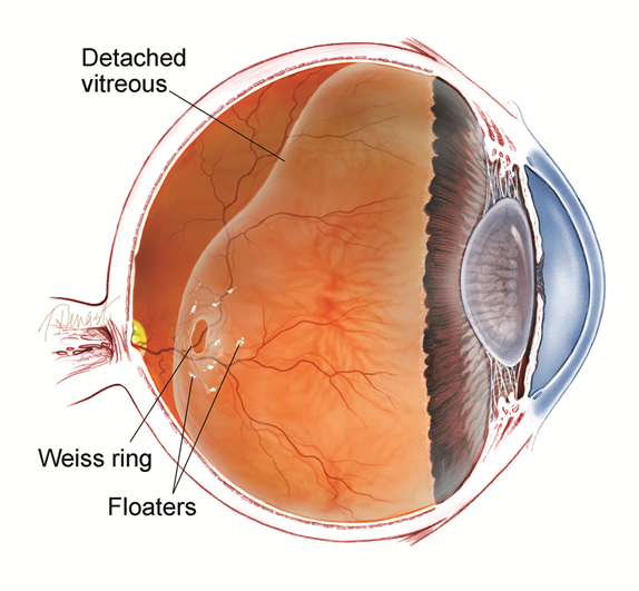
| Vitreous humor | -transparent, gelatinous mass comprising 80% of the volume of eyeball & surrounded by the vitreous membrane -not replenished -often liquefies and collapses with age, creating visual sensation of floaters |
|
Accumulation of liquefied vitreous may cause posterior vitreous detachment from the retina
Image:
Image (binary/octet-stream)
|
-results in rapid onset of increased cobweb-like floaters -areas with strong vitreoretinal adhesion may develop a retinal tear -common between ages 50-75 |
| Retinal pigment epithelium | -overlies (externally) the neurosensory retina -absorbs scattered light, forms blood-retinal barrier (among other functions) |
| Retinal detachment | -separation of neurosensory retina from retinal pigment epithelium -myopia, posterior vitreous detachment, surgery, trauma, lattice degeneration major risk factors |
| Choroidal circulation supplies... Separation results in... | -outer 1/3 of neurosensory retina (including photoreceptors) -ischemia & rapid/progressive photoreceptor degeneration |
| Regmatogenous retinal detachment | Retinal detachment w/ tear -posterior vitreous detachment (most common cause) -ocular trauma/injury |
| Non-rhegmatogenous retinal detachment | Retinal detachment w/out tear -choroidal tumors, malignant hypertension, diabetes |
| Clinical Presentation of Retinal Detachment | -spontaneous PVD -Flash of light (photopsia) lasting less than 1sec -black spots/shadows in visual field -pain -rapid diagnosis/surgical treatment required to minimize vision loss |
| Retinal vascular disease | diseases include hypertension/atherosclerosis, diabetes |
| Hypertension/atherosclerosis | -vessels may appear narrowed with blood column changing from bright red to copper/silver wire -malignant hypertension may damage retinal/choroidal vessels |
| Choroidal vessel damage may produce... | focal choroidal infarct (Elshnig spot) -overlying RPE damaged with potential visual field loss -accumulation of inflammatory exudate between RPE/neurosensory retina may produce retinal detachment |
| Diabetes and the eye | diabetics are at an increased risk for visual loss due to a variety of factors: -increased risk of cataracts -increased risk of glaucoma (b/c formation of neovascular membrane over iris) -retinopathy |
| Non-proliferative Diabetic retinopathy | -thickened basement membrane (as in diabetic microangiopathy in general) of retinal blood vessels -microaneurysms -leaky microcirculation leading to macular edema (common cause of vision loss) |
| Proliferative diabetic reinopathy | -characterized by appearance of newly sprouting vessels -retinal neovascularization may extend along potential plane between retina/vitreous >>Posterior vitreous detachment w/ massive hemorrhage >>fibrous scarring may lead to visual distortion and retinal detachment |
| Retinal artery occlusions: Atherosclerosis | -significant narrowing -risk of thrombosis |
| Retinal artery occlusion: Atheroembolism | -originating from thrombi in heart or atheromas in carotid arteries |
| Branch retinal artery occlusion | (BRAO) produces segmental infarct of retina |
| Central retinal artery occlusion | (CRAO) produces diffuse infarct of retina -sudden, painless, complete loss of vision -entire retina (except fovea) becomes swollen/opaque; fovea shows through as cherry-red spot -medical emergency |
| Age-related macular degeneration (AMD) | -most common cause of irreversible blindness in US -Smoking major modifiable risk factor -patients may complain of gradual, central vision loss in one or both eyes |
| Macula | focus of retina required for central and high-acuity vision |
| Dry (atrophic) AMD | -deposits in Bruch membrane, RPE atrophy result in severe vision loss -no effective treatment exists |
| Wet (neovascular) AMD | -characterized by choroidal neovascularization (occurs in conditions such as myopia, trauma, infection in genetically predisposed individuals) -Currently treated by injection VEGF (vascular endothelial growth factor) antagonists into vitreous of affected eye |
| Retinitis pigmentosa | inherited condition resulting from mutations that affect rods, cones, or RPE Retinal pigment accumulates around blood vessels -characterized by varying degrees of visual impairment >>Loss of rods may lead to early night blindness >>loss of cones leads to impaired central visual acuity -total blindness in some cases |
| Retinoblastoma | -most common primary intraocular malignancy of children -tends to present as abnormal white reflex to light (leukocoria) in children under 2yr -chemotherapy & radiotherapy followed by surgery |
| Anterior ischemic optic neuropathy | spectrum of stroke-like injuries to the optic nerve -transient ischemia produces episodic vision loss -total interruption of blood flow to optic nerve may result in infarction and blindness |
| temporal arteritis | inflammation of vessels supplying optic nerve may result in acute, total blindness |
| Primary neoplasms of the optic nerve | -pilocytic astrocytoma -meningioma |
| Papilledema | -optic nerve head swelling -due to intracranial pressure -optic nerve compression from primary neoplasm |
| Multiple sclerosis | -optic nerve demyelinization (optic neuritis) may lead to loss of vision |
Want to create your own Flashcards for free with GoConqr? Learn more.