2343511

| Question | Answer |
| 1.2 | 1.2 |
| What substances do organisms need and why | - Oxygen for aerobic respiration - Glucose as energy source - Proteins for growth and repair - Fats as energy source and to make membranes - Minerals to maintain water potential and help enzyme action |
| How can organisms absorb these substances | - From the surrounding environment - Make them inside their cytoplasm as part of cell metabolism |
| What waste products from their metabolic reactions within the cytoplasm do organisms need to remove | - CO2 - O2 - Ammonia or urea (excess nitrogen) |
| What type of organisms have a large surface area to volume ratio | Single-celled or small organisms |
| What type of SA to V ratio do multi celluar organisms have | Small SA to V ratio |
| Why do larger organisms need special exchange surfaces | - Cant exchange gases, nutrients or waste across their outer surfaces - Need larger area to exchange - Nutrients and gases have to travel further distance to center of organism |
| Features of a good exchange surface | Large surface area Thin barrier, small diffusion distance Fresh supply of molecules on one side to keep concentration high Removal of required molecules on other side to keep concentration low |
| What do all these features maintain | A steep diffusion gradient |
| What mechanism do some exchange surfaces use to increase exchange | Active transport |
| Example of a specialized exchange surface | - Walls of alveoli - Small intestine - Liver |
| What are the lungs | A large pair of inflatable structures lying in chest cavity |
| Describe the path of the airway through the lungs and where gas is exchanged | Air- Nose- Lungs- Trachea- Bronchi- Bronchioles- Alveoli- Gas exchanged on surface |
| How do people ventilate | Ribs covering lungs move together with action of diaphragm |
| Where does all gaseous exchange in the lungs take place | Alveoli |
| How does O2 get exchanged | Passes from air in alveoli to blood in capillaries |
| How does CO2 get exchanged | Passes from blood to alveoli |
| Features of a good exchange surface | - Large surface area - Barrier permeable to O2 and CO2 - Thin barrier to reduce diffusion distance - Maintained diffusion gradient (steep) |
| Why do exchange surface need a large surface area | To provide more space to molecules to pass through |
| Why are the alveoli sufficient despite being very small | They are numerous |
| Why is a steep diffusion gradient necessary | For diffusion to be rapid |
| How is a steep diffusion gradient maintained | Fresh supply of molecules on one side (keep concentration high) , removal of some molecules on other side (keep concentration low) |
| Summary of blood transport system | CO2 from tissue- Blood- Lungs - Concentration of CO2 always higher than that in air of alveoli - Carries O2 from lungs Ensures concentration is always lower in blood than in air of alveoli |
| How does the heart pump blood through to the lungs | Via the pulmonary artery |
| how are the lungs adapted to reduce diffusion distance | - Alveolus wall and capillary wall one cell thick - Squamous walls - Capillaries in close contact with alveolus walls - Total barrier only two cells thick |
| Summary of how breathing movements of the lung ventilate the lungs | - Replace used air with fresh hair - Brings more O2 into lungs and ensures concentration of O2 in air of alveolus remains higher than in the blood - Removes air containing CO2 from alveoli, lower concentration in alveolus |
| Describe inspiration (inhaling) | D E V P A -Diaphragm contracts- flattens and pushes digestive organs down - External intercostal muscles contract to raise ribs -Volume of chest cavity increases -Pressure in chest cavity drops (below atmospheric pressure) -Air moves into lungs |
| Describe expiration (exhaling) | D E V P A -Diaphragm relaxes, pushed up by displaced organs underneath -External intercostal muscles relax and ribs fall -Volume of chest cavity decreases -Pressure in chest cavity increases (above atmospheric pressure) -Air moves out of lungs |
| What do the trachea, bronchi and bronchioles pass air into | The lungs |
| What requirements must the airways meet in order to be effective | - Large to allow sufficient air flow without obstruction - Divide into smaller airways to deliver air to all the alveoli - Strong to prevent collapsing when there is low pressure inside - Flexible to allow movement - Able to stretch and recoil |
| Which two airway structures have similar structures but differ in size | Bronchi (narrower) and trachea |
| Which airway structures have several layers of tissue | Bronchi Trachea |
| Structure and features of trachea | - Mainly cartilage walls - Cartilage form incomplete or c-shaped rings - Loose tissue on inner surface of wall, made of glandular and connective tissue, elastic fibres, smooth muscle and blood vessel - Central inner lining of epithelium; cilia epithelium and goblet cells |
| Structure and features of Bronchi | - Mainly cartilage walls - Cartilage less regular - Loose tissue on inner surface of wall, made of glandular and connective tissue, elastic fibres, smooth muscle and blood vessel - Central inner lining of epithelium; cilia epithelium and goblet cells |
| Structure and features of bronchioles | - Much narrower than bronchi - Only larger ones have cartilage - Wall made mostly of smooth muscles and elastic fibres - Smallest ones have clusters of alveoli at their ends |
| What is the role of the cartilage tissue | - Structural role - Supports trachea and bronchi, holding them open - Prevents collapse, when pressure inside is low during inhalation - Allows flexibility in trachea without constricting the airways - |
| Role of smooth muscle Role of Elasitic fibres | - Can contract and restrict airways - Can make lumen of airway narrower - Control flow of air to alveoli (e.g. when harmful substance is present) - Not voluntary - Airways constrict, elastic fibres in loose tissues deform - As smooth muscle relaxes, elastic fibres recoil to original size and shape - Widening (dilating) the airway |
| Role of Goblet cells and Glandular tissue | - Under epithelium - Secrete mucus - Trap tiny particles from air - Trap bacteria so they can be removed and reduce risk of infection |
| Role of Ciliated epithelium | - Consists of ciliated cells - Cilia waft away mucus up airway to back of throat so it can be swallowed and killed by stomach acid |
| What is breathing | Air moving in and out of your lungs as diaphgram and intercostal muscles contract and relax (12 times a minute) |
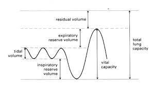
| Tidal Volume definition | Volume of air moved in and out at rest |
| Vital capacity | Largest volume of air that can be moved into and out of lungs in one breath |
| Residual Volume | Volume of air that always remains in lungs |
| Dead space | Air in the bronchioles, bronchi and trachea (where no exchange occurs with blood) |
| Inspiratory reserve volume | Volume of air that can be inhaled above tidal volume |
| Expiratory reserve volume | Volume of air that can breathed out above amount breathed in tidal volume breath |
| What does the spirometer trace look like | |
| How is a spirometer used to measure lung volume | - Chamber filled with oxygen floats on tank of water - Person breathes through mouthpiece attached to tube connected to O2 chamber. - Breathing in takes O2 from chamber so tank sinks and vice versa |
| How can the different elements of lung volume be shown using spirometer | Person can be asked to breathe in different ways or do some exercises to show different patterns of breathing |
| What gas can build up when breathing into and out of a spirometer | CO2 |
| What type of lime is used to absorb the CO2 | Soda lime |
| What do all animal cells need a supply of in order to survive | Oxygen and nutrients |
| What are the three main factors that affect the need for a transport system | 1) Size 2) SA to Volume ratio 3) Level of activity |
| How does size affect the need for a transport system | Animals with lots of layers of cells won't be able to supply cells in central region as the nutrients will be used up by outer layer of cells |
| How does the SA to Volume ratio affect the need for a transport system | In larger animals surface area isn't large enough to supply to internal cells |
| How does level of activity affect the need for a transport system | More active animals need a sufficient supply of energy from respiration which requires O2 (e.g mammals for warmth) |
| What are the features of a good transport system | - Fluid or medium to carry nutrients and O2 around body -Pump to create pressure to push fluid around body (heart) - Exchange surface - Tubes or vessels - Two circuits (one to pick up and one to deliver O2 to tissues |
| Example of an animal with a single circulatory system | Fish |
| Example of an animal type with a double circulatory system | Mammal |
| How does the fish single circulatory system work | Heart - Gills - Body - Heart |
| How does the mammal double circulatory system work | Two separate circuits Heart - Body - Heart - Lungs - Heart |
| What are the two circuits for the double circulatory system | 1) Carries blood to lungs to pick up O2 (Pulmonary circulation) 2) Carries O2 and nutrients around body to tissues (Systemic circulation) |
| How are S.C.Systems efficient for fish | -Fish are less active and don't maintain their own body temperature - Require less energy - Delivers O2 and nutrients quickly enough |
| How are D.C.Systems efficient for mammals | - Active animals, maintain their body temperature - Require energy from food in process of respiration for heat and activity - Need good supply of both nutrients and O2 to release a lot of energy |
| Conditions in fish S.C.System | - Blood pressure is reduced as blood passes through tiny capillaries of the gills. - Blood doesn't flow very quickly to rest of the body - Rate at which O2 and nutrients are delivered to respiring tissues is limited |
| Conditions in mammal D.C.System | - After blood is passed through lungs, heart can increase its pressure so blood flows more quickly to tissues - S circulation can carry blood at a higher pressure - Blood pressure cant be too high in P circulation as it may damage capillaries in lungs |
| 2 | 2 |
| What is the mammalian heart and how is it divided | - Muscular double pump - Divided into two sides |
| What does the right hand side pump | Deoxygenated blood to lungs to be oxygenated |
| What does the left hand side pump | Oxygenated blood to rest of body |
| How is blood moved along the arteries | Heart squeezes the blood putting it under pressure which then forces the blood along |
| External features of heart | - Atria (thin-walled chambers) - Ventricles (main pumping chambers) - Coronary arteries (lie over surface, carry oxygenated blood to heart muscle) - Tubes: veins and arteries |
| Internal features of heart | - Two upper atria - Vena cava (deoxy blood to right atria) - Pulmonary vein (Oxy blood from lungs to left atrium) - AV valves - Tendinous cords (inside ventricles, attach valves to walls and prevent back flow) - Septum (wall of muscle separating ventricles from each other) - Semilunar valves |
| What happens when the muscle of each chamber contracts | Pressure in blood is increased |
| Why is the muscle of the atria thin | Chambers don't need to create much pressure Function is to push blood into ventricles |
| Which ventricle is thicker and why | Left ventricle, have to pump blood around whole body, more pressure |
| Stages of contraction in cardiac cycle | - Filling phase - Atrial contraction - Ventricular contraction |
| Why is important that the chambers of the heart all contract in a coordinated fashion | To avoid inefficient pumping |
| What is the cardiac cycle | A sequence of events involved in one heartbeat (as it is a cycle there is no clear end or beginning) |
| What happens during the Filling phase and what is it known as | - Atria and ventricles are relaxing - Internal volume increases and blood flows into heart from major veins - Blood flows into atria, through AV valves and into ventricles Diastole |
| What happens during the Atrial contraction and what is it known as | - Heart beat starts - Right and left atria contract together - Small increase in pressure by contraction - Helps push blood into ventricles - Walls of ventricles stretched, full of blood - Once full, ventricles contract - Blood fills AV valves causing them to snap shut - Prevents backflow Atrial Systole |
| What happens during ventricular contraction and what is it known as | - All four heart valves are closed for a short period - Walls of ventricles contract Ventricular Systole - Raises pressure in ventricles very quickly contraction starts at apex of heart - blood pushed upwards towards arteries - semilunar valves open - blood pushed out of heart - Contractions last for short time Ventricle walls relax, heart starts filling again |
| How do valves work | - Ensure blood flows in correct direction - Opened and closed by changes in blood pressure in various chambers of the heart |
| What causes the AV Valves to open | When pressure in the ventricles drops below pressure in the atria. (When ventricular walls relax and recoil after contracting) |
| Where does blood entering the heart flow straight through | Through the atria and into the ventricles |
| What happens to the pressure in the atria and ventricles as they fill | Slowly rises |
| Do the valves remain open or closed while the atria contract | Open |
| As the ventrticles begin to contract does the pressure of blood in the ventricles rise or fall | Rise |
| In what direction does the blood start to move, when the pressure in the ventricles rises above that in the atria | Upwards |
| How does this upwards movement prevent blood flowing back into the atria | Fills valve pockets and keeps them closed |
| When the ventricles start to contract, is the pressure higher in the major arteries or in the ventricles | In the major arteries |
| What effect does this have on SV valves (ventricles contracting) | Semilunar valves are closed |
| Why does the pressure inside the ventricles rise quickly as they contract | Blood cannot escape |
| What leads to the semilunar valves being pushed open | Pressure in the ventricles being higher than the pressure in the aorta and pulmonary arteries |
| In what state of pressure is blood forced out of the ventricles | Under very high pressure |
| What happens to the heart muscle once the ventricle walls have finished contracting | Heart muscle starts to relax |
| Role of elastic tissues in ventricular walls after contraction & as a result what does it cause | - Recoils to stretch muscle out again and return ventricles to original size - This causes the pressure in pressure to drop quickly |
| What happens once the pressure in the ventricles drops below pressure in the major arteries | SV valves pushed closed By blood starting to flow back towards the ventricles & collecting in valves pockets |
| What does the closing of the SV valves prevent | Back flow to the ventricles (blood) |
| What sound does the heart make | Lub-dup |
| What is the first sound 'Lub' a result of | AV valves closing as ventricles start to contract |
| What is the second sound 'Dup' a result of | SV vales closing as ventricles start to relax |
| The shutting of which valve is louder (not due to blood accumulating in their pockets) | AV valves |
Want to create your own Flashcards for free with GoConqr? Learn more.