2695221

| Question | Answer |
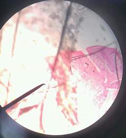
| [DON'T MOVE].. folliate papillae (rabbit tongue) | |
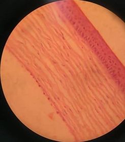
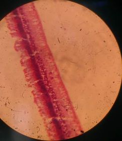
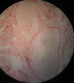
| [DON'T MOVE] cornea..see the parallel collagen fibers | |
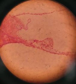
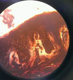
| [DON'T MOVE] retina.. see the pigmented epithelium | |
| [DON'T MOVE] organ of corti see the tectorial membrane | |
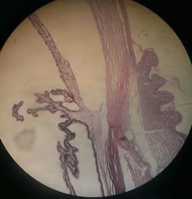
| [DON'T MOVE] corneo-scleral junction see cornea, iris and ciliary boby | |
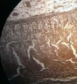
| [DON'T MOVE] motor end plate gold chloride | |
| [DON'T MOVE] paccinian corpuscle.. onion like shaped | |
| [DON'T MOVE] Miessner's corpuscles | |
| [DON'T MOVE] muscle spindles.. don't see any thing, so special :D | |
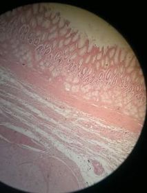
| tongue see the papillae | |
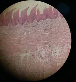
| esophegus see stratified non keratinized epith. and esophgeal gland in submucosa | |
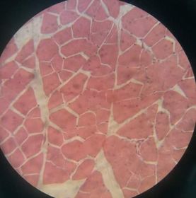
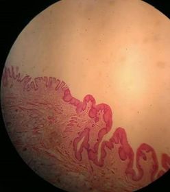
| fundus of stomach there's a distinct line of epith. coming up and down but still distinct (unlike jejunum).. :)) | |
| pylorus see pyloric glands | |
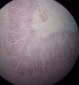
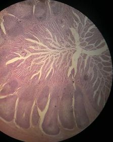
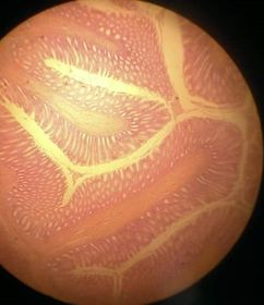
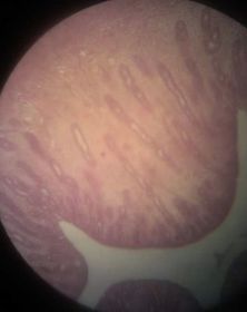
| jejunum see long villi, crypts NO BRUNNER'S GLANDS | |
| duodenum see brunner's glands | |
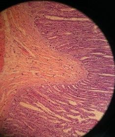
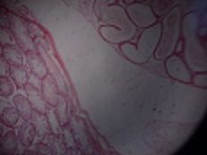
| ileum see peyer's patch like branches of tree ^_^ | |
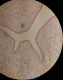
| colon (large intestine) | |
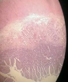
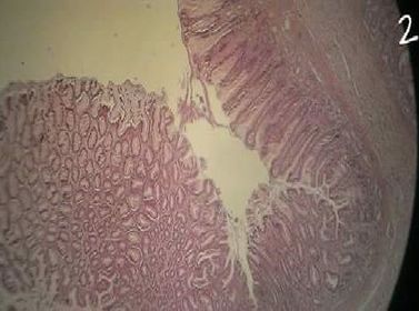
| gastro-esophageal junction see the stratified squamus epith. becoming cardiac glands. | |
| pyloroduodenal junction see the gland become shorter and pyloric glands appear. | |
| rectoanal junction see the crypts of rectum being stratified squamous epith. of anal | |
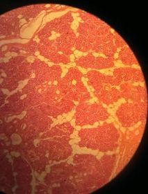
| parotid gland only serous acini .. see the ducts | |
| submandibular gland see serous and mucous acini and ducts inbetween | |
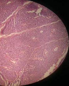
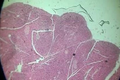
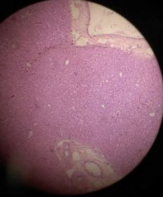
| pancreas see islets of langerhans (pale purple) | |
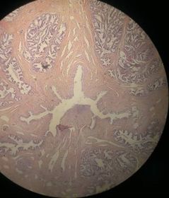
| human liver central veins (white dots) hepatocytes radiating from it ( like a sun :D ) | |
| liver silver | |
| liver pig | |
| pituitary gland see the cleft and pale circle in the center (pars nervosa) | |
| thyroid gland see the colloid, it may be with parathyroid . move the slide all around :D | |
| suprarenal gland silver see parallel cords of cells in zona fasciculata | |
| urinary bladder transitional epith. | |
| ureter muscle layer is small and wide lumen NOT like Vas deferns | |
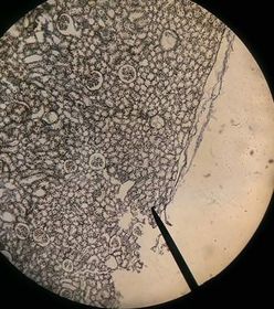
| kidney (Gelatin Carmin) see the glomeruli | |
| kidney silver | |
| testis & epididymis mooove the slide all around :D | |
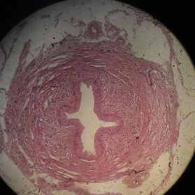
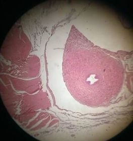
| vas deferns small lumen, large muscle area | |
| spermatic cord see vas and anything else xD moooove the slide all around :)) | |
| prostate arranged glands | |
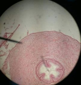
| penis 2 eyes and mouth :D | |
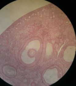
| ovary see Graffian, primary and secondary follicules | |
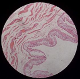
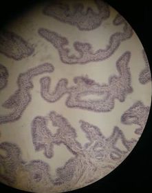
| Fallobian tube see folds folds everywhere xD | |
| uterus see the uterine glands , if u saw "8" shaped muscles, u would be awesome :)) | |
| vagina see nothing except stratified squamous non keratinized epith. ^^ if u saw glands... it would be esophegus | |
| placenta so unique .. just red dots everywhere | |
| resting mammary gland C.T and ducts | |
| lactating mammary gland alveoli with milk | |
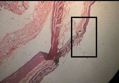
| eye lid see the tarsal plate |
Want to create your own Flashcards for free with GoConqr? Learn more.