16856536
Description
Mind Map by Fatima Alkhateeb, updated more than 1 year ago
|
|
Created by Fatima Alkhateeb
almost 6 years ago
|
|
Skin Ulcers
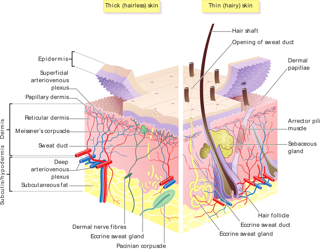
- Histology of the skin
- Epidermis
- Cells
- Keratinocytes
- Melanocytes
- Langerhan's cells
- Merkel cells
- Keratinocytes
- Layers
- Stratum Basale
- Stratum spinosum
- Stratum granulosum
- Stratum Lucidum
- Stratum Corneum
- Found in
thick
skin only
- Stratum Corneum
- Stratum Lucidum
- Stratum granulosum
- Stratum spinosum
- Stratum Basale
- Cells
- Dermis
- Layers
- Papillary
- Reticular
- Papillary
- Layers
- Hypodermis
- Subcutaneous layer deep
to the skin, composed of
adipose and areolar
connective tissue
- Subcutaneous layer deep
to the skin, composed of
adipose and areolar
connective tissue
- Glands
- Eccrine (sweat glands)
- Apocrine (after puberty)
- Holocrine (sebaceous glands)
- Eccrine (sweat glands)
- Hair
- Medulla
- Cortex
- Cuticle
- Medulla
- Epidermis
- Skin Functions
- Protection
- Sensation
- Thermo-regulation
- Secretion
- Protection
- Types of skin lesions
- Macule:Flat lesion with
well circumscribed
change in color < 1cm
- Patch : Macule >1cm
- Papule: Raised solid skin lesion < 1cm
- Plaque:Papule > 1cm
- Vesicle:Small blister containing fluid < 1cm
- Bulla:Large blister containing fluid > 1cm
- Pustule:Vesicle containing pus
- Wheal:Transient smooth papule/plaque
- Scale:Scaling off the corneum
- Crust:Dry exudate
- Macule:Flat lesion with
well circumscribed
change in color < 1cm
- Dermatological changes
- Hyperkeratosis:
Increased corneum
thickness
- Parakeratosis:Retention
of nuclei in corneum
- Hypergranulosis:
Increased granulosum
thickness
- Spongiosis: Transient
smooth papule/plaque
- Acantholysis:Separation
of epidermal cells
- Acanthosis: Epidermal
hyperplasia
- Hyperkeratosis:
Increased corneum
thickness
- Skin cancer
- Squamous cell
carcinoma
- Ulcer with a necrotic base
and everted edges, keratin
pearls on histopathology
- Ulcer with a necrotic base
and everted edges, keratin
pearls on histopathology
- Basal cell
carcinoma
- Rodent ulcer with rolled in
edges, basal cells with
pallisading pattern
- Rodent ulcer with rolled in
edges, basal cells with
pallisading pattern
- Melanoma
- significant metastasis,
ABCDE's to diagnose it
- significant metastasis,
ABCDE's to diagnose it
- Squamous cell
carcinoma
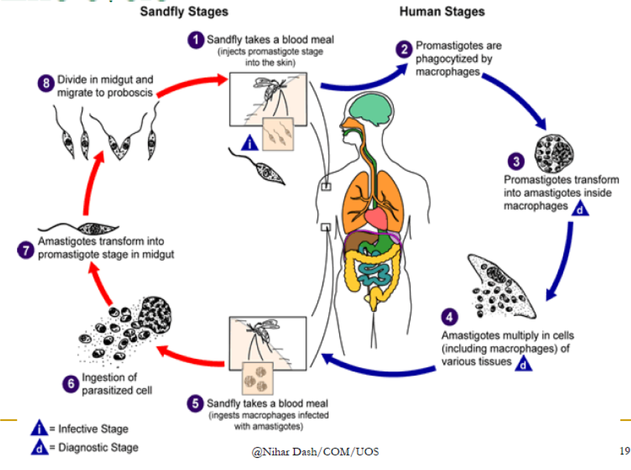
- Leishmeniasis
- Investigations
- Amastigotes in tissues using
giemsa stain
- Bone Marrow aspirate test (Sensitivity 54-86%)
- Splenic aspirate is most sensitive (96-98%)
- Amastigotes in tissues using
giemsa stain
- Management
- Sodium stingluconate
- Meglumine antimoniate
- Miltefosine
- Amphotericin
- Paromomycin
- Sodium stingluconate
- Types
- Cutaneous
- The lesions typically evolve from
papules to nodular plaques to
ulcerative lesions, with a raised
border and central depression
- The lesions typically evolve from
papules to nodular plaques to
ulcerative lesions, with a raised
border and central depression
- Visceral
- Symptoms range from: severe
hepatosplenomegaly, irregular bouts
of fever, hyperpigmented
granulomatus areas on the skin,
weight loss (Kala-Azar)
- Symptoms range from: severe
hepatosplenomegaly, irregular bouts
of fever, hyperpigmented
granulomatus areas on the skin,
weight loss (Kala-Azar)
- Mucocutaneous
- lesions can lead to partial or total
destruction of the mucous membranes of
the nose, mouth and throat cavities and
surrounding tissues
- lesions can lead to partial or total
destruction of the mucous membranes of
the nose, mouth and throat cavities and
surrounding tissues
- Cutaneous
- Investigations
Media attachments
Want to create your own Mind Maps for free with GoConqr? Learn more.