21971285
Descrição
FlashCards por Evian Chai, atualizado more than 1 year ago
|
|
Criado por Evian Chai
mais de 4 anos atrás
|
|
| Questão | Responda |
| During inhalation which two muscles contract? | 1. External intercostals move ribcage up and out 2. Diaphragm contracts and moves down Results in drop in pressure below atmospheric |
| During inhalation which two muscles contract? | 1. Internal/innermost intercoastals contract and move ribcage down and in 2. Abdominal muscles contract and push diaphragm back up Results in raise in pressure above atmospheric |
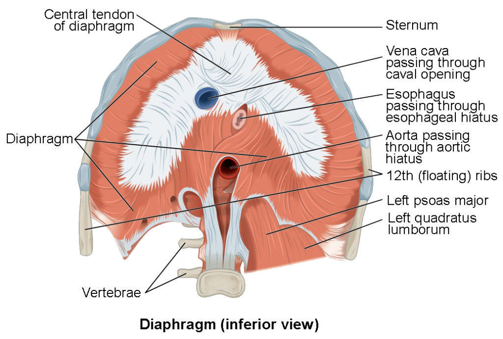
| What are the 5 attachments of the Diaphragm? | 1. Lumbar vertebrae and accurate ligaments - Right crus stretches down and attaches to L1, L2 & L3 - Left crus stretches down and attaches to L1 &L2 2. Coastal cartilages of ribs 7-10 3. Direct attachments of floating ribs 11 & 12 4. Xiphoid process of sternum 5. Central tendon of Diaphragm which is an aponeurosis (pearly white fibrous tissue) - inferior to fibrous pericardium |
| What are the 3 major tubular structures passing through the diaphragm? | 1) Caval Hiatus/Opening - Inferior Vena Cava (T8) - Passes through the central tendon 2) Oesophagus Hiatus - Oesophagus (T10) - Oesophageal branches of left gastric artery/vein 3) Aortic hiatus - Aorta (T12 - behind the diaphragm, sitting next to the vertebrae) |
| Which opening does the right phrenic nerve pass through in the oesophagus? | Caval Hiatus with the inferior vena cava |
| Which opening do the vagus nerves pass through in the oesophagus? | Oesophagus Hiatus behind the root of the lung |
| Which opening do the Thoracic duct and Azygous vein pass through in the oesophagus? | Aortic hiatus |
| What 2 things innervate the diaphragm? | 1. C3 4 5 keeps the diaphragm alive! Phrenic Nerve (somatic PNS) 2. Intercoastal Nerves |
| Motor neurons are located in the | Ventral root ganglion |
| Sensory neurons are located in the | Dorsal root ganglion |
| What are the primary muscles of respiration? | Intercostal muscles |
| What are the secondary muscles of respiration? | 1. Pectoralis Major 2. Pectoralis Minor 3. Sternocleidomastoid 4. Scalene muscles 5. Serratus Anterior 6. Subcostal muscles 7. Transversus thoracis 8. but MAINLY ABDOMINAL MUSCLES |
| What facilitates quiet expiration? | Is a passive process facilitated by the elastic recoil of lungs |
| Which 2 muscles are primarily involved in forced expiration? | 1. Internal intercostals 2. Abdominals |
| What is the function of the larynx? | Produce sound Breathe Protect trachea from food aspiration Opens during coughing/sneezing |
| Where is the pharynx? What does it do? | In the throat right behind the nasal cavity, above the larynx and oesophagus Plays a role in vocalisation |
| What are the three parts of the pharynx? | 1. Nasopharynx 2. Oropharynx 3. Laryngopharynx |
| What type of cartilage lines the trachea? What smooth muscle? | Hyaline cartilage The trachealis |
| What is the carina? Where is it located? | A ring of cartilage between main division of bronchi (in line w sternal angle/T4) Mucous membrane triggers cough reflex/prevents things from entering |
| Which bronchus is more vertical? What is a consequence of this? | The right one (left is more angled because of the heart) Foreign objects more likely to fall into right bronchus |
| What part of the lungs are the bronchopulmonary segments in? What is unique about them? | In the tertiary branch of the bronchioles Independent units with own artery/vein |
| The left lobe has how many lobes? Which fissure (s)? | 2 (smaller bc heart) Oblique fissure |
| The right lobe has how many lobes? Which fissure (s)? | 3 (bigger bc no heart) Oblique+horizontal fissure |
| What are the 3 surfaces and borders of the lungs? | Surfaces: costal, diaphragmatic, mesdiastinal Borders: anterior, inferior, posterior |
| What 6 things do the root of the lung contain? | 1. Bronchi 2. Pulmonary artery (superior) 3. Pulmonary vein (inferior) 4. Oesophagus 5. Aorta 6, Superior vena cava and inferior vena cava |
| What does the superior vena cava branch into? | Right/left brachiocephalic veins |
| What are the pleuras of the lung from inside to out? | 1. Visceral pleura (covers surface of lung) 2. Pleural cavity (fluid filled) 3. Parietal pleura (Membrane outside) |
| What are three functions of the pleura? | 1. reduce friction 2. reduce surface tension between layers 3. create negative pressure |
| The visceral pleura is innervated by | 1. Vagus nerve (pns) 2. sympathetic nerves |
| The parietal pleura is innervated by | 1. Intercostal nerves 2. Phrenic nerve |
| What are the two separate circulations to the lungs? | 1. Pulmonary (for reoxygenation) 2. Bronchial (for nutritive) |
| What does the phrenic nerve innervate? | 1. Diaphragm 2. Parietal pleura 3. Pericardium |
| What are the 4 subdivisions of the parietal pleura? | 1. Cervical (at apex) 2. Costal (at side) 3. Mediastinal (near heart) 4. Diaghpramatic (at bottom) |
| The posterior limit of the diaphragm is at? The anterior limit of the diaphragm is at? The inferior limit is at? The superior limit is at? | 1. T12 2. Costal margin 3. 4th intercostal for right 5th intercostal for left xiphisternum for central tendon |
Quer criar seus próprios Flashcards gratuitos com GoConqr? Saiba mais.