20493484
Descrição
Quiz por Charlotte Jakes, atualizado more than 1 year ago
|
|
Criado por Charlotte Jakes
aproximadamente 5 anos atrás
|
|
Questão 1
Questão
Skeletal muscle cells fuse to form giant multinucleate cells with several nuclei located on the periphery that control a common cytoplasm - they form a syncitium.
Responda
- True
- False
Questão 2
Questão
What name is given to the primary site of force transmission at the muscle-tendon interface?
Responda
-
Myotendinous junction
-
Neuromuscular junction
-
Myotendinous point
-
Myocollagenous junction
Questão 3
Questão
The myotendinous junction arises from the fusion of what structures? Check all that apply
Responda
-
Epimyisum
-
Perimysium
-
Endomysium
-
Fascicles
-
Scarpa's fascia
-
Compact bone
-
Articular cartilage
Questão 4
Questão
Fill in the blanks to give the names of each classification of muscle according to its function.
[blank_start]Flexor[blank_end] muscles bend a joint, decreasing its angle.
[blank_start]Extensor[blank_end] muscles straighten a joint, increasing its angle.
The [blank_start]prime mover[blank_end] or agonist muscle performs concentric contraction to bring about movement.
The [blank_start]antagonist[blank_end] muscle opposes the action of the primer mover to return it to its normal position.
The [blank_start]fixator[blank_end] muscle steadies the position produced by the prime mover by isometric contraction.
The [blank_start]synergist[blank_end] muscle complements the action of the prime mover by performing the same contraction.
Responda
-
Flexor
-
Extensor
-
prime mover
-
antagonist
-
fixator
-
synergist
Questão 5
Questão
Blood vessels and nerves are embedded in the fascicles.
Responda
- True
- False
Questão 6
Questão
Fill in the blanks below to describe the structure of muscle based on the divisions by the different layers of connective tissue.
The [blank_start]epimysium[blank_end] is the tough outermost layer surrounding the entire muscle.
The [blank_start]perimysium[blank_end] surrounds bundles of muscle fibres to create fascicles.
The [blank_start]endomysium[blank_end] surrounds each muscle fibre within the fascicles.
Responda
-
epimysium
-
perimysium
-
endomysium
Questão 7
Questão
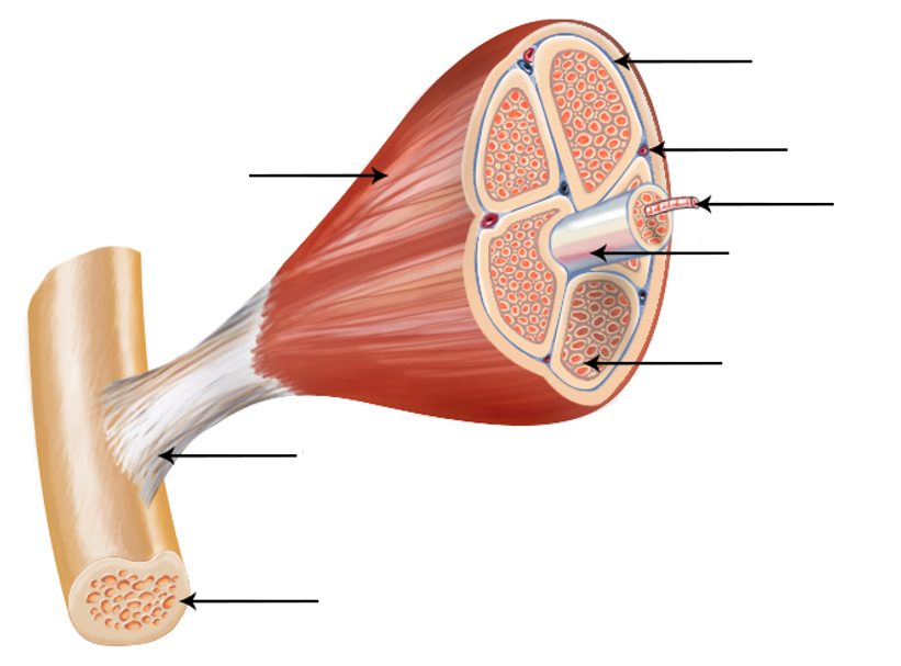
Label this image to show the general structure of a skeletal muscle.
Responda
-
Epimysium
-
Perimysium
-
Blood vessel
-
Muscle fibre
-
Fascicle
-
Endomysium
-
Tendon
-
Bone
Questão 8
Questão
Fill in the blanks below to describe the formation of skeletal muscle.
1. A single [blank_start]myoblast[blank_end] cell exists in a medium containing growth factor.
2. The cell [blank_start]proliferates[blank_end].
3. Multiple cells fuse to form [blank_start]myotubes[blank_end].
4. Myotubes gather to form [blank_start]myofibres[blank_end].
Responda
-
myoblast
-
proliferates
-
myotubes
-
myofibres
Questão 9
Questão
Muscle fibres are post-mitotic cells - myonuclei cannot re-enter the cell cycle and divide.
Responda
- True
- False
Questão 10
Questão
What type of cell exists on the surface of muscle fibres that divide in response to stimuli and differentiate into new myoblasts?
Responda
-
Stem cells
-
Satellite cells
-
Mesenchymal cells
-
Mast cells
Questão 11
Questão
Satellite cells cannot self-renew.
Responda
- True
- False
Questão 12
Questão
What processes do we need satellite cells for? Check all that apply.
Responda
-
Muscle growth after birth
-
Muscle repair
-
Muscle hypertrophy (growth in response to exercise)
-
Muscle formation in utero
-
Muscle degradation during starvation
-
Tendon formation
-
Ossification
Questão 13
Questão
What is the most basic unit of skeletal muscle?
Responda
-
Sarcomeres
-
Muscle fibres
-
Fascicles
-
Myonuclei
Questão 14
Questão
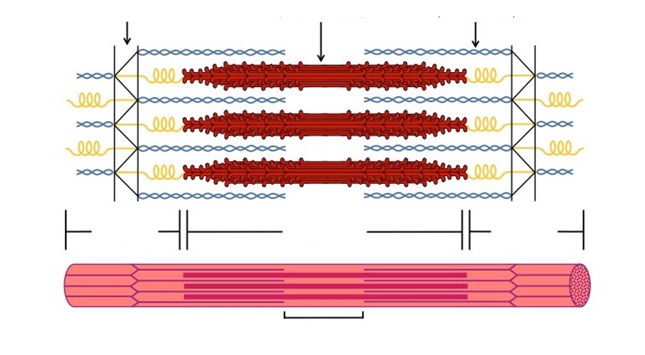
Label this image to give the structure of a sarcomere.
Responda
-
Myosin
-
Actin
-
Z disk
-
I band
-
A band
-
I band
-
H zone
Questão 15
Questão
What does the speed of contraction of a muscle fibre depend on?
Responda
-
Amount of myosin heavy chain isoform present
-
Amount of myosin light chain isoform present
-
Number of sarcomeres present
-
Number of mitochondria present
Questão 16
Questão
Which muscle fibre isoform is described as a white muscle fibre as it only contains a small amount of myoglobin?
Responda
-
Fast isoform (Type II)
-
Slow isoform (Type I)
Questão 17
Questão
Which muscle fibre isoform is described as a red muscle fibre as it contains a lot of myoglobin?
Responda
-
Slow (type I)
-
Fast (type II)
Questão 18
Questão
Type I slow muscle fibres are suited to [blank_start]long[blank_end]-term contraction for [blank_start]endurance[blank_end] work. They are red and contract with slow [blank_start]twitch[blank_end] velocity. They have a higher density of [blank_start]mitochondria[blank_end] so are better suited to aerobic respiration. They are therefore fatigue [blank_start]resistant[blank_end]. However, they produce [blank_start]less[blank_end] instantaneous force so are described as 'weak'. They store energy in the form of [blank_start]triglycerides[blank_end].
Responda
-
long
-
endurance
-
twitch
-
mitochondria
-
resistant
-
less
-
triglycerides
Questão 19
Questão
Type II muscle fibres are better suited to [blank_start]short[blank_end]-term contraction. They are [blank_start]white[blank_end] in colour due to a lack of myoglobin. They contract with [blank_start]fast[blank_end] twitch velocity and are more prone to aerobic respiration. They therefore fatigue easily, but produce more instantaneous force. They store energy in the form of ATP and creatine phosphate.
Responda
-
short
-
white
-
fast
Questão 20
Questão
Type II muscle fibres are better suited to [blank_start]short[blank_end]-term contraction. They are [blank_start]white[blank_end] in colour due to a lack of myoglobin. They contract with [blank_start]fast[blank_end] twitch velocity and are more prone to [blank_start]anaerobic[blank_end] respiration. They therefore fatigue easily, but produce more [blank_start]instantaneous[blank_end] force. They store energy in the form of ATP and [blank_start]creatine phosphate[blank_end].
Responda
-
short
-
white
-
fast
-
anaerobic
-
instantaneous
-
creatine phosphate
Questão 21
Questão
The Type IIa fast [blank_start]oxidative[blank_end] muscle fibres have higher concentrations of [blank_start]myoglobin[blank_end] and mitochondria. They are therefore the most resistant to [blank_start]fatigue[blank_end] of the Type II fibres and are used in [blank_start]long[blank_end] term anaerobic activities.
Type IIb fast [blank_start]glycolytic[blank_end] muscle fibres have low concentrations of myoglobin and [blank_start]mitochondria[blank_end] so fatigue easily but contract [blank_start]quickly[blank_end] with high power. They are therefore used in short term [blank_start]anaerobic[blank_end] activities.
Type IIx super-fast [blank_start]glycolytic[blank_end] muscle fibres have the lowest concentrations of muscle fibres so are very vulnerable to [blank_start]fatigue[blank_end]. They do, however, contract the fastest with the largest amount of [blank_start]power[blank_end].
Responda
-
oxidative
-
glycolytic
-
glycolytic
-
myoglobin
-
fatigue
-
long
-
mitochondria
-
quickly
-
anaerobic
-
fatigue
-
power
Questão 22
Questão
Each muscle fibre receives innervation from one motor neuron.
Responda
- True
- False
Questão 23
Questão
What name is given to the group of muscle fibres from a single neuron innervating multiple fibres?
Responda
-
Motor unit
-
Motor nerve
-
Neuromuscular junction
-
Motor end plate
Questão 24
Questão
Larger motor units have greater degrees of control.
Responda
- True
- False
Questão 25
Questão
What is dystrophin?
Responda
-
Cytoplasmic protein that connects the cytoplasm of muscle fibres to the extracellular membrane
-
Cytoskeletal protein that myosin binds to in the sarcomere
-
Voltage-gated calcium ion channel found in the sarcolemma
-
Voltage-gated calcium ion channel found on the sarcoplasmic reticulum
Questão 26
Questão
Why are males more prone to muscular dystrophies (disorders of the dystrophin protein)?
Responda
-
The gene for dystrophin is X-linked
-
The gene for dystrophin is autosomally linked
-
Males tend to have higher lean body mass
-
Testosterone can mutate dystrophin
Questão 27
Questão
What is the function of dystrophin?
Responda
-
Stabilises the sarcoplasm and sarcolemma during contraction
-
Stabilises the position of the myonuclei during contraction
-
Helps filter material through the basal lamina
-
Transports Ca2+ out of the sarcoplasmic reticulum for contraction
Questão 28
Questão
The C-terminus of dystrophin binds to the [blank_start]dystrophin associated protein complex[blank_end] (DAPC) found at the [blank_start]sarcolemma[blank_end].
The N-terminus of dystorphin binds to filamentous [blank_start]actin[blank_end] in the cytoskeleton.
Responda
-
dystrophin associated protein complex
-
sarcolemma
-
actin
Questão 29
Questão
What disease is characterised by necrosis and fibrosis of the muscles causing later-stage walking, toe walking, Gower's manoeuvre often leading to death in 20s-30s?
Responda
-
Duchenne muscular dystrophy
-
Creutzfeldt Jakob disease
-
Patau syndrome
-
Sickle-cell anaemia
Questão 30
Questão
The limb and trunk muscles are derived from embryonic structures known as [blank_start]somites[blank_end] - buds of the mesoderm. The [blank_start]somites[blank_end] are divided into sections which will give rise to different parts of the body.
The [blank_start]dermomyotomes[blank_end] will give rise to the skeletal muscle, the satellite cells and the overlying dermis.
The [blank_start]sclerotome[blank_end] will give rise to cartilage, joints and tendons.
The [blank_start]myotome[blank_end] will form differentiated myocytes.
Responda
-
somites
-
somites
-
dermomyotomes
-
sclerotome
-
myotome
Questão 31
Questão
Muscle precursor cells that end up in medial locations will initially migrate into the limb bud periphery before moving back towards the midline to their final location.
Responda
- True
- False
Quer criar seus próprios Quizzes gratuitos com a GoConqr? Saiba mais.