8844605
Descrição
Quiz por Ella Middlemiss, atualizado more than 1 year ago
|
|
Criado por Ella Middlemiss
aproximadamente 7 anos atrás
|
|
Questão 1
Questão
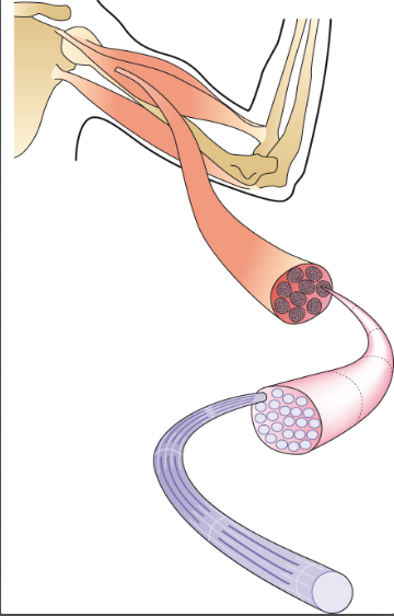
What is muscle made up of?
Responda
-
neurones
-
bundles of muscle fibres
-
endothelium
Questão 2
Questão
Each muscle fibre is a single muscle cell
Responda
- True
- False
Questão 3
Questão
What does multinucleate mean and why are cells multinucleate?
Responda
-
each cell has several nuclei - because a single nucleus couldn't effectively control the metabolism of such a long cell
-
each cell has many mitochondria - because muscle fibres carry out large amounts of aerobic respiration
Questão 4
Questão
During [blank_start]prenatal[blank_end] development, several cells [blank_start]fuse[blank_end] together forming an [blank_start]elongated[blank_end] muscle fibre. Muscle cells are also [blank_start]striped[blank_end] which is related to their ability to [blank_start]contract[blank_end].
Responda
-
prenatal
-
fuse
-
elongated
-
striped
-
contract
Questão 5
Responda
-
tendon
-
ligament
-
connective tissue
-
myosin
-
bundle of muscle fibres
-
myofibrils
-
one muscle fibre
-
bundle of muscle fibres
-
myofibrils
-
myosin
-
sarcomere
-
actin
Questão 6
Questão
muscle fibres are bound together by connective tissue, which is continuous with the tendons
Responda
- True
- False
Questão 7
Questão
Each muscle [blank_start]fibre[blank_end] may be up to 2cm long, but less than 0.1mm in diameter. Inside the muscle fibre is the cytoplasm containing [blank_start]mitochondria[blank_end] and the other organelles found in a cell. Within each muscle fibre there are also numerous [blank_start]myofibrils[blank_end]; each is composed of [blank_start]repeated contractile units[blank_end] called sarcomeres.
Responda
-
fibre
-
bundle
-
mitochondria
-
vacuole
-
myofibrils
-
myosins
-
repeated contractile units
-
blocks
Questão 8
Questão
Sarcomeres are made up of myofibrils
Responda
- True
- False
Questão 9
Questão
What is the name of the protein that makes up the thinner filament in a sarcomere?
Responda
-
actin
-
myosin
-
myofibril
Questão 10
Questão
What is the name of the protein that makes up the thicker filament in a sarcomere?
Responda
-
myosin
-
actin
-
myofibril
Questão 11
Questão
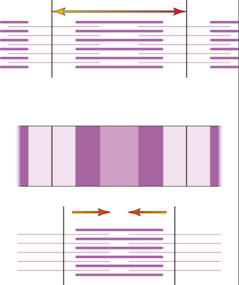
[blank_start]Contractions[blank_end] are brought about by co-ordinated sliding of these protein filaments within the muscle cell [blank_start]sarcomeres[blank_end]. The proteins overlap and give the muscle fibre its characteristic [blank_start]striped[blank_end] (striated) appearance under the microscope. When the muscle contracts, the [blank_start]actin[blank_end] moves between the [blank_start]myosin[blank_end] - this [blank_start]shortens[blank_end] the length of the sarcomere and hence the length of the [blank_start]muscle[blank_end].
Responda
-
Contractions
-
Extensions
-
sarcomeres
-
tendons
-
striped
-
spotted
-
actin
-
myosin
-
myosin
-
actin
-
shortens
-
lengthens
-
muscle
-
ligament
Questão 12
Responda
-
one sarcomere
-
half a sarcomere
-
actin
-
myosin
-
actin
-
myosin
Questão 13
Questão
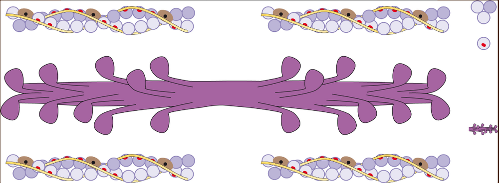
How are myosin molecules shaped?
Responda
-
like golf clubs
-
like sweet chilli flavour sunbites
Questão 14
Questão
What are troponin and tropomyosin?
Responda
-
protein molecules
-
fatty acid chains
-
polysaccharides
Questão 15
Responda
-
myosin binding site
-
troponin molecule
-
myosin head
-
myofibril
-
troponin molecule
-
tropomyosin
-
tropomyosin
-
troponin molecule
-
myosin molecule
-
actin molecule
-
actin molecule
-
myosin molecule
Questão 16
Questão
What is sarcoplasm?
Responda
-
the name given to cyotplasm in a muscle cell
-
specialised type of endoplasmic reticulum, a system of membrane-bound sacs around the myofibrils
-
cell surface membrane
Questão 17
Questão
What is sarcoplasmic reticulum?
Responda
-
specialised type of endoplasmic reticulum, a system of membrane-bound sacs around the myofibrils
-
cell surface membrane
-
the name given to cytoplasm in a muscle cell
Questão 18
Questão
What is the sarcolemma
Responda
-
cell surface membrane
-
specialised type of endoplasmic reticulum, a system of membrane-bound sacs around the myofibrils
-
the name given to cytoplasm in a muscle cell
Questão 19
Responda
-
neuromuscular junction
-
sarcolemma
-
sarcolemma
-
neuromuscular junction
-
motor neurone
-
relay neurone
-
myofibril
-
myosin
-
transverse tubule
-
microtubule
-
sarcoplasmic reticulum
-
sarcoplasm
-
route of nerve impulse
-
release of calcium ions
-
route of nerve impulse
-
release of calcium ions
Questão 20
Questão
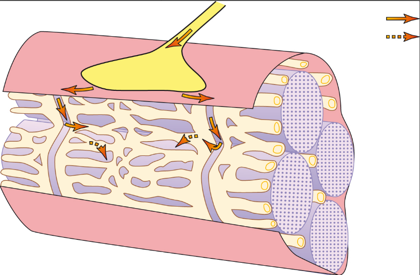
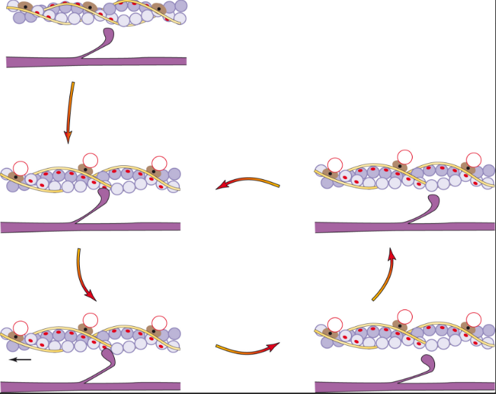
When a [blank_start]nerve impulse[blank_end] arrives at a neuromuscular junction, [blank_start]calcium[blank_end] ions are released from the [blank_start]sarcoplasmic[blank_end] reticulum. The calcium ions [blank_start]diffuse[blank_end] through the sarcoplasm. This initiates the movement of protein filaments. Calcium ions attach to the [blank_start]troponin[blank_end] molecule causing it to move. As a result, the [blank_start]tropomyosin[blank_end] on the actin filament shifts its position, exposing [blank_start]myosin-binding sites[blank_end] on the actin filaments. Myosin heads bind with myosin-binding sites on the actin filament, forming [blank_start]cross-bridges[blank_end]. When the myosin heads bind to the actin, [blank_start]ADP and P[blank_end] on the myosin head [blank_start]are[blank_end] released. The [blank_start]myosin[blank_end] changes shape, causing the [blank_start]myosin head[blank_end] to nod forward. This movement results in the relative movement of filaments; the attached [blank_start]actin[blank_end] moves over the myosin. An [blank_start]ATP[blank_end] molecule binds to the myosin head - this causes the myosin head to [blank_start]detach from[blank_end] the actin. An [blank_start]ATPase[blank_end] on the myosin head hydrolyses the ATP, forming ADP and P. This [blank_start]hydrolysis[blank_end] causes a change in the shape of the myosin head. It returns to its upright position. This enables the cycle to start again
Responda
-
calcium
-
sodium
-
sarcoplasmic
-
endoplasmic
-
nerve impulse
-
hormone
-
diffuse
-
are actively transported
-
troponin
-
tropomyosin
-
tropomyosin
-
troponin
-
myosin-binding sites
-
myosin heads
-
cross-bridges
-
ionic interactions
-
ADP and P
-
ATP
-
are
-
is
-
myosin
-
myosin head
-
myosin head
-
myosin
-
actin
-
myofibril
-
detach from
-
attach to
-
ATP
-
DNA
-
ATPase
-
ATP synthase
-
hydrolysis
-
synthesis
Questão 21
Responda
-
ADP + Pi
-
ATP
-
calcium ion
-
myosin- binding site
-
ADP + Pi
-
ATP
-
ATP
-
ADP + Pi
-
ADP + Pi
-
ATP
-
ADP + Pi released
-
ATPase causes hydrolysis
-
Myosin head moves forward - actin moves
-
ADP + Pi released
-
cross-bridge forms
-
ATPase causes hydrolysis
-
Myosin head nods forward - actin moves
-
ATPase causes hydrolysis
-
Myosin head nods forward - actin moves
-
ADP + Pi released
-
cross-bridge forms
-
cross-bridge forms
-
ADP + Pi released
-
myosin head nods forward - actin moves
-
ATPase causes hydrolysis
-
myosin head detaches
-
lwngnwarg
-
myosin head returns to upright position
-
;oawingopn
Questão 22
Questão
When a muscle [blank_start]relaxes[blank_end], it's no longer being stimulated by nerve impulses. Calcium ions are [blank_start]actively pumped[blank_end] out of the muscle [blank_start]sarcoplasm[blank_end], using [blank_start]ATP[blank_end]. The troponin and tropomyosin move back, once again blocking the [blank_start]myosin-binding sites[blank_end] on the actin.
Responda
-
relaxes
-
actively pumped
-
sarcoplasm
-
ATP
-
myosin-binding sites
Questão 23
Questão
What happens in the absence of ATP?
Responda
-
the cross-bridges remain attached
-
the cross-bridges still break
Quer criar seus próprios Quizzes gratuitos com a GoConqr? Saiba mais.