7858705
| Frage | Antworten |
| 1 1/2 times larger than the normal proximal segment | Aneurysm |
| T/F tortuosity of the abdominal aorta means that it could be crooked or possibly kinked | true |
| T/F hepatopedal flow is a term used to describe normal flow in the portal vein | true |
| T/F a uterine fibroid may be mistakenly diagnosed as an abdominal aneurysm | true |
| T/F a ruptured aneurysm would cause hypotension & a decreased hematocrit | true |
| T/F less than 50% of aneurysms are located inferior to the renal arteries | false {95% are infrarenal} |
| T/F approximately 75% of the patients w/ an abdominal aortic aneurysm are asymptomatic | false {30-60% are asymptomatic} |
| T/F sonographers should measure the abd aorta from outer wall to inner wall | false {outer-to-outer} |
| What is the easiest & quickest way to determine if a cystic structure is truly cystic & not a vascular structure? a. look for ascites b. use grayscale imaging c. turn on colorflow d. take a BP | c. turn on colorflow |
| All of the following arteries produce a low resistance waveform EXCEPT: a. CCA b. renal artery c. hepatic artery in a fasting patient d. femoral artery | d. femoral artery (high resistance = extremities, things you can live w/o) |
| A percentage of patients w/ abd aneurysms are also at risk for developing aneursyms in an artery of the lower extremity. What is the artery that may be involved? a. femoral b. profunda femoral c. posterior tibial d. popliteal | d. popliteal |
| The #1 risk factor/cause for developing an abd aneurysms is: | atherosclerosis |
| The clinical signs of leg edema, low back pain, pelvic pain, gastrointestinal complaints, renal & liver problems may represent: a. abd rupture b. retroperitoneal tumor c. IVC thrombosis d. superior mesenteric thrombus | c. IVC thrombosis |
| In patients w/ lower trunk & leg edema and dilated IVC, a(n) ___ should be suspected. a. retroperitoneal tumor b. arteriovenous fistula c. rupture d. infection | b. arteriovenous fistula |
| Compression technique can be used to treat which kind of aneurysm? | pseudoaneurysm |
| Ultrasound is __ accurate in detecting aneurysms. | 98.9% |
| The normal diameter of the aorta is less than | 3cm |
| Clinical symptoms for dissection include all of the following EXCEPT: a. back pain b. shock c. hematuria d. chest pain | c. hematuria |
| The most common cause of portal hypertension is: | intrinsic liver disease, such as cirrohosis |
| Berry-shaped aneurysms primarily affect which of the following arteries? a. abd aorta b. cerebral c. hepatic d. splenic | b. cerebral |
| Most surgeons recomment surgery to repair aneurysms between _-_cm if that patient has risk factors for rupture such as hypertension, smoking, and COPD. | 5-6cm |
| The most common type of aneurysm is: | fusiform |
| Of the following, in which group of people are aneurysms more commonly found. a. women b. men c. children d. 30-40 yr olds | b. men |
| The umbilical vein changes into the _ postnatally. | Ligamentum Teres |
| _ is a condition in which the aorta is uniformly but not focally enlarged. | Ectasia |
| Dissection of the abdominal aorta is linked to all of the following EXCEPT: a. renal cell carcinoma b. dissection of the thoracic aorta c. trauma d. Marfan's syndrome | a. renal cell carcinoma |
| When a patient develops Portal Hypertension, what is the most common collateral pathway that the body develops to compensate for increased pressure in the liver? | coronary & gastroesophogeal veins |
| The most common tumor to fill the IVC is: | renal cell carcinoma usually from the right kidney |
| Which of the following abd arteries is crucial to evaluate in heart transplant patients? a. SMA b. renal c. phrenic d. hepatic | d. hepatic |
| The sonographic evidence of dissection is: a. increased velocity b. swirling bloodflow on colorflow c. reversal of flow d. intimal flap & 2 channels of bloodflow | d. intimal flap & 2 channels of bloodflow |
| What is the most common imaging modality used to examine a thoracic aortic aneurysm? | CT |
| Most commonly results from intrinsic liver disease; however, it also arises from obstruction of the portal vein, hepatic veins, IVC or prolonged congestive heart failure. | portal venous hypertension |
| Pulsatile hematoma that results from leakage of blood into soft tissues abutting the punctured artery with fibrous encapsulation & failure of the vessel wall to heal. | pseudoaneurysm |
| A disease of arterial vessels marked by thickening, hardening, & loss of elasticity in the arterial walls | ateriosclerosis |
| Hereditary disorder of connective tissues, bones, muscles, ligaments, and skeletal structures | Marfan's syndrome |
| A communication between two blood vessels w/o any intervening capillary network | anastomosis |
| Periportal collateral channels in patients w/ choronic portal vein obstruction | cavernous transformation of the the portal |
| Circumferential enlargement of a vessel with tapering at both ends | fusiform aneurysm |
| thrombosis of the hepatic veins | Budd-Chiari syndrome |
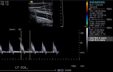
| Image above represents what kind of flow: high resistance or low resistance | high resistance |
| What is a tear in the intima or media of the aortic wall demonstratiing a linear echo within the lumen of the aorta called? | dissection |
| What is the surgical procedure used to relieve portal hypertension? | TIPS |
| Surgical & angiographic placement of a _ into the IVC has been used to prevent recurrent embolization in patients who cannot tolerate anticoagulants. | IVC filter |
| After birth the umbilical vein constricts and changes into the _. | ligamentum teres |
| Class symptoms of gallbladder disease include all of the following except: a. hematuria b. right upper quad pain c. right shoulder pain d. nausea and vomiting | a. hematuria |
| A patient presents with a history of weight loss & pancreatic cancer. Multi target-shaped lesions are demonstrated within the liver & also a polypoid mass is demonstrated within the gb. Given the histroy, the echogenic foci within the gb are most suspicious for: | metastatic gallbladder disease |
| Sonographic findings in metastatic disease of the gallbladder include all of the following except: a. poypoid or irregular intraluminal mass b. indistinct walls c. focal thickening of the gallbladder wall d. intraluminal soft tissue mass on thin stalk | d. intraluminal soft tissue mass on thin stalk |
| inflammation of the gallbladder is: | cholecystitis |
| the most frequent cause of metastic disease to the gallbladder is: | melanoma |
| In the case of a porcelain gallbladder, all of the following facts are true except: a. 90% associated w/ stones b. higher incidence in females than males c. dense wall w/o shadowing d. associated incidence of gb carcinoma | c. dense wall w/o shadowing |
| A condition in which cholesterol stones/ crystals become lodged between diverticula (Rokitansky-Aschoff sinuses) causing comet tail artifacts is called: | adenomyomatosis |
| Causes of non-inflammatory gallbladder wall thickening include all of the following except: a. AIDS b. cirrhosis c. ascites d. cholecystitis | d. cholecystitis |
| The term used to define inflammation of the gb in absence of the cholelithiasis is: | acalculous cholecystitis |
| The following term defines a soft tissue mass pedunculating from the gallbladder wall protruding into the lumen, It does not shadow and does not move with patient position. | adenoma (polyp) |
| Gallstone appearance on ultrasound examination is: | dependent, mobile, echogenic |
| A hyperplastic change in the gallbladder wall that is sometimes referred to as a "strawberry gallbladder" is called: | cholesterolosis |
| A patient presents w/ a postitive Murphy's sign and an elevated bilirubin level. Based on this clinical history, the sonogram is most suspicious for: | Wall-echo-shadow (WES) |
| An impacted stone in the cystic duct, cystic duct remnant or gb describes which condition? | Mirizzi's syndrome |
| Gallbladder perforation may appear in patients with any of the following diseases except: a. gangrenous cholecystitis b. acute cholecystitis c. negative Murphy's sign d. gallbladder carcinoma | c. negative Murphy's sign |
| Thickening of the gallbladder wall may be caused by all of the following except: a. ascites b. nephritis c. AIDS d. congestive heart failure | b. nephritis |
| Nonshadowing, low-amp echoes that sometimes layer out in a dependent gallbladder & move with patient position is most characteristic of: | sludge |
| A 30 yr old asymptomatic patient presents with a history of Hep B. A sonogram is ordered to rule out pathology. The gallbladder demonstrates multi echogenic foci demonstrating comet-tail artifacts. The sono findings are most consistent with: | adenomyomatosis |
| small, well defined soft tissue projection from the gallbladder wall | polyp |
| excessive bilirubin accumulation causes yellow pigmentation of the skin; first seen in the whites of the eyes | jaundice |
| variant of adenmyomatosis; cholesterol polyps | cholesterolosis |
| gallstones in the gallbladder | cholelithiasis |
| small polypoid projections from the gallbladder wall | adenomyomatosis |
| sono pattern found when the gb is packed w/ stones | Wall echo shadow (WES) |
| calcification of the gallbladder wall | porcelain gallbladder |
| hormone secreted into the blood by the mucosa of the upper small intestine; stimulates contraction of the gallbladder & pancreatic secretion of enzymes. | cholecystokinin |
| inflammation of the gb; may be acute or choronic | cholecystitis |
| low-level echoes found along the posterior margin of the gallbladder; move w/ change in position | sludge |
| An enlarged gallbladder w/ a diameter >5cm is called? | hydrops |
| What two lab values will be elevated in cases of biliary obstruction? | alkaline phosphatase & bilirubin |
| cystic dilation within the biliary tract that are congenital anomalies. | choledochal cysts |
| congential segmental saccular cystic dilation of major intrahepatic bile ducts (beaded appearance) found in young adult or pediatric population; may be associated w/ renal disease or congenital hepatic fibrosis | Caroli's Disease |
| inflammation of the bile ducts | cholangitis |
| sono appearance of dilated biliary system | shotgun sign, parallel channel sign |
| stones within the biliary system | choledocholithiasis |
| Acute cholecystitis is associated with what clinical sign? | Positive Murphy's sign |
| Most common cause of acute pancreatitis | cholelithiasis |
| Pancreas lab value that increase 1st (within 1st 24 hrs) | Serum Amylase |
| 2nd most common cause of acute pancreatitis | alcohol abuse |
| most common location for a pseudocyst to occur. | lesser sac |
| sonographic appearance of chronic pancreatits | hyperechoic w/ possible calcifications |
| fluid collection in the pancreas caused by inflammatory process necrosis or hemorrhage due to excretion of enzymes outside the duct. | pseudocyst |
| most common primary neoplasm of the pancreas | adenocarcinoma |
| hydrops of the gb caused by mass in the head of the pancreas compressing CBD | Courvoisier's sign |
| Most common functioning islet cell tumor | insulinoma |
| Where do islet cell tumors occur within the pancreas? | Islets of Langerhans |
| One of the most common clinical signs of adenocarcinoma of the pancreas is _. | painless jaundice |
| What is the disease that is congenital and indicates an iron overload in the liver? | hemochromatosis |
| Results from thrombus within the hepatic veins. | Budd-Chairi |
| Most common type of glycogen storage disease | Type 1 or von Gierke disease |
| Lab value elevated with hepatocellular carcinoma. | Alpha fetoprotein |
| Lab values elevated with cirrhosis | AST & ALT Bilirubin Alkaline Phosphatase |
| Most common cause of intrahepatic portal hypertension | cirrhosis |
| Most common cause of transmission of hepatitis in healthcare workers is hepatitis _. | B |
| Condition in which the hepatocytes have accumulated an increase of lipids, tryglycerides are elevated, can be reversed w/ diet and meds | Fatty Infiltration |
| Which two liver masses appear very similar on sonography & may require a biopsy to confirm the diagnosis? | Focal nodular hyperplasia/Adenoma |
| A liver mass seen in sheepherding countries call echinococcal cyst or hydatid cyst is associated w/ which sonographic sign? | waterlily |
Möchten Sie mit GoConqr kostenlos Ihre eigenen Karteikarten erstellen? Mehr erfahren.