22701133
Description
Quiz by Charlotte Jakes, updated more than 1 year ago
|
|
Created by Charlotte Jakes
about 4 years ago
|
|
Question 1
Question
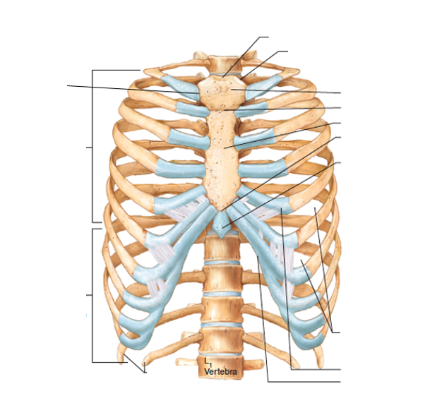
The following questions will be about the thoracic cage. First, drag and drop the correct labels to describe the bones of the thoracic cage.
Answer
-
1st sternocostal joint
-
True ribs (1-7)
-
False ribs (8-12)
-
Jugular notch
-
Clavicular notch
-
Manubrium of the sternum
-
Sternal angle
-
Body of the sternum
-
Xiphisternal joint
-
Xiphoid process
-
Intercostal spaces
-
Costal cartilage
-
Costal margin
-
Floating ribs (11 and 12)
Question 2
Question
On the lateral edges of the manubrium, there is a facet for articulation with...
Answer
-
The first rib
-
The second rib
-
The clavicle
-
The body of the sternum
Question 3
Question
What occurs on the manubrium which will articulate with the second rib?
Answer
-
Facet
-
Demi-facet
-
Tubercle
-
Spine
Question 4
Question
The sternal angle marks the articulation of the manubrium with the body of the sternum. What else does it mark?
Answer
-
The level of the second rib's costal cartilage
-
The level of the first rib's costal cartilage
-
The level of the clavicle
-
The level of the carina
Question 5
Question
Which ribs articulate with the body of the sternum?
Answer
-
Ribs 2-7
-
Ribs 1-2
-
Ribs 3-6
-
Ribs 1-12
Question 6
Question
At what level is the tip of the xiphisternum found?
Answer
-
T10
-
T11
-
T12
-
L1
Question 7
Question
The xiphisternum is a largely cartilaginous structure which ossifies completely around the age of 40.
Answer
- True
- False
Question 8
Question
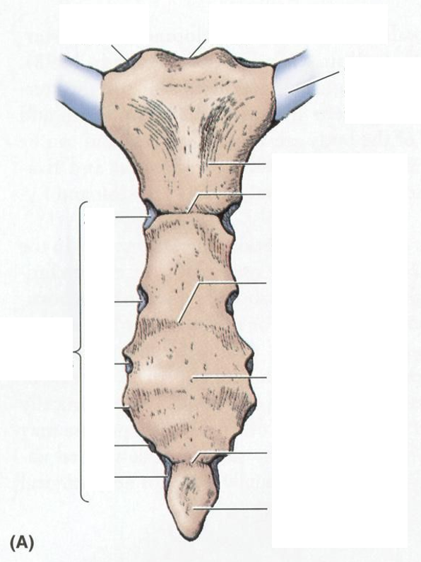
Drag and drop the correct labels to describe the sternum.
Answer
-
Clavicular notch
-
Jugular notch
-
Costal cartilage of 1st rib
-
Manubrium
-
Sternal angle
-
Body of sternum
-
Xiphisternal joint
-
Xiphoid process
-
Costal notches
-
2nd
-
3rd
-
4th
-
5th
-
6th
-
7th
Question 9
Question
What is meant by a 'true rib'?
Answer
-
A rib that attaches to the sternum directly via a synovial joint
-
A rib that attaches to the costal cartilage of another rib by a synovial joint and thus does not articulate with the sternum directly
-
A rib with a complete lack of anterior attachment
-
A rib that attaches to the costal cartilage of another rib by a secondary cartilaginous joint and thus does not articulate with the sternum directly
Question 10
Question
What is meant by a 'false' rib?
Answer
-
A rib that attaches to the costal cartilage of another rib via a synovial joint with its costal cartilage and thus does not articulate with the sternum directly
-
A rib that attaches directly to the sternum via a synovial joint
-
A rib that attaches to the costal cartilage of another rib via a secondary cartilaginous joint with its costal cartilage and thus does not articulate with the sternum directly
-
A rib that has a complete lack of anterior attachment
Question 11
Question
Floating ribs...
Answer
-
Lack any anterior attachment
-
Lack any posterior attachment
-
Only attach to the superior ribs
-
Form synovial joints directly with the sternum
Question 12
Question
Which ribs are the true ribs?
Answer
-
1-7
-
1-10
-
1-8
-
1-5
Question 13
Question
Which ribs are the false ribs?
Answer
-
8-12
-
10-12
-
11 and 12
-
7-12
Question 14
Question
Which ribs are the floating ribs?
Answer
-
1 and 2
-
11 and 12
-
8-12
-
1-7
Question 15
Question
The typical rib consists of a head, neck and body/shaft. Where does the tubercle of the typical rib occur?
Answer
-
Where the neck meets the body
-
Where the neck meets the head
-
Where the body meets the head
-
At the end of the head
Question 16
Question
How many articular facets does the typical rib have on its head?
Answer
-
2
-
1
-
3
-
4
Question 17
Question
Which is true of the articular facets on the heads of the typical ribs?
Answer
-
One articulates with the numerically corresponding vertebra and the other articulates with the vertebra above
-
One articulates with the numerically corresponding vertebra and the other articulates with the vertebra below
-
One articular facet articulates with the numerically corresponding vertebra
-
One articulates with the numerically corresponding vertebra and the other articulates with the transverse process of the same vertebra
Question 18
Question
What does the tubercle of the typical rib articulate with?
Answer
-
The transverse process of the corresponding vertebra
-
The superior costal facet of the corresponding vertebra
-
The inferior costal facet of the corresponding vertebra
-
The spinous process of the corresponding vertebra
Question 19
Question
What occurs on the internal surface of the typical rib to house the intercostal vein, artery and nerve?
Answer
-
Costal groove
-
Costal notch
-
Costal sulcus
-
Intercostal groove
Question 20
Question
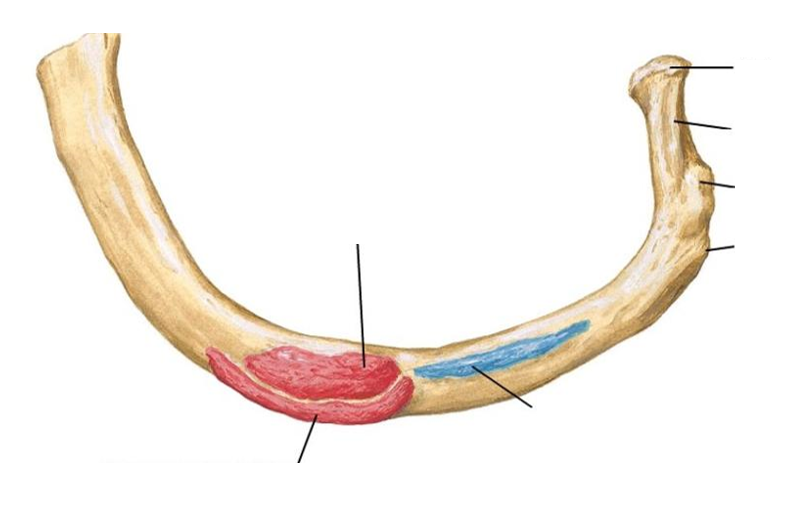
Drag and drop the correct labels to describe this typical rib.
Answer
-
Superior articular facet
-
Inferior articular facet
-
Head
-
Neck
-
Tubercle
-
Facet
-
Angle
-
Costal groove
Question 21
Question
Which ribs are described as 'atypical' as they have features that are not common to all the ribs?
Answer
-
1
-
2
-
3
-
4
-
11
-
12
-
7
-
8
-
9
-
10
Question 22
Question
How many facets occur on the head of the atypical rib 1?
Answer
-
1
-
2
-
4
-
3
Question 23
Question
There are two grooves on the superior surface of the 1st rib. Why?
Answer
-
Houses the subclavian vessels
-
Houses the intercostal nerve, artery and vein
-
Attaches the middle scalene muscle
-
Attaches the anterior scalene muscle
Question 24
Question
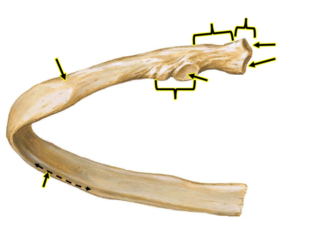
Drag and drop the correct labels to describe the anatomy of the atypical first rib and its muscular attachments.
Answer
-
Subclavius muscle
-
Grooves for subclavian vessels
-
Anterior scalene muscle
-
Serratus anterior muscle
-
Middle scalene muscle
-
Tubercle
-
Neck
-
Head
Question 25
Question
How many articular facets does the atypical rib 2 have on its head?
Answer
-
1
-
2
-
3
-
4
Question 26
Question
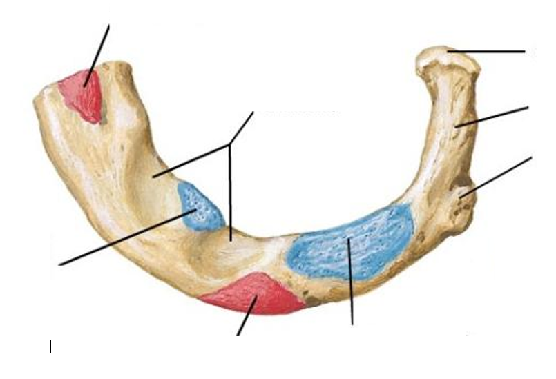
Drag and drop the correct answers to describe the anatomy of the second rib and its muscular attachments.
Answer
-
Head
-
Neck
-
Tubercle
-
Angle
-
Posterior scalene muscle
-
Serratus anterior
Question 27
Question
Why is the 10th rib described as atypical?
Answer
-
The 10th rib only has one facet on its head
-
The 10th rib has no tubercle
-
The 10th rib has no subcostal groove
-
The 10th rib has three facets on its head
Question 28
Question
The 11th and 12th ribs have no neck.
Answer
- True
- False
Question 29
Question
How many articular facets occur on the heads of the 11th and 12th ribs?
Answer
-
1
-
2
-
3
-
4
Question 30
Question
Which ribs operate via a 'pump-handle' motion during ventilation?
Answer
-
True ribs
-
False ribs
Question 31
Question
Which ribs operate via a 'bucket-handle' motion during ventilation?
Answer
-
True ribs
-
False ribs
Question 32
Question
Drag and drop the correct answers to show how the ribs articulate with the thoracic vertebrae.
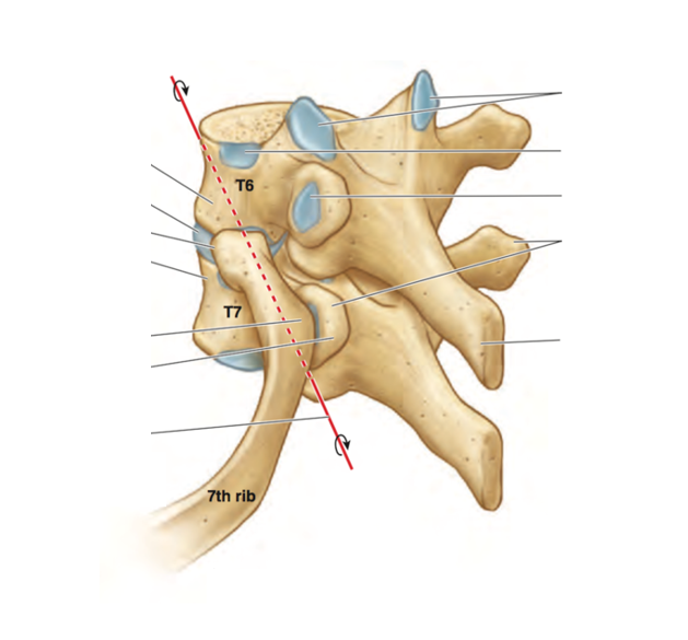
Answer
-
Superior articular facets
-
Costal demifacet for 10th rib
-
Articular facet of 6th rib's tubercle
-
Transverse processes
-
Spinous process
-
Axis of rib rotation
-
Tubercle of rib
-
Vertebral body
-
Head of rib
-
Intervertebral disc
Question 33
Question
Which thoracic vertebra's superior facet will not be a demifacet?
Answer
-
T1
-
T10
-
T11 and 12
-
T7
Question 34
Question
Which vertebrae has a single pair of whole facets across the vertebral body and pedicle?
Answer
-
T10
-
T11 and T12
-
T1
-
T2
Question 35
Question
Which vertebra has a single pair of entire costal facets located on the pedicles?
Answer
-
T11 and T12
-
T10
-
T1
-
T7
Question 36
Question
Drag and drop the correct labels to describe the ligaments of the ribs.
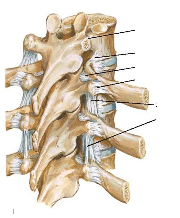
Answer
-
Transverse process (cut)
-
Radiate ligament
-
Costotransverse ligament
-
Lateral costotransverse ligament
-
Superior costotransverse ligament
-
Intertransverse ligament
Question 37
Question
Drag and drop the correct labels to describe the ligaments of the rib shown in this transverse section.
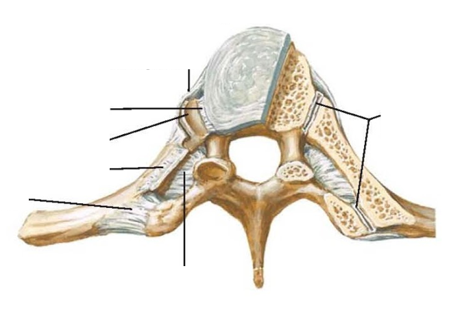
Answer
-
Synovial cavities
-
Radiate ligament
-
Interarticular ligament
-
Superior articular facet
-
Superior costotransverse ligament (cut)
-
Costotransverse ligament
-
Lateral costotransverse ligament
Question 38
Question
The following questions will be about the muscles of the chest wall and thorax. First, drag and drop the correct labels to describe the layers of the chest wall.
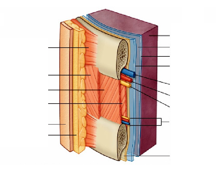
Answer
-
Lung
-
Pleural cavity
-
Visceral pleura
-
Parietal pleura
-
Serratus anterior
-
External intercostal muscle
-
Internal intercostal muscle
-
Innermost intercostal muscle
-
Skin
-
Superficial fascia
-
Intercostal vein
-
Intercostal artery
-
Intercostal nerve
-
Collateral branches
-
Endothoracic fascia
Question 39
Question
The following questions will be on the diaphragm. Which of the following are the peripheral attachments of the diaphragm?
Answer
-
Costal cartilages of ribs 7-10
-
Direct articulation with ribs 11 and 12
-
Xiphisternum
-
Lumbar vertebrae and arcuate ligaments
-
Direct articulate with ribs 7-10
-
Body of the sternum
-
Thoracic vertebrae
Question 40
Question
The right and left crura of the diaphragm are tendinous in structure. Where does the right crus of the diaphragm arise from?
Answer
-
L1-L3 and their intervertebral discs
-
L1 and L2 and their intervertebral discs
-
Transverse processes of L1
-
Xiphisternum
Question 41
Question
The right and left crura of the diaphragm are tendinous in structure. Where does the right crus of the diaphragm arise from?
Answer
-
L1 and L2 and their intervertebral discs
-
L1-L3 and their intervertebral discs
-
Xiphisternum
-
Ribs 1 and 2
Question 42
Question
The fibres of which crus of the diaphragm surround the oesophageal opening to form a physiological sphincter?
Answer
-
Right crus
-
Left crus
Question 43
Question
What do the fibres of the diaphragm combine to form, which ascends to fuse with the interior surface of the fibrous pericardium?
Answer
-
Central tendon
-
Right and left crura
-
Diaphragmatic pericardium
-
Right and left domes
Question 44
Question
Why does the right dome of the diaphragm sit higher than the left?
Answer
-
Presence of the liver
-
There is no heart on the right side
-
Presence of the stomach
-
Different embryological origins
Question 45
Question
The caval hiatus (T8) acts as a conduit for...
Answer
-
Inferior vena cava
-
Terminal branches of right phrenic nerve
-
Oesophagus
-
Right and left vagus nerve
-
Oesophageal branches of left gastric artery/vein
-
Aorta
-
Thoracic duct
-
Azygous vein
Question 46
Question
The oesophageal hiatus (T10) in the diaphragm acts as a conduit for...
Answer
-
Oesophagus
-
Right and left vagus nerves
-
Oesophageal branches of left gastric artery/vein
-
Inferior vena cava
-
Terminal branches of right phrenic nerve
-
Aorta
-
Thoracic duct
-
Azygous vein
Question 47
Question
The aortic hiatus (T12) acts as a conduit for...
Answer
-
Aorta
-
Thoracic duct
-
Azygous vein
-
Oesophagus
-
Right and left vagus nerves
-
Oesophageal branches of left gastric artery/vein
-
Inferior vena cava
-
Terminal branches of right phrenic nerve
Question 48
Question
The diaphragm is the primary muscle of respiration.
Answer
- True
- False
Question 49
Question
When does the diaphragm contract and flatten?
Answer
-
Inspiration
-
Expiration
Question 50
Question
The left and right hemidiaphragms receive innervation from left and right phrenic nerves respectively.
Answer
- True
- False
Question 51
Question
What spinal roots supply the phrenic nerves?
Answer
-
C3, C4, C5
-
C1 and C2
-
C6 and C7
-
C2, C3, C4
Question 52
Question
The majority of the arterial supply to the diaphragm is achieved by the inferior phrenic arteries. Where do these arise from?
Answer
-
Abdominal aorta
-
Thoracic aorta
-
Renal arteries
-
Coeliac trunk
Question 53
Question
Drag and drop the correct answers to describe the diaphragm from a posterior view.
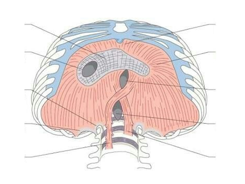
Answer
-
Central tendon
-
Vena caval opening
-
Medial arcuate ligament
-
Lateral arcuate ligament
-
Right crus
-
Left crus
-
Aortic opening
-
Oesophageal opening
-
Costal fibres
-
Sternal fibres
Question 54
Question
Drag and drop the correct labels to describe the diaphram and surrounding neurovasculature.
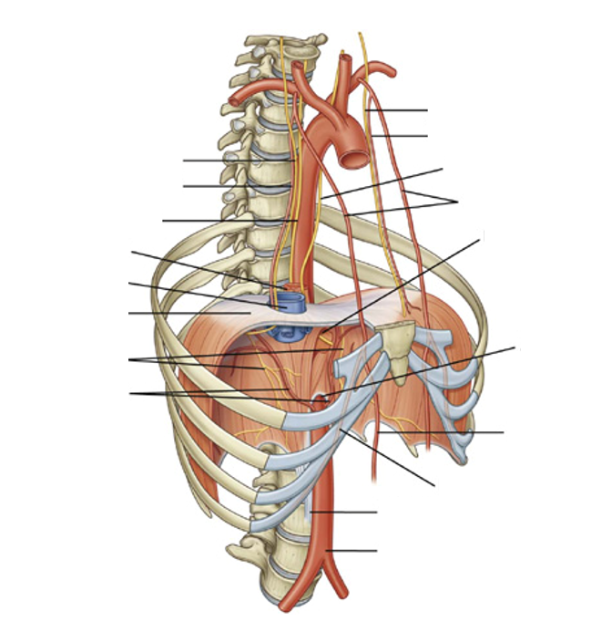
Answer
-
Left phrenic nerve
-
Left pericardiacophrenic artery
-
Right phrenic nerve
-
Right pericardiacophrenic artery
-
Right vagus nerve
-
Oesophagus
-
Inferior vena cava
-
Central tendon
-
Phrenic nerves
-
Inferior phrenic arteries
-
Left vagus nerve
-
Internal thoracic arteries
-
Oesophageal hiatus
-
Aortic hiatus
-
Superior epigastric artery
-
Musculophrenic artery
-
Right crus
-
Abdominal aorta
Question 55
Question
The following questions will be on the muscles of the thoracic wall. How many external intercostal muscle pairs do we have?
Answer
-
11
-
12
-
13
-
14
Question 56
Question
The external intercostal muscles are continuous with the external oblique of the abdomen
Answer
- True
- False
Question 57
Question
What is the function of the external intercostal muscles?
Answer
-
Elevation of the ribs during inspiration
-
Depression of the ribs during expiration
Question 58
Question
In what orientation do the fibres of the external intercostals run from the superior rib to the rib below?
Answer
-
Inferoanteriorly
-
Inferoposteriorly
Question 59
Question
What are the internal intercostal muscles continuous with?
Answer
-
External oblique
-
Internal oblique
-
Transversus abdominis
Question 60
Question
In what orientation do the fibres of the internal intercostal muscles run from the superior rib to the rib below?
Answer
-
Inferoposteriorly
-
Inferioanteriorly
Question 61
Question
The interosseus part of the internal intercostals elevates the ribcage whilst the interchrondral part depresses the ribcage.
Answer
- True
- False
Question 62
Question
What separates the innermost intercostal muscles from the internal intercostal muscles?
Answer
-
The intercostal neurovascular bundle
-
The external intercostal muscles
-
The serratus anterior muscle
-
The musculophrenic arteries
Question 63
Question
What does the interosseus part of the innermost intercostals do?
Answer
-
Depresses the ribcage
-
Elevates the ribcage
Question 64
Question
What does the interchondral part of the innermost intercostal muscles do?
Answer
-
Elevates the ribcage
-
Depresses the ribcage
Question 65
Question
What is the function of the transversus thoracis?
Answer
-
Weak depression of the ribs
-
Weak elevation of the ribs
Question 66
Question
The transversus thoracis are continuous with the transversus abdominis.
Answer
- True
- False
Question 67
Question
The transversus thoracis arise from...
Answer
-
Posterior surface of inferior sternum
-
Anterior surface of inferior sternam
-
Internal surfaces of costal cartilages 2-6
-
External surfaces of costal cartilages 2-6
Question 68
Question
The transversus thoracis inserts at...
Answer
-
The internal surface of costal cartilages 2-6
-
The external surface of costal cartilages 2-6
-
The posterior surface of the inferior sternum
-
The anterior surface of the inferior sternum
Question 69
Question
Which muscles run from the internal surface of one rib to the second and third ribs below?
Answer
-
Subcostals
-
Transversus thoracis
-
Internal intercostals
-
Innermost intercostals
Question 70
Question
The subcostal muscles share the direction of fibres of the innermost intercostal muscles and the action of the intercostal muscles.
Answer
- True
- False
Want to create your own Quizzes for free with GoConqr? Learn more.