6665027
Descripción
Fichas por Ashutosh Kumar, actualizado hace más de 1 año
|
|
Creado por Ashutosh Kumar
hace alrededor de 8 años
|
|
| Pregunta | Respuesta |
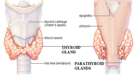
| Describe the location of the parathyroid gland: Describe the anatomy of the parathyroid gland: Describe the embryological origin of the glands: Describe the hormone secreted: | The parathyroid glands are small oval glands located within the thyroid capsule on the posterior surface of the thyroid gland. There are usually two pairs of gland although some individuals can have five or even six parathyroid glands. Embryologically, the parathyroid glands are derived from the 3rd and 4th branchial arches. They secrete parathyroid hormone, a hormone involved in the regulation of plasma calcium and phosphate. |
| Describe the arrangement of cells in the parathyroid gland: List the cell types found in the parathyroid gland: | The epithelial cells are arranged in cords with a branching networks of capillaries in between cells. There are 3 main cell types within the parathyroid gland; chief cells, oxyphil cells and adipocytes. |
| Describe the chief cells: | Chief or principal cells are the most abundant, identified as small cells with a prominent nucleus and little cytoplasm. These cells secrete parathyroid hormone, a peptide hormone and therefore have the typical appearance of a peptide secreting cell. In this stain, the black dots are the nuclei, the cytoplasm is stained pink and the spaces represent blood vessels. |
| Describe the oxyphil cells: | Oxyphil cells are less numerous but are found in clumps; they are larger cells with a pink, eosinophilic cytoplasm and small, densely stained nuclei. These cells are thought to be chief cells gone wrong. This is supported by the idea that there are more in an older individual and therefore increase with age. Moreover, their cytoplasm is full of mitochondria but they do not contain any stored hormone. |
| Describe the embryological development of the thyroid gland: | The thyroid gland is a down growth of the endodermal floor of the pharynx that then migrates caudally to become the thyroid gland. |
| Describe the embryological development of the parafollicular/C cells of the thyroid gland: | The parafollicular/C cells develop from neural crest cells from the ectoderm which invade the ventral part of pharyngeal pouch 4 and then enter the thyroid gland as part of the ultimobranchial body. |
| Describe the embryological development of the parathyroid gland: | The distal ends of pharyngeal pouches 3 and 4 bud off and attach to the posterior part of the developing thyroid gland to form the parathyroid gland. |
| Describe the arterial and venous drainage of the parathyroid glands: | The superior parathyroid glands receive their blood supply from the inferior thyroid arteries. The inferior parathyroid glands receive their blood supply from either the inferior thyroid arteries or an anastomotic branch between superior and inferior thyroid artery. The inferior thyroid artery arises from the subclavian artery via the thyrocervical trunk. The arteries drain into either the superior, middle or inferior thyroid veins. The superior and middle thyroid veins drain into the internal jugular vein whereas the inferior thyroid vein drains into the brachiocephalic vein. |
¿Quieres crear tus propias Fichas gratiscon GoConqr? Más información.