6706356
Descripción

Fichas por RadTech Fairy, actualizado hace más de 1 año
|
|
Creado por RadTech Fairy
hace alrededor de 8 años
|
|
| Pregunta | Respuesta |
| A. proximal radioulnar joint B. olecranon process C. coronoid process D. coronoid tubercle E. body/shaft F. ulna G. ulnar notch H. head I. styloid process J. distal radioulnar joint K. styloid process L. radius M. body/shaft N. radial tuberosity O. neck P. head Q. radial notch (on ulna) | |
| a. radius b. ulna c. trapezium d. trapezoid e. capitate f. hamate g. scaphoid h. lunate i. triquetrium j. pisiform k. capitulum l. olecranon process m. medial epicondyle n. radial head o. coronoid tubercle p. radial tuberosity | |
| A. humerus B. coronoid fossa C. medial epicondyle D. humeral condyle E. trochlea F. trochlear sulcus G. radial head H. capitulum I. lateral epicondyle J. radial fossa | |
| A. medial epicondyle B. trochlea C. coronoid tubercle D. radial head E. capitulum F. lateral epicondyle H. olecranon process | |
| A. radial and coronoid fossas B. capitulum C. trochlea D. coronoid process E. radial tuberosity F. radial neck G. radial head H. trochlear sulcus I. trochlear notch J. olecranon process K. epicondyle L. olecranon fossa | |
| G. superimposed epicondyles of humerus I. trochlear sulcus J. trochlear notch K. outer ridges of capitulum and trochlea L. coronoid process of ulna M. radial head N. radial neck | |
| PROXIMAL ULNA A. olecranon process B. trochlear notch C. coronoid process D. radial notch (lateral) | |
| A. ginglymus (hinge) joint B. trochoidal (pivot) joint | |
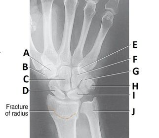
| A. trapezoid B.. trapezium C. capitate D. scaphoid E. hamulus F. hamate G. triquetrium H. pisiform I. lunate J. ulna | |
| A. scaphoid B. lunate C. capitate E. trapezium | |
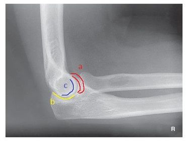
| a. outer ridges of capitulum and trochlea b. trochlear notch c. trochlear sulcus | |
| 1. trochlear sulcus 2. outer ridges of capitulum and trochlea 3. trochlear notch | |
| C. anterior fat pad D. posterior fat pad E. supinator fat stripe (not visible) | |
| AP no rotation radius and ulna partially superimposed | |
| AP Lateral Rotation separation of radius and ulna | |
| AP Medial Rotation superimposed radius and ulna | |
| A. Supination B. Pronation | |
| 1. Bennett's Fx 2. Boxer's Fx 3. Barton's Fx 4. Colle's Fx | |
| Smith's Fx | |
| Incomplete Fx | |
| Baseball (Mallet) Fx | |
| Nursemaid's (Jerked) Elbow | |
| Epiphyseal Fx | |
| Osteoarthritis | Degenerative Joint Disease deterioration of the articular cartilage with hypertrophic bone formation |
| Osteomyelitis | Infection of bone or bone marrow |
| Osteopetrosis | hereditary disease abnormally dense bone |
| Osteoporosis | Atrophy of skeletal tissues |
| Bursitis | Inflammation of the bursae |
| Bone Metastases | the spread of cancer from bone and bone marrow to another body part |
| carpal tunnel syndrome | compression of the median nerve |
| joint effusion | accumulated synovial or hemorrhagic fluid in the joint cavity |
| rheumatoid arthritis | chronic inflammatory changes throughout connective tissues of the wrist and metacarpals most common in women rather than men |
| Paget's Disease | destruction of bone followed by reparative process of overproduction of very dense yet soft bones that fx easily |
| Increase your technique __ - __ kV for small - medium dry plaster casts | 5 - 7 kV |
| increase your technique __ - __ kV for large or wet plaster casts | 8 - 10 kV |
| increase your technique by __ - __ kV for fiberglass casts | 3 - 4 kV |
| Dislocation | Displacement of bone from a joint |
| Subluxation | Partial dislocation |
| Sprain | Rupture or tearing of connective tissues |
| Contusion | bruise without Fx |
| Simple Fx | closed |
| Compound Fx | open, breaks through skin |
| Comminuted Fx | Splintered or crushed |
| Impacted Fx | fragments driven into each other |
| What are the routine exams of the forearm? | AP Lateral |
| What is wrong if there is excessive separation between the radius and ulna on an AP forearm projection? | Excessive Lateral Rotation |
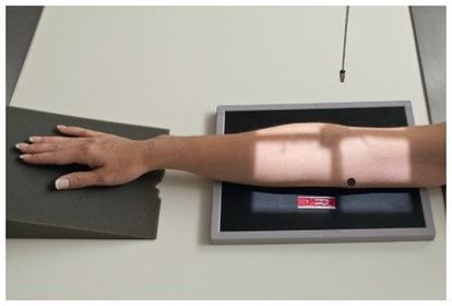
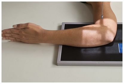
| AP Forearm | 60-75 kV 40 SID 14x17 IR nongrid CR @ mid forearm MUST SEE entire radius/ulna humeral epicondyles slight superimposition of distal radioulnar joint |
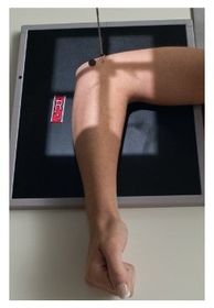
| Lateral Forearm | 60-75 kV 40 SID 14x17 IR nongrid CR @ mid forearm MUST SEE 90 degree elbow flex entire radius/ulna head of ulna superimposed over radius |
| What are the routine exams of an elbow? | AP Lateral Oblique Medial Oblique Lateral |
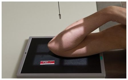
| AP Elbow Fully Extended | 60-75 kV 40 SID 14x17 IR nongrid CR @ mid-elbow joint MUST SEE entire elbow joint very slight superimposition of radial head by the ulna |
| For the two AP Elbow partial flex projections, if the patient cannot extend elbow, and it remains at almost 90 degrees, what should you do? | angle the CR 10-15 degrees into the elbow |
| AP Elbow Partial Flex with humerus parallel (patient cannot fully extend elbow - 2 projections) | 60-75 kV 40 SID 14x17 IR nongrid CR @ mid-elbow MUST SEE distal humerus |
| AP Elbow partial flex forearm parallel (2nd projection) | 60-75 kV 40 SID 14x17 IR nongrid CR @ mid-elbow MUST SEE proximal radius/ulna |
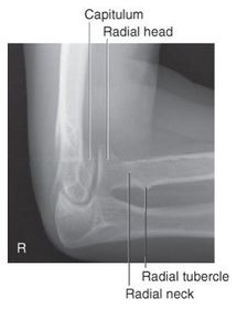
| Which elbow projection/position best demonstrates the radial neck-head with only slight superimposition? | AP Oblique - lateral rotation |
| Which elbow projection/position best demonstrates the coronoid process? | AP Oblique - medial rotation |
| Which fat pad of the elbow will only appear on x-ray if there's a joint pathologic process? | Posterior Fat Pad |
| Why does the elbow need to be in 90 degree flexion to determine whether the posterior fat pad is visible or not? | When elbow is flexed more or less than 90 degrees it pushes the fat pad into different position, it can show up on x-ray then when there's no pathologic issue |
| for an AP forearm, the distal humeral epicondyles are _____ to the IR | parallel |
| For the lateral forearm, the distal humeral epicondyles are _____, or _____ over the IR | Superimposed Perpendicular |
| Epicondyles must be _____ to the IR for an AP fully extended elbow projection. | Parallel |
| AP Oblique Lateral (External) Rotation | 60-75 kVp 40 SID 10x12 IR nongrid CR @ midelbow joint MUST SEE Radial head, neck, and tuberosity , lateral epicondyle and capitulum in profile |
| AP Oblique Medial (Internal) Rotation | 60-75 kVp 40 SID 10x12 IR nongrid CR @ midelbow joint MUST SEE coronoid process, trochlea, and medial epicondyle |
| AP Lateral Elbow | 60-75 kVp 40 SID 10x12 IR nongrid CR @ midelbow joint MUST SEE 3 concentric arcs, olecranon process |
| What are some special projections of the elbow? | Acute Flexion Trauma Axial (Coyle Method) Radial Head Laterals |
| Acute Flexion (1st Projection) CR perpendicular to Humerus | 60-75 kVp 40 SID 10x12 IR nongrid CR perpendicular to IR and distal humerus, midway between epicondyles |
| Acute Flexion (2nd Projection) CR Perpendicular to proximal forearm | 60-75 kVp 40 SID 10x12 IR nongrid CR perpendicular to forearm - 2 in superior to olecranon process |
| Why would you perform an acute flexion projection? | Looking for moderate dislocations of elbow and patient cannot move elbow |
| Trauma Axial Lateral *Coyle Method* for radial head | elbow flexed 90 degrees* CR angled 45 degrees toward the shoulder |
| Trauma Axial Lateral *Coyle Method* for coronoid process | elbow flexed 80 degrees* CR angled 45 degrees away from the shoulder |
| Why would you perform the Coyle Method? | When there's trauma to the radial head or coronoid processes |
| COYLE METHOD alternative view supine, CR angled 45 degrees to the shoulder, elbow flexed 90 degrees | to view Radial Head |
| COYLE METHOD alternative view supine, CR angled 45 degrees away from the shoulder, elbow flexed 80 degrees | to view Coronoid Process |
| What is the only difference among the four radial head lateral projections of the elbow? | the rotational positioning of the wrist exposure factors stay the same |
| Why would you perform any of the 4, or all 4 of the radial head lateral views? | To view occult Fx's of the radial head or neck |
| Some sites use this projection as part of their routine elbow series. Which one is it? | "Radial Head View" Trauma Axial Lateral - Coyle Method - Radial Head |
| According to the textbook, the CR is perpendicular to the IR and humerus, directed to a point midway between epicondyles for ______ when the elbow is in acute flexion. | Acute Flexion of Distal Humerus |
| How many exposures are required for the AP Acute Flexion series? | 2 projections: Acute Flexion of Distal Humerus Acute Flexion of Proximal Forearm |
| How much is the elbow flexed for a Trauma Axial Lateral Projection (Coyle Method) to demonstrate the coronoid process? | 80 Degrees |
| Which oblique elbow projection best demonstrates the coronoid process? | AP Oblique projection - Medial rotation |
| Which oblique elbow projection best demonstrates the radial head and neck? | AP Oblique projection - Lateral rotation |
| Which elbow projection best demonstrates pathologic issues such as osteomyelitis and arthritis? | Lateral Elbow projection (lateromedial) |
| According to the textbook, the CR perpendicular to the forearm (angling CR as needed), directed to a point 2 in proximal or superior to the olecranon process, with the elbow in acute flexion, indicates which projection/position? | Acute Flexion Elbow - Proximal Forearm |
| Which trauma axial elbow projection has the elbow flexed 80 degrees? And what specific anatomy is this projection demonstrating? Which way is the tube angled for this projection? | Trauma Axial Lateral - Coronoid Process 45 degrees away from the elbow |
| Which Trauma Axial Lateral projection has the elbow in 90 degree flexion with the hand pronated? What specific anatomy does this projection demonstrate? Which way is the tube angled for this? | Trauma Axial Lateral - Radial Head 45 degrees towards the shoulder |
| Which projection/position best demonstrates occult fractures or the radial head or neck? | any 4 of the Radial Head Laterals |
| How is the CR angled for the radial head lateral view? | 45 degrees towards the shoulder |
| What is in profile for a lateral oblique elbow? | radial head radial neck capitulum |
| What anatomy is in profile for a medial oblique elbow? | trochlea coronoid semilunar notch |
| For the axial radial head view, how is the hand positioned? | pronated |
¿Quieres crear tus propias Fichas gratiscon GoConqr? Más información.