5158578
| Questão | Responda |
| State 4 cell fate options covered thus far and to be covered | to divide, to become quiescent, to differentiate or to die |
| define 'proliferation' | the growth or production of cells by multiplication of parts |
| define 'apoptosis' | a process of programmed cell death that occurs in multicellular organisms. |
| what processes is apoptosis essential to? | 1)normal metezoan development 2)function of adult systems 3)defence against cancer |
| define 'necrosis' | widespread cell death that results from exposure of cells to harmful influences (eg heat or toxins) |
| how was apoptosis discovered? | by pathologists who noticed rare small densely staining nuclei scattered through tissues that were undergoing physiological regression. |
| Describe a property of cells that are described as 'undergoing physiological regression'. | -shedding of endometrium during normal development, such as in the rat adrenal cortex |
| state the changes in cell appearance of apoptotic cells in tissue (ie changes seen) | 1) cell withdraws from its neighbours and nucleus changes it's appearance 2) nucleus disintergrates, cell cytoplasm is disrupted (blebs form) 3)cell fragments 4) fragments engulfed (phagocytosed) by neighbouring cells and tissue macrophages |
| state another name for tissue macrophages | histiocytes |
| give a role for apoptosis in adults | regulating the immune system- turning off the immune response once an infection is over. |
| describe a specific role of apoptosis in development | development of fingers requires the separation of digits which is enabled through apoptosis of cells between each digit. |
| how are the changes in cell appearance during apoptosis best viewed? | changs first seen via light microscopy but can be seen more clearly under a scanning electron microscope. key steps in the pathway can be visualised by fluorescence microscopy |
| State the first 3 key steps in apoptosis | 1)mitochondria become permeable 2)they release proteins including cytochrome c into the cytoplasm. 3)cytochrome c contributes to the activation of enzymes called 'caspases' |
| state the roles of caspases in apoptosis INSIDE the cell. | caspases drive subsequent events in the apoptotic process these include: breakdown of nuclear envelope, fragmentation of genomic DNA (cut into nucleosome sized fragmemts) and destruction of the cytoskeleton |
| how do caspases effect the cell membrane structure | structure of cell membrane changes- phosphatidyl serine, usually only appears on inner surface of membrane, appears on extracellular surface. this self signals the cell for engulfment. |
| what is the mechanism through which phosphatidyl serine is able to appear on the extracellular surface. | loss of aminophospholipid translocase activity and with non specific flip flop of phospholipids. aminophospholipid translocase usually actively transports phosphatidyl serine inwards |
| Describe how the process of 'blebbing' occurs. | - actinmyosin core constantly under pressure -proteins attaching membrane to actin cytoskeleton degrade -actin contracts "squeezing" cytoplasm out causing blebs to form |
| why does blebbing not occur during necrosis? | necrosis not "contained", so cell does not have time to bleb- blebbing is an intermediate stage |
| why is the detection of apoptotic cells difficult? | apoptosis is rapid, may only occur in a few cells in a population and the cell debris is rapidly disposed of |
| describe 2 methods used to visualise apoptotic cells despite these difficulties. | 1)live cell imaging, looking for the characteristic appearance of apoptotic cells 2) immunofluorescent staining with antibodies specific for activated caspase |
| describe 3 more methods | 3, identifying cells which are irreversibly committed to apoptosis. 4) DNA fragmentation can be seen in phase and DNA stained images. 5) fluorescent Annexin 5 binds to phosphatidyserine, revealing apoptotic cells. |
| describe the method used to create a nucleosomal ladder, and when this technique is used. | can be used when a large proportion of cells undergo apoptosis. ladder can be detected when genomic DNA is isolated and run on an ethidium bromide gel electrophoresis- ladder is of multiples of 120 base pair fragments |
| what does the ladder of 120bp multiples represent? | corresponds to one turn around a nucleosome. |
| why was C.elegans specifically a useful contribution to understanding apoptosis. | because it develops according to a pre-set plan. ie every cell division follows the same 'fate map'. apoptosis occurs exactly 131/947 germ cells. made it easy to identify mutants. |
| describe the discoveries made through research on apoptosis in C elegans (nematode worm) | genes such as Ced-3 and Ced-4 identified (C elegans death genes) (if genes mutated all germ cells survive) genes highly conserved so led to identification of homologues in other species |
| state the 3 groups into which apoptotic genes can be classified into | 1)Pro apoptotic genes 2) Anti Apoptotic genes 3)Executioner genes |
| Describe pro apoptotic genes | Unless inhibited, drive the apoptotic pathway. These genes have to be actively repressed by survival signals known as 'trophic factors'. In the absence of such signals, a cell will undergo apoptosis. |
| Describe Anti apoptotic genes. | Repress the activity of pro apoptotic genes: examples are worm Ced-9 and its mammalian homologue Bcl2 |
| describe executioner genes | they excecute the apoptotic pathway ie they include enzymes that initiate the apoptotic cascade and their downstream targets that will destroy cellular organelles |
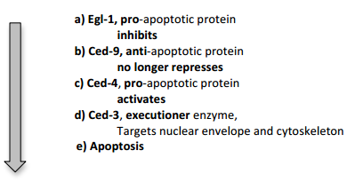
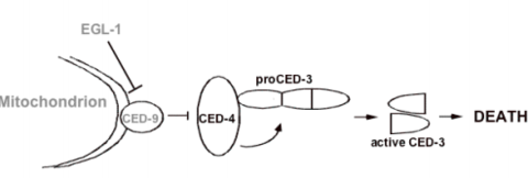
| Give a simplified model of the apoptotic pathway in C elegans. | |
| Draw a diagram representing this | |
| state how these steps are achieved. | 1) protein-protein interactions 2)Proteolytic activation of pro enzymes |
| give an example of protein protein interactions in the C elegans pathway | Egl-1 binds Ced-9 displacing Ced-4, which is inert while bound to Ced-9 |
| Give an example of the proteolytic activation of proenzymes in the C elegans apoptotic pathway | Ced-4 cleaves inert precursor Ced-3 converting it into an active protease, called a caspase. the critical interaction between Egl-1, Ced-9 and Ced4 occurs on the surface of the mitochondria. |
| Describe the very basic differences in principles of apoptotic pathways in C elegans and mammals. | principles similar but with more genes involved at each step. eg) the worm has 1 major executioner caspase, Ced-9, while human cells have 15 caspases. |
| give a diagram displaying the key apoptotic processes in mammals | |
| Name and describe the origins and action of the key regulator of apoptosis in mammals | Bcl2. The protein gets its name due to being highlu expressed in B cell lymphoma in humans. Expressing Bcl2 protects the cancer cells from apoptosis. To trigger apoptosis Bcl2 must be limited. |
| where is this protein found? | Bcl2 is located on and maintains the integrity of the mitochondrial outer membrane, keeping proteins within the mitochondria (such as cytochrome c) from entering the cytoplasm. |
| describe the pro- apoptotic proteins in mammals | Bim, Bad and Bid. found upstream of Bcl2. The proteins function as Bcl2 inhibitors, effectively 'pull the trigger' on apoptosis |
| What process occurs once Bcl2 is inhibited? | pro-apop proteins Bax and Bak are derepressed. They then assemble into channels allowing cytochrome c to be released from the mitochondria. At this point, they cell is committed to undergo apoptosis. |
| describe the next sequence of events in apoptosis | cytochrome c activated the 'initiator caspase', caspase 9, which in turn activates the 'executioner caspase', caspase 3, after which the cascade of apoptotic events proceeds rapidly. |
| describe how the functions of both proteins have been demonstrated in transgenic mice | individual mice lacking both Bax and Bak have webs between digits, enlarged livers and spleens due to excess lymphocytes, which fail to undergo physiological apoptosis and accumulations of neurons in the brain that would normally die. |
| Describe the inhibition of bcl2 | Non-phosphorylated, activated Bad inhibits Bcl2 (allows Bak and Bax to form channels ect) this eventually leads to apoptosis |
| where is caspase 9 found in the genome? | downstream of Bak and Bax, part of a large multiprotein complex called the apoptosome. |
| State the 2 overall routes which can trigger apoptosis. | -the intrinsic pathway -the extrinsic pathway. |
| describe, generally, the mechanism used to trigger apoptosis in the intrinsic pathway. | apoptosis is triggered by inhibition of Bcl2 as described earlier. There are various signals which can trigger intrinsic apoptosis. |
| State the various signals that trigger intrinsic apoptosis. | 1)genome 'quality control' 2)lack of 'survival' or 'trophic' factors 3) loss of attachment to the underlying basement membrane |
| describe how intrinsic apoptosis can be signalled from genome quality control | 1)DNA damage resulting in mutation 2)leads to p53 protein activation 3)triggers the induction of proapoptotic protein PUMA. 4) PUMA binds to and activates Bak or Bax. |
| Describe how a loss of attachment to the underlying basement membrane can induce intrinsic apoptosis | via disruption of integrin signalling. This frees the pro-apoptotic protein Bim from the cytoskeleton, where it migrates to the mitochondria and promotes Bak or Bax channel formation. |
| give an example of how lack of trophic factors effects apoptosis in an organism | mice lacking nerve growth factor or its receptor TrkA have no pain sensitive neurones in the skin. |
| how do trophic factors function? | 1)activate protein kinase PKB. 2) PKB phosphorylates the proapoptotic protein Bad 3) Bad is then inhibited by binding a 14-3-3 protein. 4) Bad is therefore no longer inhibiting Bcl2 5) Bcl2 blocks mitochondrial channels, apoptosis does not occur |
| What occurs in the absence of trophic factors? | 1) loss of PKB activity 2) leads to the accumulation of non-phosphorylated Bad 3) which binds to Bcl2 4)relieves inhibition of Bak and Bax and triggers apoptosis |
| Give a general pathway for extrinsic signalling of apoptosis. | 1) Binding of a death ligand (ie Fas ligand) to a death receptor (ie Fas) 2) recruitment of a death domain protein 3) results in caspase 8 activation 4) casp8 activates downstream caspases directly + activates Bid 5)Bid inhibits Bcl2 triggering cytochrome c release. 6)casp8 also triggers cell death |
| More in detail- how is extrinsic signalling initiated? | through ligands from immune cells binding to cell surface receptors triggering cell death. |
| Give examples of receptors and ligands | best characterised of the receptors = Tumour Necrosis Factor receptor family. Examples: Fas and TNF-R1. Ligands for receptors = Fas-ligand and TNF(alpha) |
| how are these ligand signalling molecules produced? | Fas-ligand = cell surface protein expressed by cell killing lymphocytes (cytotoxic T cells) TNF(alpha) = released by macrophages |
| what happens once the ligand has bound to the receptors? | receptors initiate the formation of an apoptosis-inducing signalling complex (ie death domain protein) which activates cell death by 2 routes |
| state these 2 routes by which the complex activates apoptosis | Bcl2 inihibition and a Bcl2 independent caspase cascade initiated by caspase 8 |
| give an example of apoptosis and autoimmune diseases | extrinsic pathway inappropriately activated in 'autoimmune' diseases. blocking the death receptor ligand TNF(alpha) with antibodies has proved to be highly effective in treating such diseases eg rheumatoid arthritis. |
Quer criar seus próprios Flashcards gratuitos com GoConqr? Saiba mais.