13158678
Descrição

Quiz por Jessica Bulley, atualizado more than 1 year ago
|
|
Criado por Jessica Bulley
mais de 6 anos atrás
|
|
Questão 1
Questão
Describe the position of the heart within the mediastinum
Responda
-
thoracic cavity
-
pleural cavity
Questão 2
Questão
Select Three functions of the CVS
Responda
-
• Assists the production of the digestive and absorbtion system
-
• Transports fluids, nutrients, waste products, gases, and hormones throughout the body.
-
• Exchange materials between blood, cells and extracellular fluid.
-
• Plays a role in the immune response, blood pressure and the regulation of body temperature.
-
• Maintains optimal body temperature
Questão 3
Questão
Select Four components which comprise of the CVS
Responda
-
heart
-
blood
-
capillary beds
-
blood vessels
-
lungs
-
larynx
Questão 4
Questão
Select Five components the CVS transports
Responda
-
fluids
-
hormones
-
gases
-
waste products
-
nutrients
-
urine
-
chyne
Questão 5
Questão
Select Five functions of the Heart
Responda
-
• Generating blood pressure – moves blood through vessels
-
• Changes to match need ie. exercise, sleeping
-
• Regulating blood supply
-
• Ensuring one-way blood flow
-
• Routing blood: separates pulmonary and systemic circulations
-
• Regulates hormones
Questão 6
Questão
The Heart – 2 pumps in 1 which are: (select two)
Responda
-
Coronal circulation
-
Systemic circulation
-
Pulmonary circulation
-
Adrenal circulation
Questão 7
Questão
The shape of the heart consists of:
[blank_start]Apex[blank_end]: Blunt rounded point of cone
[blank_start]Base[blank_end]: Flat part at opposite of end of cone
Responda
-
Apex
-
Base
Questão 8
Questão
The [blank_start]pericardial[blank_end] sac has two layers, a [blank_start]serous[blank_end] layer and a [blank_start]fibrous[blank_end] layer. It encloses the pericardial cavity which contains [blank_start]pericardial[blank_end] fluid.
Responda
-
pericardial
-
myocardium
-
serous
-
parietal
-
fibrous
-
phrenic
-
pericardial
-
plasma
Questão 9
Questão
The [blank_start]Serous[blank_end] portion of Pericardium, consists of [blank_start]two[blank_end] layers, [blank_start]visceral[blank_end] and [blank_start]parietal[blank_end]. The space between the layers is the pericardial cavity.
Responda
-
Serous
-
Fibrous
-
two
-
three
-
visceral
-
inner
-
parietal
-
myocardial
Questão 10
Questão
The Visceral Serous pericardium is situated to the [blank_start]Myocardium[blank_end] of the Heart.
Responda
-
Myocardium
-
Epicardium
-
Endocardium
Questão 11
Questão
Walls of the Heart:
Three layers of tissue -
1. [blank_start]Epicardium[blank_end] : Serous membrane; smooth outer surface of heart
2. [blank_start]Myocardium[blank_end] : Middle layer composed of cardiac muscle cells – contractility
3. [blank_start]Endocardium[blank_end] : Smooth inner surface of heart chambers
Responda
-
Epicardium
-
Myocardium
-
Endocardium
Questão 12
Questão
The Endocardium is the smooth inner surface of heart chambers
Responda
- True
- False
Questão 13
Questão
[blank_start]Pectinate muscles[blank_end] : muscular ridges in auricles and right atrial wall
[blank_start]Trabeculae carnae[blank_end] : muscular ridges and columns on inside walls of ventricles
Responda
-
Pectinate muscles
-
Trabeculae carnae
Questão 14
Questão
Trabeculae carnae: muscular ridges and columns on inside walls of ventricles
Responda
- True
- False
Questão 15
Questão
Pectinate muscles: muscular ridges in auricles and right atrial wall
Responda
- True
- False
Questão 16
Questão
Pectinate muscles: muscular ridges and columns on inside walls of ventricles
Responda
- True
- False
Questão 17
Questão
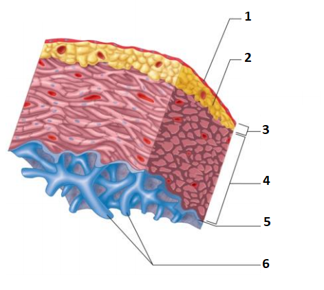
Walls of the Heart Diagram:
1. [blank_start]Simple Squamous Epithelium[blank_end]
2. [blank_start]Loose connective and adipose tissue[blank_end]
3. [blank_start]Epicardium (Visceral)[blank_end]
4. [blank_start]Myocardium[blank_end]
5. [blank_start]Endocardium[blank_end]
6. [blank_start]Trabeculae carneae[blank_end]
Responda
-
Simple Squamous Epithelium
-
Loose connective and adipose tissue
-
Epicardium (Visceral)
-
Myocardium
-
Endocardium
-
Trabeculae carneae
Questão 18
Questão
The Heart chambers:
[blank_start]Atrioventricular canals[blank_end]: openings between atria and respective ventricles
[blank_start]Right ventricle[blank_end]: opens to pulmonary trunk
[blank_start]Left ventricle[blank_end]: opens to aorta – very muscular wall.
[blank_start]Interventricular septum[blank_end]: between the two ventricles.
Responda
-
Atrioventricular valves
-
Right ventricle
-
Left ventricle
-
Interventricular septum
Questão 19
Questão
Right ventricle: opens to pulmonary trunk
Responda
- True
- False
Questão 20
Questão
Atrioventricular valves: openings between atria and their respective ventricles
Responda
- True
- False
Questão 21
Questão
Left ventricle: opens to aorta – very muscular wall
Responda
- True
- False
Questão 22
Questão
Blood Vessels - overview.
[blank_start]Arteries[blank_end] :
Elastic, Muscular, Arterioles
Take blood away from the heart
Contain blood under pressure
[blank_start]Capillaries[blank_end] :
site of exchange with tissues (interstitial fluid)
[blank_start]Veins[blank_end] :
Large, medium, small, venules
Take blood to the heart
Thinner walls than arteries, contain less elastic tissue less
smooth muscle
Valves to prevent backflow
Responda
-
Arteries
-
Capillaries
-
Veins
Questão 23
Questão
Blood vessel diagram:
1. [blank_start]Tunica Adventitia[blank_end]
2. [blank_start]Tunica Media[blank_end]
3. [blank_start]Tunica Intima[blank_end]
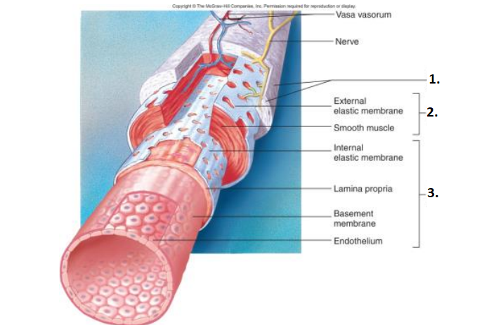
Responda
-
Tunica Adventitia
-
Tunica Media
-
Tunica Intima
Questão 24
Questão
Blood Vessels – arteries & veins:
- [blank_start]Tunica intima[blank_end]: Endothelium
- [blank_start]Tunica media[blank_end]: smooth muscle cells arranged circularly around the blood vessel.
- [blank_start]Vasoconstriction[blank_end]: smooth muscles contract, decrease in blood flow
- [blank_start]Vasodilation[blank_end]: smooth muscles relax, increase in blood flow
- [blank_start]Tunica externa (adventitia)[blank_end]: connective tissue
Responda
-
Tunica intima
-
Tunica media
-
Vasoconstriction
-
Vasodilation
-
Tunica externa (adventitia)
Questão 25
Questão
Select Five functions of blood
Responda
-
Clot formation
-
Protection against foreign substances
-
Maintenance of body temperature
-
Regulation of pH and osmosis (normal pH 7.4)
-
Transport: gases, nutrients, waste products, processed molecules, hormones, enzymes
-
Absorption of nutrients
Questão 26
Questão
Blood consists of [blank_start]55%[blank_end] Plasma and [blank_start]45%[blank_end] formed elements
Responda
-
55%
-
50%
-
45%
-
55%
Questão 27
Questão
Plasma consists of [blank_start]7%[blank_end] Proteins, [blank_start]91%[blank_end] Water and [blank_start]2%[blank_end] Other solutes
Responda
-
7%
-
91%
-
91%
-
7%
-
2%
-
7%
Questão 28
Questão
The Proteins in Plasma consist of (select Three)
Responda
-
Albumins 58%
-
Globulins 38%
-
Fibrinogen 4%
-
Neutrophils 4%
Questão 29
Questão
Other solutes in Blood consist of (select Five)
Responda
-
Ions
-
Nutrients
-
Waste products
-
Gases
-
Regulatory substances
-
Globulins
-
Neutrophils
Questão 30
Questão
Hemoglobin is a
Responda
-
protein which attaches to Oxygen
-
carbohydrate which attaches to Oxygen
Questão 31
Questão
Cardiac cycle –
[blank_start]Systole[blank_end] - contraction of the ventricles, causes the ejection of blood into the aorta and pulmonary trunk
[blank_start]Diastole[blank_end] – when the heart muscle relaxes and allows the chambers to fill with blood, to refill each atrium and each ventricle
Responda
-
Systole
-
Diastole
Questão 32
Questão
Stroke volume - the volume of blood pumped from the left ventricle in one contraction
Responda
- True
- False
Questão 33
Questão
The heart [blank_start]can[blank_end] generate it’s own action potentials.
Responda
-
can
-
can't
Questão 34
Questão
The Sinoatrial node (SA) node is the heart's natural pacemaker. The SA node consists of a cluster of cells that are situated in the upper part of the wall of the [blank_start]right atrium[blank_end].
Responda
-
right atrium
-
left atrium
Questão 35
Questão
[blank_start]Atrioventricular node[blank_end]: The electrical relay station between the upper and lower chambers of the heart. The [blank_start]AV[blank_end] node, which controls the heart rate, sends electrical signals from the atria which must pass through the [blank_start]AV[blank_end] node to reach the ventricles.
Responda
-
Atrioventricular node
-
Sinoatrial node
-
AV
-
SA
-
AV
-
SA
Questão 36
Questão
The mode of Capillary exchange is via [blank_start]Diffusion[blank_end]
Responda
-
Diffusion
-
Osmosis
Questão 37
Questão
Left Atrium: one of the four chambers of the heart, located on the left posterior side. Its primary roles are to act as a holding chamber for blood returning from the lungs
Responda
- True
- False
Questão 38
Questão
Right atrium: one of the four chambers of the heart, located on the left posterior side. Its primary roles are to act as a holding chamber for blood returning from the lungs
Responda
- True
- False
Questão 39
Questão
Deoxygenated blood enters the right atrium through the inferior and superior vena cava.
Responda
- True
- False
Questão 40
Questão
Deoxygenated blood enters the left atrium through the inferior and superior vena cava.
Responda
- True
- False
Questão 41
Questão
The Fibrous pericardium: tough fibrous outer layer, prevents over distention; acts as anchor.
Responda
- True
- False
Questão 42
Questão
Serous pericardium: thin, transparent, inner layer, simple squamous epithelium.
- Parietal pericardium: lines the fibrous outer layer
- Visceral pericardium: covers heart surface
Responda
- True
- False
Questão 43
Questão
Serous pericardium: thin, transparent, inner layer, simple squamous epithelium.
- Visceral pericardium: lines the fibrous outer layer
- Parietal pericardium: covers heart surface
Responda
- True
- False
Questão 44
Questão
The aortic valve is a valve in the human heart between the left ventricle and the aorta.
Responda
- True
- False
Questão 45
Questão
The bicuspid valve is a valve in the human heart between the left ventricle and the aorta.
Responda
- True
- False
Questão 46
Questão
The pulmonic valve is one of two valves that allow blood to leave the heart via the arteries. It is located in the right ventricle of the heart.
Responda
- True
- False
Questão 47
Questão
The tricuspid valve forms the boundary between the right ventricle and the right atrium.
Responda
- True
- False
Questão 48
Questão
The tricuspid valve forms the boundary between the left ventricle and the left atrium.
Responda
- True
- False
Questão 49
Questão
The bicuspid valve is situated between the left atrium and the left ventricle.
Responda
- True
- False
Questão 50
Questão
REMEMBER THIS FOR VALVES: This Assists Pushing Blood (from left to right)
Responda
- True
- False
Questão 51
Questão
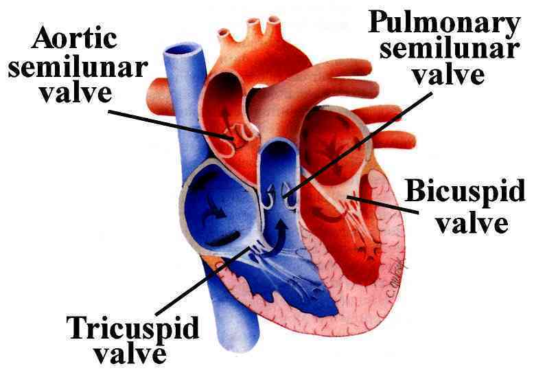
Valves of the Heart:
1. [blank_start]Tricuspid[blank_end]
2. [blank_start]Aortic Semilunar[blank_end]
3. [blank_start]Pulmonary[blank_end]
4. [blank_start]Bicuspid[blank_end]
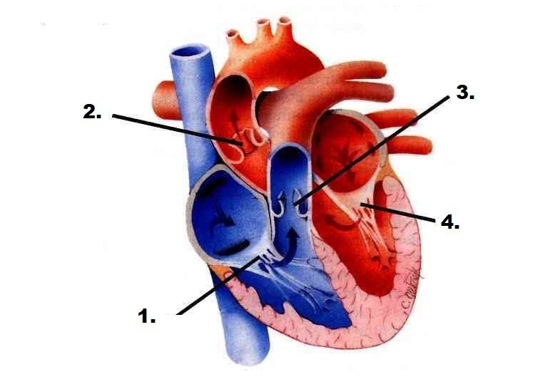
Responda
-
Tricuspid
-
Aortic Semilunar
-
Pulmonary
-
Bicuspid
Questão 52
Responda
-
Tricuspid valve
-
Aortic semilunar valve
Questão 53
Responda
-
Aortic semilunar valve
-
Tricuspid valve
Questão 54
Responda
-
Aortic semilunar valve
-
Pulmonary semilunar valve
Questão 55
Responda
-
Pulmonary semilunar valve
-
Bicuspid valve
Questão 56
Questão
The pectinate muscles (musculi pectinati) are parallel ridges in the walls of the atria of the heart.
Responda
- True
- False
Questão 57
Questão
Tunica External is the external layer of the artery wall
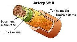
Responda
- True
- False
Questão 58
Questão
The SA node is the heart's natural pacemaker. The SA node consists of a cluster of cells that are situated in the upper part of the wall of the right atrium
Responda
- True
- False
Questão 59
Questão
The NV node is the heart's natural pacemaker. The NV node consists of a cluster of cells that are situated in the upper part of the wall of the right atrium
Responda
- True
- False
Questão 60
Questão
The AV node, which controls the heart rate, is one of the major elements in the cardiac conduction system. The AV node serves as an electrical relay station, slowing the electrical current sent by the sinoatrial (SA) node before the signal is permitted to pass down through to the ventricles.
Responda
- True
- False
Questão 61
Questão
Tunica externa (adventitia): connective tissue
Responda
- True
- False
Questão 62
Questão
Tunica intima: smooth muscle cells arranged circularly around the blood vessel.
Responda
- True
- False
Questão 63
Questão
Tunica media: Endothelium
Responda
- True
- False
Quer criar seus próprios Quizzes gratuitos com a GoConqr? Saiba mais.