383093
Beschreibung
Mindmap von melian.yates, aktualisiert more than 1 year ago
|
|
Erstellt von melian.yates
vor etwa 11 Jahre
|
|
Neuropathology
- Anatomy
- Brain
- Grey Matter (Outside)
- Cerebral & Cerebellar cortex
- Contains neuronal cell bodies
- Contains neuronal cell bodies
- Cerebral & Cerebellar cortex
- White Matter (Inside)
- Contains axons, myelin
sheaths, astrocyte fibers
- Contains axons, myelin
sheaths, astrocyte fibers
- Nuclei
- Foci of grey matter also
occur w/in the white matter
- Ex. Olivary, red, cuneate, etc.
- Ex. Olivary, red, cuneate, etc.
- Foci of grey matter also
occur w/in the white matter
- Meninges
- Protect CNS
- Encase CSF
- Support blood vesels
- Support blood vesels
- Encase CSF
- Dura (Outside -thick (Packy)) -> Arachnoid
-> Pia Matter (Inside - thin (Lepto))
- Sunbarachnoid space (Blood vessels)
- Sunbarachnoid space (Blood vessels)
- Protect CNS
- Grey Matter (Outside)
- CNS Cell Types
- 1) Neurons
- Response to Injury
- Extremely vulnerable to injury
- High metabolic rate
- Little NRG storage
- An Axon to care for
- Axon has no Nissl
(ribosomes)
- Can't make protein
- Can't dispose of own waste
- Can't dispose of own waste
- Can't make protein
- Neurons CANNOT regenerate
- Axon has no Nissl
(ribosomes)
- An Axon to care for
- Little NRG storage
- High metabolic rate
- Acute Necrosis
- Most common response
- Ischaemia
- O2 required for oxidative phosphorylation
- Cell's ATP drops & ion channels leak
- Ca 2+ enters cells =>
membrane damage
- Increased
intracytoplasmic
Ca2+ =>
- ATPase => Decreased ATP
- Phospholipase =>
Decreased phospholipids
- Protease => Disruption of
membrane & cytoskeletal
proteins
- Endonuclease => Nucleus
chromatin damage
- Endonuclease => Nucleus
chromatin damage
- Protease => Disruption of
membrane & cytoskeletal
proteins
- Phospholipase =>
Decreased phospholipids
- ATPase => Decreased ATP
- Increased
intracytoplasmic
Ca2+ =>
- Ca 2+ enters cells =>
membrane damage
- Cell's ATP drops & ion channels leak
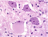
- Red dead cells (Red cytoplasm)
- Pyknosis
- Pyknosis
- O2 required for oxidative phosphorylation
- Hypoxia
- Hypoglycaemia (toy breed
puppies -> seizures)
- Nutritional Deficiency (e.g.
thiamine deficiency)
- Toxins (e.g. Pb & Hg)
- Laminar cortical necrosis
- Polioencephalomalacia
- Outer cortex "Melting" =>
Gelatinous material
- Ruminants
- Outer cortex "Melting" =>
Gelatinous material
- Acute neuronal necrosis
in a distinct pattern
- Causes:
- Salt poisoning (Pigs)
- Water deprivation
- Pb & Hg toxicity
- Polioencephalomalacia
- Polioencephalomalacia
- Pb & Hg toxicity
- Water deprivation
- Salt poisoning (Pigs)
- Polioencephalomalacia
- Polioencephalomalacia
- Cerebrocortical necrosis (CCN)
- Pathogenesis unclear
- Thiamine deficiency vs. sulfide toxicity
- Thiamine required for
glucose metabolism =>
Hypoglycaemia
- Thiamine required for
glucose metabolism =>
Hypoglycaemia
- Cerebrocortical necrosis (CCN)
- Most common response
- Chromatolysis
- Swelling of the Neuron
- Powdery appearence => Nissel
substances dispersed ( unraveling ER)
- Powdery appearence => Nissel
substances dispersed ( unraveling ER)
- NOT necrosis
=> Adaptive
change
- Dissolution of
Nissel bodies
- Ex. Equine dysautonomia
(grass sickness)
- Swelling of the Neuron
- Wallerian degeneration
- Response of Axon to injury
(Normally seen in white matter)
- Healing => Axoplasm (Myelinated - built
up at point of injury => swollen axon)
- Neuroma => conglomeration of axons,
axoplasm ("abortive attempt" at repair)
- Ex. tail dock neuroma
- Ex. tail dock neuroma
- No renegeration of basal lamina, inefficent
clearance of debris & re-myelination
(oligodendrocyte), inhibits axonal sprouting =>
Chances of repair low
- Healing => Axoplasm (Myelinated - built
up at point of injury => swollen axon)
- Breakdown of the nerve
fiber DISTAL to the point
of initiating injury
- Myelin sheath
breaks up
- Myelin sheath
breaks up
- Response of Axon to injury
(Normally seen in white matter)
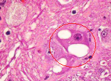
- Vacuolation
- Transmissable spongioform
encephalopathies (BSE, scrapie)
- Common artefact
(Not fixed
properly)
- Normal finding
- Lysosomal storage disease
- Early cell injury
- Some toxins
- Transmissable spongioform
encephalopathies (BSE, scrapie)
- Extremely vulnerable to injury
- Response to Injury
- 2) Glial cells (Neural glue)
- Astrocytes
- " Star-shaped",
multiple dendritic
extensions
- CNS equivalent
of fibroblast
- Functions:
- Support
- Insulating synapses
- Detoxification
- BBB
- BBB
- Produces cytokines
- Detoxification
- Insulating synapses
- Support
- Response to Injury
- Necrosis
- Astrocytosis
(astrocyte hyperplasia)
- Astrogliosis (astrocyte
hypertrophy & hyperplasia)
- Starting to
produce glial
fibers ("Fibrosis")
- Starting to
produce glial
fibers ("Fibrosis")
- Necrosis
- " Star-shaped",
multiple dendritic
extensions
- Oligodendrocytes
- Look like circular nuclei
(Circular cells),
Myelinate axons
- Inefficient at
re-myelinating in
response to injury
- Myelinate axons
- Oligodendrocyte injury
=> De-myelination
- Oligodendrocyte injury
=> De-myelination
- Look like circular nuclei
(Circular cells),
Myelinate axons
- Microglial cells
- " Macrophage"
- " Macrophage"
- Ependymal cells
- Line the ventricles
- Line the ventricles
- Choroid plexus epithelial cells
- Produce CSF
- Produce CSF
- Astrocytes
- Order of vulnerability to injury:
- Neurons > Oligodendrocytes
> Astrocytes > Microglia >
Blood vessels
- Neurons > Oligodendrocytes
> Astrocytes > Microglia >
Blood vessels
- 1) Neurons
- Brain
- General Responses to Injury (CNS & PNS)
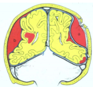
- Oedema
- Vasogenic
- Most common form
in animals
- Follows vascular injury
- Breaches BBB ->
Increased permeability
- Fluid is extracellular
- Ex. Oedema
disease in Pigs
- E.coli (bacterial
infection of GI tract)
- Bacteria produce toxins in
the intestine (endotoxaemia)
- Toxin enters bloodstream =>
Brain => Vasculitis, Leaky vessels
- Toxin enters bloodstream =>
Brain => Vasculitis, Leaky vessels
- Bacteria produce toxins in
the intestine (endotoxaemia)
- E.coli (bacterial
infection of GI tract)
- Most common form
in animals
- Interstitial
- Usually follows
hydrocephalus
- Ventricles dilate & increased
hydrostatic pressure => Fluid forced
into parenchyma
- Fluid is Extracellular
- Usually follows
hydrocephalus
- Cytotoxic
- ATP deficit & Failure of
Na+/K+ pump => Na+ &
H2O build up in cell
- Fluid in Intracellular
- Hypoxia, Ischaemia, toxins
(anything that inhibits ATP
production)
- => Hydrophobic
degeneration in other cells
- ATP deficit & Failure of
Na+/K+ pump => Na+ &
H2O build up in cell
- Consequences
- Brain swells, becomes
compressed => Herniation
- 1) Through the Foramen Magnum
- Most common
- 2) At Cingulate gyrus under the falx
- 3) Transtentorial herniation
- 3) Transtentorial herniation
- Most common
- Ex. Cerebellar herniation
- Compresses Medulla (Resp.
centers) => Sudden death
- Compresses Medulla (Resp.
centers) => Sudden death
- 1) Through the Foramen Magnum
- Brain swells, becomes
compressed => Herniation
- Vasogenic
- De-myelination
- Primary: Myelin forms normally & then is selectively destroyed
- Impaired maintenance (i.e.
infectious destruction of
oligodendrocytes)
- Nutritional (Cu, Vit. B12)
- Toxins (Cyanide)
- Cytotoxic oedema
- Immune mediated (i.e.
multiple sclerosis, canine
distemper (Most common))
- Proposed Pathogenesis:
- Oligodendrocytes die as a
result of immune response
against viral Ag on their surface
- Molecular mimicry
- Incorporation of neural
elements into the virus
during assembly resulting
in anti-self response
- Incorporation of neural
elements into the virus
during assembly resulting
in anti-self response
- Molecular mimicry
- Oligodendrocytes die as a
result of immune response
against viral Ag on their surface
- Proposed Pathogenesis:
- Immune mediated (i.e.
multiple sclerosis, canine
distemper (Most common))
- Cytotoxic oedema
- Toxins (Cyanide)
- Nutritional (Cu, Vit. B12)
- Impaired maintenance (i.e.
infectious destruction of
oligodendrocytes)
- Secondary: Loss of Myelin following axonal damage (i.e.
Wallerian degeneration)
- Vs. Dysmyelination =>
Myelin doesn't form
properly in first place
- Primary: Myelin forms normally & then is selectively destroyed
- Vascular Disturbances
- Reduced blood flow (or complete
blockage) leads to ischaemia
- Consequences of ischaemia depend on:
- It's degree & duration
- Size & type of
vessel involved
- Relative susceptibility
of tissue to hypoxia
- Relative susceptibility
of tissue to hypoxia
- Size & type of
vessel involved
- It's degree & duration
- Consequences of ischaemia:
- Neuronal necrosis
- Vasogenic oedema
- The ischaemia is not a direct cause
of the oedema, but the two could
occur together if the ischaemia was
the result of vascular damage
- The ischaemia is not a direct cause
of the oedema, but the two could
occur together if the ischaemia was
the result of vascular damage
- Infarct(s)
- Necrosis of a tissue
following obstruction of
a supplying blood
vessel
- Often well demarcated
- Causes:
- Thrombosis/thromboembolism
(Uncommon in the CNS of animals, can
see with DIC or sepsis)
- Emboli: FCEM (fibrocartilaginous
embolic myelopathy), bone marrow
embolism following fracture
- Vasculitis: Classical swine fever (hog cholera
- pestivirus), MCF (malignant catarrhal fever
-herpesvirus), Oedema disease (vasculitis
caused by E.coli toxin)
- Vasculitis: Classical swine fever (hog cholera
- pestivirus), MCF (malignant catarrhal fever
-herpesvirus), Oedema disease (vasculitis
caused by E.coli toxin)
- Emboli: FCEM (fibrocartilaginous
embolic myelopathy), bone marrow
embolism following fracture
- Thrombosis/thromboembolism
(Uncommon in the CNS of animals, can
see with DIC or sepsis)
- Necrosis of a tissue
following obstruction of
a supplying blood
vessel
- Malacia
- Grossly: appreciable softening of
brain/spinal cord (usually resulting
from necrosis)
- Histologically: Necrosis
- Occurs in infarcts, but there
are other causes (toxicosis,
hypoxia, nutritional, infectious
or metabolic disease)
- The pattern & location of Malacia
are often more Dx helpful than the
lesion itself
- Symmetrical Malacia:
- Nutritional, Toxic, Metabolic, Genetic
- Nutritional, Toxic, Metabolic, Genetic
- Asymmetrical Malacia:
- Vascular, Infectious, Traumatic
- Vascular, Infectious, Traumatic
- Symmetrical Malacia:
- Grossly: appreciable softening of
brain/spinal cord (usually resulting
from necrosis)
- Neuronal necrosis
- Reduced blood flow (or complete
blockage) leads to ischaemia
- Oedema
- Inflammation of the Nervous System
- Defence (against infection)
- Skin
- Skull/vertebrae
- Meninges & CSF
- Barriers (CSF/ECF,
CSF/blood, BBB)
- Microglia (monocyte/
macrophage system)
- Immunologic responses
(innate & adaptive)
- Immunologic responses
(innate & adaptive)
- BBB
- Endothelial cell
- Not fenestrated
- Tight junctions
- Fewer transporting vesicles
- P glycoprotein
- P glycoprotein
- Fewer transporting vesicles
- Tight junctions
- Not fenestrated
- Astrocytes
- Pericytes
- Endothelial cell
- Microglia (monocyte/
macrophage system)
- Barriers (CSF/ECF,
CSF/blood, BBB)
- Meninges & CSF
- Skull/vertebrae
- Skin
- Inflammation is usually
the result of infection
- Bacteria, Fungi,
Parasites (protozoa)
- Main Routes of Entry
- Haemotogenous
(most common)
- Direct
- Spread from adjacent
structure (inner ear,
skull sinuses)
- Injection
- Spread from adjacent
structure (inner ear,
skull sinuses)
- Penetrating trauma
- Via peripheral nerves
- Leukocyte trafficking
("Trojan horse")
- Haemotogenous
(most common)
- Bacteria, Fungi,
Parasites (protozoa)
- Sites of Inflammation
- Epidural or subdural space
- Tends to result in
abscess (Uncommon)
- Tends to result in
abscess (Uncommon)
- Meninges
- Leptomeninges - Pia &
Arachnoid Matter
(Leptomeningitis)
- Suppurative:
- Most common
- Bacterial (E.coli,
Streptococcus, Haemophilus)
- Swollen brain, meninges
opaque, OFTEN NO GROSS
LESIONS
- Neutrophils predominate
initially
- Usually fatal
- Usually of haemotogenous origin
- Most common
- Eosinophilic
meningoencephalitis:
- Porcine salt poisoning/
H2O deprivation
- Perivascular eosinophilic cuffing
in the cerebrum & meninges
- Porcine salt poisoning/
H2O deprivation
- Lymphocytic:
- Usually viral
- Usually viral
- Granulomatous:
- Fungal disease &
mycobacteriosis
- Idiopathic forms (Mainly Dogs)
- Idiopathic forms (Mainly Dogs)
- Fungal disease &
mycobacteriosis
- Suppurative:
- Pachymeninges -
Dura matter
(Pachymeningitis)
- Leptomeninges - Pia &
Arachnoid Matter
(Leptomeningitis)
- Parenchyma
- Encephalitis
- Inflammation of the
cerebral parenchyma)
- Classified based on
nature of Exudate:
- Suppurative
- Fibrinous
- Granulomatous
- Lymphoplasmacytic
- Eosinophilic
- Haemorrhagic
- Haemorrhagic
- Eosinophilic
- Lymphoplasmacytic
- Granulomatous
- Fibrinous
- Suppurative
- Bacterial:
- Typically result in Abscesses
(Suppurative inflammation - i.e.
localized collections of neutrophils)
- Single or multiple
depending on route
- Vary in size
with a central,
liquefied cavity
- Ex. E.coli, Streptococcus,
Stapyhlcoccus, Pasteurella &
Fusobacterium necrophorum
- Listeria
monocytogenes (Listeriosis)
- Sheep, cattle goats
- 3 forms (CNS,
abortion, sepsis)
- Compared to other
bacteria, pathogenesis
in CNS disease is a
little different
- Oral mucosa ->
Trigeminal nerve ->
Trigeminal ganglion in
brain
- Source: Silage (pH > 5.4
allows bacterial growth)
- Sxs: Circling, Facial nerve
paralysis (unilateral), Drooling,
Pharyngeal paralysis,
Recumbancy, paddling, death
- Usually NO
GROSS lesions
- Sheep, cattle goats
- Listeria
monocytogenes (Listeriosis)
- White/grey matter
junction predisposed
- Typically result in Abscesses
(Suppurative inflammation - i.e.
localized collections of neutrophils)
- Viral:
- Lymphoplastic inflammation
- Haematogenous &
neural routes mainly
- Hallmark lesions
(regardless of virus type):
- 1) Neuronal necrosis
- 2) Gliosis (glial nodules)
- 3) Vascular changes -lymphoplasmacytic cuffing
- 3) Vascular changes -lymphoplasmacytic cuffing
- 2) Gliosis (glial nodules)
- 1) Neuronal necrosis
- Types of Virus:
- Neurtropic
- Viruses which can
overcome innate
immunity of brain
- Rabies (rhabdovirus)
- Encephalomyelitis &
ganglionitis
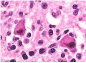
- Lesions are macroscopic
- Intracytoplasmic, oval, pink
inclusion bodies (Negri bodies)
- Intracytoplasmic, oval, pink
inclusion bodies (Negri bodies)
- Lesions are macroscopic
- Encephalomyelitis &
ganglionitis
- Aujesky's disease
(Herpesvirus)
- Visna (Ovine lentivirus)
- Viruses which can
overcome innate
immunity of brain
- Endotheliotropic
- EHV1 (Equine
herpesvirus type 1)
- Classical swine
fever (pestivirus)
- Infectious canine
hepatitis (canine
adenovirus)
- Endothelial cell damage
leads to vascular injury &
secondary parenchymal
necrosis & haemorrhage
- EHV1 (Equine
herpesvirus type 1)
- Pantropic
- Canine distemper
(morbillivirus)
- Infectious bovine
rhinotracheitis (Bovine
herpesvirus type 1)
- Respiratory,
urogenital & CNS
- Virus can be
latent in nerve
ganglia
- Canine distemper
(morbillivirus)
- Neurtropic
- Lymphoplastic inflammation
- Prion Diseases:
- Transmissable spongiform
encephalopathies (TSEs)
- BSE, scrapie,
chronic wasting
disease
- A group of fatal
neurodegenerative diseases
which occur in a number of
species
- BSE, scrapie,
chronic wasting
disease
- Proteinaceous infectious particle
- Abnormal isoform of prion
protein (PrP) in the brain
-> PrPsc (common to all cases)
- PrPsc accumulates in nervous
tissue as amyloid fibrils
- Long incubation period, difficult to Dx
(no immune response)
- Agent is highly resistant
- Agent is highly resistant
- Long incubation period, difficult to Dx
(no immune response)
- Alpha helix -> beta
pleated sheet
- Abnormal isoform of prion
protein (PrP) in the brain
-> PrPsc (common to all cases)
- No gross lesions;
Characteristic
microscopic lesions
- Spongioform change,
Amyloid plaques,
Astrogliosis
- Spongioform change,
Amyloid plaques,
Astrogliosis
- Transmissable spongiform
encephalopathies (TSEs)
- Fungal/ Protozoa:
- Cryptococcus
- Opportunistic -Aspergillus
- Neospora
- Toxoplasma gondii
- Toxoplasma gondii
- Neospora
- Opportunistic -Aspergillus
- Cryptococcus
- Inflammation of the
cerebral parenchyma)
- Myelitis
- Encephalitis
- Epidural or subdural space
- Defence (against infection)
- Trauma
- Less common in
animals than man
- Less risk, Better
protection
- Less risk, Better
protection
- Main forms of injury
- Concussion
- Contusion
- Pathogenesis:
- Head is moving, but suddenly
stopped by solid object
- 1st point of impact = where
object hits the head
- Head stops, brain doesn't
- Brain strikes inside of
skull at first point of impact
(Coup)
- Brain moves in cranium,
stretching & tearing vessels &
nerves on the opposite side to
the original impact (Contrecoup)
- Brain strikes inside of
skull at first point of impact
(Coup)
- Head stops, brain doesn't
- 1st point of impact = where
object hits the head
- Head is moving, but suddenly
stopped by solid object
- Pathogenesis:
- Laceration
- Haemorrhage
- Locations:
- a) Epidural
- b) Subdural
- c) Leptomeningeal
- d) Subpial
- e) Intramedullary
- e) Intramedullary
- d) Subpial
- c) Leptomeningeal
- b) Subdural
- a) Epidural
- Locations:
- Concussion
- Less common in
animals than man
- Response of Spinal Cord to Injury
- Like the brain, can be affected by infectious,
inflammatory, toxic, nutritional & neoplastic
disease, & lesions can occur in its parenchyma
- Traumatic injuries:
- Concussion,
contusion,
haemorrhage &
Compression
- Compression
- May arise within
(Intra-medullary) or
outside (Extra-medullary)
the spinal cord
- Causes:
- Neoplasia
- Abscess
- Extradural
- Vertebral
- Intervertebral
- Intervertebral
- Vertebral
- Extradural
- Fracture: Traumatic
or pathological
- Vertebral bodies (e.g. due to
abscess, metabolic disease,
neoplasia)
- Vertebral bodies (e.g. due to
abscess, metabolic disease,
neoplasia)
- Invertebral disk disease (IVDD)
- Prolapsed disk can cause
acute or chronic compression
- Chrondrodystrophic
- Nucleus pulposus replaced
by chondroid tissue
- Degeneration of annulus
fibrosus is 2ndary
- Young Dogs
- Young Dogs
- Degeneration of annulus
fibrosus is 2ndary
- Nucleus pulposus replaced
by chondroid tissue
- Non-chrondrodystrophic
- Degeneration begins in
annulus fibrosis
- Nucleus undergoes fibrosis
- Middle aged dogs (TL)
- Age-related & not
breed dependent
- Degeneration begins in
annulus fibrosis
- Prolapsed disk can cause
acute or chronic compression
- Malformations (esp. vertebral)
- Wobbler horses (stenotic myelopathy):
Vertebral canal narrows due to malformation &
malarticulation of the cervical vertebrae (usually
C3-C4)
- Cervical vertebral
malformation-malarticulation (Dogs) -
similar pathogenesis to wobbler horses
- Alantoaxial subluxation of toy Dogs
(Hypoplastic dens leads to
subluxation)
- Wobbler horses (stenotic myelopathy):
Vertebral canal narrows due to malformation &
malarticulation of the cervical vertebrae (usually
C3-C4)
- Neoplasia
- Lesions are similar
regardless of the cause
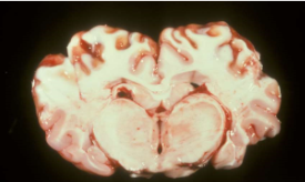
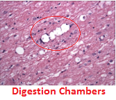
- Gross: Spinal cord may
be indented or flattened
- Histologically: Myelin sheath
ballooning in all funiculi w/
axonal swelling/loss, Myelin
digestion chambers, Neuronal
loss, gliosis, malacia (due to
vascular damage), oedema
- Gross: Spinal cord may
be indented or flattened
- May arise within
(Intra-medullary) or
outside (Extra-medullary)
the spinal cord
- Compression
- Concussion,
contusion,
haemorrhage &
Compression
- Like the brain, can be affected by infectious,
inflammatory, toxic, nutritional & neoplastic
disease, & lesions can occur in its parenchyma
- Congenital Malformations
- Abnormal
development of
neural tube
- May involve Spinal
cord, Brain, Calvaria,
Meninges &/or
Vertebral column
- Morphological
- Hydrocephalus
- Increased
accumulation of
fluid within the
cranial cavity
- Types:
- Internal (fluid
w/in ventricles)
- Congenital form:
- Obstruction NOT found
- Common in brachycephalic dogs
(mesenphalic aqueduct may be
stenotic)
- Sporadic in cattle
- Sporadic in cattle
- Obstruction NOT found
- Acquired form:
- Causes: Obstruction
due to inflammation (of
meninges &/or
ependymal cells) or
space occupying lesions
compression
(neoplasms, abscesses
& cholesteatomas)
- Causes: Obstruction
due to inflammation (of
meninges &/or
ependymal cells) or
space occupying lesions
compression
(neoplasms, abscesses
& cholesteatomas)
- Common
- Congenital form:
- External (w/in arachnoid space)
- Communicating (w/in both locations)
- Hydrocephalus ex vacuo (ventricles
dilate 2ndary to replace lost tissue)
- Hydrocephalus ex vacuo (ventricles
dilate 2ndary to replace lost tissue)
- Communicating (w/in both locations)
- Internal (fluid
w/in ventricles)
- Increased
accumulation of
fluid within the
cranial cavity
- Others
- Anencephaly (absence of brain)
- Lissencephaly (normal in mice,
rats, rabbits & birds)
- Meningoencephalocoele
- Spinal: spina bifida,
hydromyelia &
syringomyelia
- Spinal: spina bifida,
hydromyelia &
syringomyelia
- Meningoencephalocoele
- Anencephaly (absence of brain)
- Cerebellar Defects
- Cerebellar
hypoplasia
- Common
- Inherited: (Arab foals,
Jersey cows, Chows,
Corriedale sheep)
- Acquired: Environmental teratogens
attack germinal cells in external
granular layer of cerebellum (source
of neurons)
- Ex. Bovine virus diarrhea
(BVD), feline parvovirus, CSF
(hog cholera), canine parvovirus,
schmallenberg virus
- Ex. Bovine virus diarrhea
(BVD), feline parvovirus, CSF
(hog cholera), canine parvovirus,
schmallenberg virus
- Acquired: Environmental teratogens
attack germinal cells in external
granular layer of cerebellum (source
of neurons)
- Common
- Cerebellar
Abiotrophy
- Premature degeneration
of nervous tissue
elements after they have
formed (form of atrophy)
- probably inherited
- Premature degeneration
of nervous tissue
elements after they have
formed (form of atrophy)
- probably inherited
- Cerebellar
hypoplasia
- Hydrocephalus
- Functional (biochemical
abnormalites - ex. lysosomal
diseases or leukodystrophies)
- Causes:
- Inherited
- Environmental
(most common)
- Toxic
- Infectious
- Nutritional
- Radiation
- Radiation
- Nutritional
- Infectious
- Toxic
- Inherited
- Abnormal
development of
neural tube
- CNS Neoplasia
- Primary
- Meninges
- Meningioma
- Most frequent
in Dogs & Cats
- Compressive, space-occupying
lesion (seldom invades)
- Most frequent
in Dogs & Cats
- Meningioma
- Glial cells
- Astrocytoma
- Most common glial tumor
- Brachycephalic breeds
- Solid, firm, grey-white (sometimes mottled
red w/ areas of necrosis/haemorrhage)
- Solid, firm, grey-white (sometimes mottled
red w/ areas of necrosis/haemorrhage)
- Brachycephalic breeds
- Most common glial tumor
- Oligodendroglioma
- Most common in Dogs
(brachycephalic breeds)
- Soft, grey to pink/red &
often gelatinous
- Soft, grey to pink/red &
often gelatinous
- Most common in Dogs
(brachycephalic breeds)
- Ependymoma
- Mainly in ventricles
(primarily lateral)
- May spread in the
ventricular system via CSF
- Expansile, but can be
invasive & destructive
- Mainly in ventricles
(primarily lateral)
- Choroid plexus
tumors
- Rare, mainly Dogs, 4th ventricle
- Rare, mainly Dogs, 4th ventricle
- Astrocytoma
- Neurons (very rare)
- Meninges
- Secondary (Metastatic)
- Haemangiosarcoma
- Mammary or Pulmonary Carcinoma
- Lymphoma
- Pituitary tumors
- Vertebral osteosarcoma
- Vertebral osteosarcoma
- Pituitary tumors
- Lymphoma
- Mammary or Pulmonary Carcinoma
- Haemangiosarcoma
- Primary
- Idiopathic Diseases
- Idiopathic Epilepsy
- Small group of neurons
periodically & spontaneously
depolarize
- Structural : Neoplasma, inflammation, trauma
- Biochemical: Hypocalcemia,
Hypoglycemia, Hepatic encephalopathy
- Idiopathic: No cause identified (indiviuduals
have a low seizure threshold)
- Idiopathic: No cause identified (indiviuduals
have a low seizure threshold)
- Biochemical: Hypocalcemia,
Hypoglycemia, Hepatic encephalopathy
- Structural : Neoplasma, inflammation, trauma
- Small group of neurons
periodically & spontaneously
depolarize
- Idiopathic Epilepsy
- Diseases of Peripheral Nerves
- Dorsal & Ventral roots, spinal
ganglia, spinal & peripheral nerves,
cranial nerves & peripheral
components of the ANS
- Trauma
- Nerves are
predisposed to
injury because of:
- Superficial position
- Proximity to bone
- Proximity to injection sites
- Proximity to injection sites
- Proximity to bone
- Superficial position
- Laceration
- Avulsion ("pull out" -
usually at roots)
- Transection
- Compression &
stretching (following
fracture or dislocation)
- => Wallerian degeneration
- => Wallerian degeneration
- Compression &
stretching (following
fracture or dislocation)
- Transection
- Avulsion ("pull out" -
usually at roots)
- Nerves are
predisposed to
injury because of:
- Infectious
- Some infectious agents
involve peripheral nerves as
well as CNS
- Ex. Rabies, Aujesky's
disease, Neospora
caninum, Toxoplasma gondii
- Neospora caninum
- Dorsal roots (Dog)
- Equine Guttural Pouch mycosis
- Inflammation in the
recurrent laryngeal nerve
-> vocal cord hemiplegia
- Macaw Wasting Disease
(Proventricular dilation syndrome)
- Non suppurative inflammation around
myenteric ganglia => gastric dysfunction
(wasting, anorexia, depression) =>
neurological signs (ataxia, seizures)
- Borna disease virus ?
- Non suppurative inflammation around
myenteric ganglia => gastric dysfunction
(wasting, anorexia, depression) =>
neurological signs (ataxia, seizures)
- Inflammation in the
recurrent laryngeal nerve
-> vocal cord hemiplegia
- Dorsal roots (Dog)
- Some infectious agents
involve peripheral nerves as
well as CNS
- Idiopathic/ Immune mediated
- Coonhound paralysis
- Polyradiculoneuritis
(racoon bites) => Quadriplegia
- Lesions in ventral roots of spinal
nerves & in peripheral nerves
- Lesions in ventral roots of spinal
nerves & in peripheral nerves
- Polyradiculoneuritis
(racoon bites) => Quadriplegia
- Coonhound paralysis
- Neuropathies
- Often breed related ->
Inherited pathogenesis
- Often breed related ->
Inherited pathogenesis
- Degenerative
- Equine laryngeal
hemiplegia ("roarers")
- Common
- Pathogenesis:
- Degeneration of left recurrent pharangeal nerve ->
Atrophy of the left dorsal cricoarytenoid muscle ->
Inability to abduct arytenoid cartilage & vocal fold =>
Airways partially obstructed on inspiration
- Cause:
- Idiopathic/ inherited, 2ndary to
guttural pouch mycosis, Trauma
- Idiopathic/ inherited, 2ndary to
guttural pouch mycosis, Trauma
- Degeneration of left recurrent pharangeal nerve ->
Atrophy of the left dorsal cricoarytenoid muscle ->
Inability to abduct arytenoid cartilage & vocal fold =>
Airways partially obstructed on inspiration
- 95% cases on Left
- Common
- Equine laryngeal
hemiplegia ("roarers")
- Toxic
- Uncommon
- Heavy metals (Pb, Hg, Thallium)
- Pb -> damages Schwann cells ->
peripheral degeneration
- Hg -> Targets neuronal cell bodies ->
2ndary axonal degeneration in
peripheral nerves
- Hg -> Targets neuronal cell bodies ->
2ndary axonal degeneration in
peripheral nerves
- Pb -> damages Schwann cells ->
peripheral degeneration
- Uncommon
- Neoplastic
- Uncommon
- Dogs
- Peripheral nerve sheath tumors
(Benign/ Malignant)
- Uncommon
- Nutritional/metabolic
- Vitamin A & B, Pantothenic acid
& Cu deficiency => Neuropathy
- Cu deficiency: Swayback (New born lambs) &
enzootic ataxia (older lambs)
- Cu deficiency: Swayback (New born lambs) &
enzootic ataxia (older lambs)
- Metabolic: Diabetes mellitus, Hypothyroidism
- Vitamin A & B, Pantothenic acid
& Cu deficiency => Neuropathy
- Dorsal & Ventral roots, spinal
ganglia, spinal & peripheral nerves,
cranial nerves & peripheral
components of the ANS
Medienanhänge
Möchten Sie kostenlos Ihre eigenen Mindmaps mit GoConqr erstellen? Mehr erfahren.