20644976
Beschreibung
Quiz von Charlotte Jakes, aktualisiert more than 1 year ago
|
|
Erstellt von Charlotte Jakes
vor mehr als 4 Jahre
|
|
Frage 1
Frage
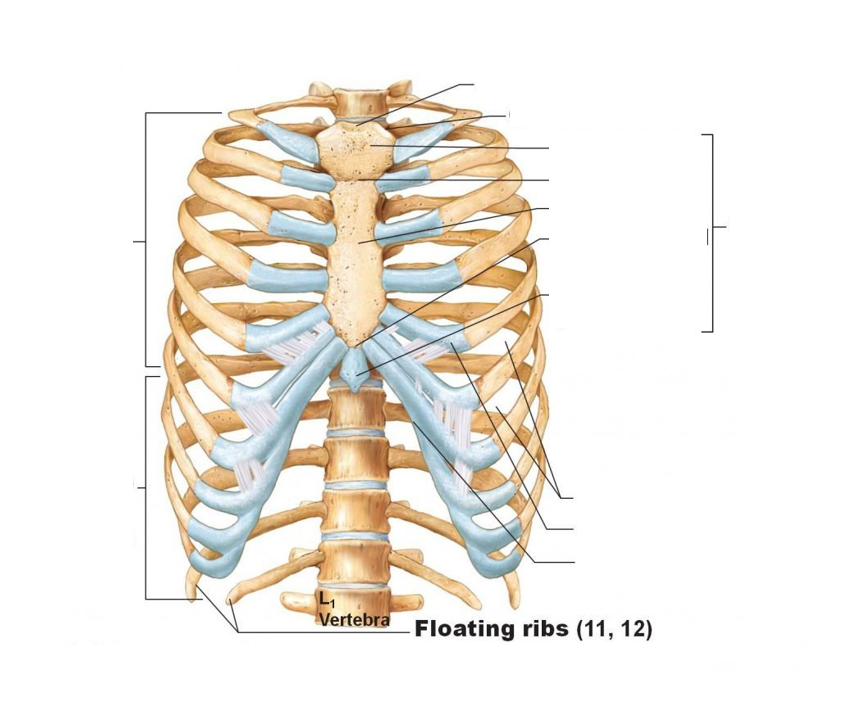
Label this image to show the oesteology of the thoracic cage.
Antworten
-
Jugular notch
-
Clavicular angle
-
Maunbrium
-
Sternal angle
-
Body
-
Xiphisternal angle
-
Xiphoid process
-
Sternum
-
Intercostal space
-
Costal margin
-
Costal cartilage
-
False ribs
-
True ribs
Frage 2
Antworten
-
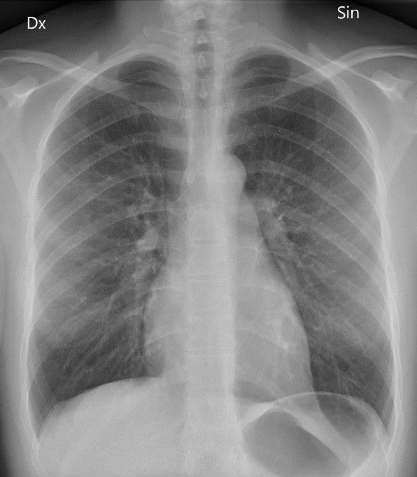
Lung tissue doesn't absorb X-rays
-
Air doesn't absorb X-rays
-
The tissue of the lungs is too thick to be shown
-
The tissue of the lungs is too thin to be shown
Frage 3
Frage
What are the wispy lines shown in the lung field, emanating from the heart?
Antworten
-
Pulmonary blood vessels
-
Bronchioles
-
Alveoli
-
Coronary blood vessels
Frage 4
Antworten
-
It is full of blood which absorbs x-rays
-
It is full of air which absorbs x-rays
-
It is full of blood which does not absorb x-rays
-
Its walls are made of muscle tissue which absorbs x-rays
Frage 5
Frage
Why do we use posterior-anterior orientation in chest x-ray?
Antworten
-
To prevent false enlargement of the heart
-
To prevent false shrinkage of the heart
-
To prevent false consolidation of the lungs
-
To get a better resolution
Frage 6
Frage
Why is the right hemidiaphragm slightly higher than the left?
Antworten
-
Due to the presence of the liver
-
The right lung is smaller
-
Due to the presence of the stomach
-
Due to the suspensory ligament of Treitz
Frage 7
Frage
How do we visualise the tubular system of the lungs?
Antworten
-
Patient inhales imaging gas
-
X-ray
-
MRI
-
CT
Frage 8
Frage
The heart appears longer and thinner on x-ray if taken during inspiration.
Antworten
- True
- False
Frage 9
Frage
What is the region of translucency in the bottom right corner of this x-ray?
Antworten
-
Gas Bubble in Stomach
-
Left hemidiaphragm
-
Liver
-
Heart
Frage 10
Frage
Which of the following are boundaries of the superior thoracic aperture?
Antworten
-
T1
-
Inside of first rib
-
Superior border of manubrium
-
Inside of second rib
-
T2
-
C7
-
Costal margin
Frage 11
Frage
The suprapleural membrane spreads over the superior thoracic aperture.
Antworten
- True
- False
Frage 12
Frage
WHere does the ligament that holds the suprapleural membrane up in a dome shape attach?
Antworten
-
Transverse process of C7
-
Spinous process of C7
-
Transverse process of T1
-
Transverse process of C6
Frage 13
Frage
Which of the following are borders of the inferior thoracic aperture?
Antworten
-
Costal margin
-
Tip of 11th rib
-
Inside of 11th rib
-
Inferior border of 12th rib
-
T12
-
T11
-
Inferior border of manubrium
Frage 14
Frage
Where does the diaphragm arch down and attach at?
Antworten
-
L1 and L2
-
L1 only
-
L2 only
-
T12
-
L3
Frage 15
Frage
The aorta passes through the central tendon of the diaphragm.
Antworten
- True
- False
Frage 16
Frage
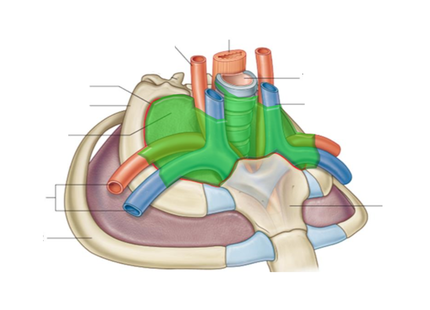
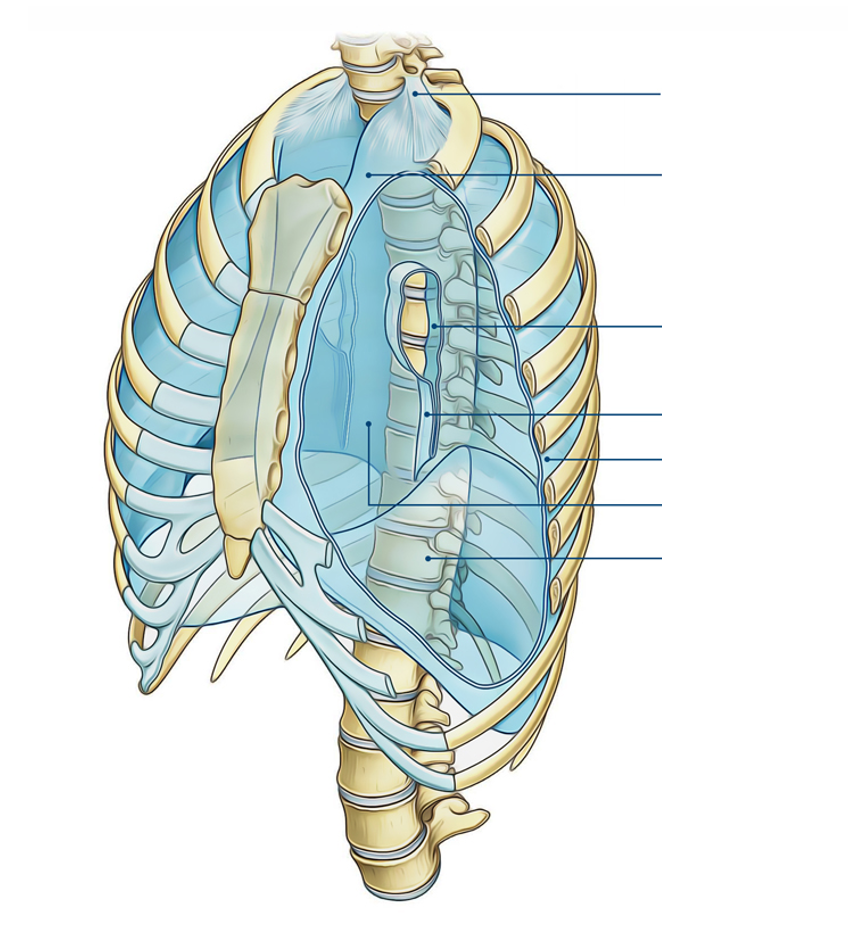
Label this diagram of the superior thoracic aperture. The green coloured area represents the suprapleural membrane.
Antworten
-
Manubrium
-
2nd rib
-
1st rib
-
Subclavian vessels
-
Suprapleural membrane
-
Internal jugular vein
-
Trachea
-
Common carotid artery
-
Oesophagus
Frage 17
Frage
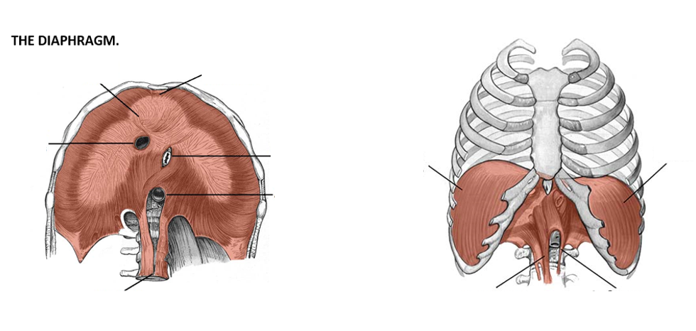
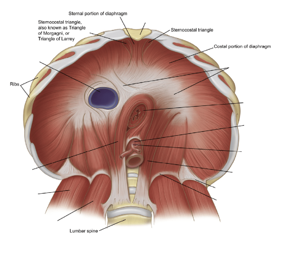
Label these images to show the anatomy of the diaphragm.
Antworten
-
Central tendon
-
Caval hiatus
-
Oesophageal hiatus
-
Aortic hiatus
-
Right hemidiaphragm
-
Left hemidiaphragm
-
Right crus
-
Left crus
-
Sternal attachment
Frage 18
Frage
The left hemidiaphragm is innervated by the left phrenic nerve and the right hemidiaphragm is innervated by the right phrenic nerve
Antworten
- True
- False
Frage 19
Frage
Where does the costodiaphragmatic recess occur?
Antworten
-
8th-11th intercostal spaces along the midaxillary line
-
7th-11th intercostal spaces along the midaxillary line
-
10th and 11th intercostal spaces along the midaxillary line
-
7th-10th intercostal spaces along the midaxillary line
Frage 20
Frage
What does the costodiaphragmatic recess allow us to do?
Antworten
-
Access the pleural cavity with less risk to lung tissue
-
Access the pleural cavity with no risk to lung tissue
-
Access the vena cava
-
Access the abdominal arota
Frage 21
Frage
What type of nerves are the phrenic nerves?
Antworten
-
Somatic
-
Autonomic
Frage 22
Antworten
-
Xiphoid
-
Vena cava
-
Central tendon
-
Oesophagus
-
Aorta
-
Coeliac trunk
-
Left crus
-
Right crus
-
Quadratus lumborum
-
Psoas major
-
Lateral arcuate ligament
-
Medial arcuate ligament
Frage 23
Frage
What is the function of the arcuate ligaments?
Antworten
-
Separate the diaphragm from the posterior trunk muscles
-
Suspend the posterior trunk muscles
-
Separate the diaphragm from the abdominal organs
-
Join the diaphragm to the lumbar spine
Frage 24
Frage
The oesophageal hiatus occurs in the muscle of the diaphragm - what does this mean?
Antworten
-
Reflux of stomach contents during breathing is prevented
-
Diaphragm as a true valve around the oesophagus
-
Prevents choking during breathing
-
Enables gastric emptying
Frage 25
Frage
The caval hiatus occurs in the [blank_start]central tendon[blank_end]. This means that during diaphragmatic [blank_start]contraction[blank_end] the walls of the [blank_start]vena cava[blank_end] are pulled apart. This reduces [blank_start]pressure[blank_end] in the vena cava which pulls venous blood up through the abdomen, thus aiding [blank_start]venous return[blank_end].
Antworten
-
central tendon
-
contraction
-
vena cava
-
pressure
-
venous return
Frage 26
Frage
Where does parietal pleura become visceral pleura?
Antworten
-
At the lung root
-
At the horizontal fissure
-
At the oblique fissure
-
At the diaphragm
Frage 27
Frage
What is the purpose of the pulmonary ligament?
Antworten
-
Provides room for bronchi to move during expiration/inspiration
-
Prevents lungs from entering abdominal cavity
-
Prevents lungs from protruding into the neck
-
Provides route for bronchi, pulmonary vessels, lymphatics and nerves to enter the lungs
Frage 28
Frage
What innervates the pleura?
Antworten
-
The phrenic nerves
-
The cranial accessory nerve XI
-
Intercostal nerves from thoracic vertebrae
-
The vagus nerve
Frage 29
Frage
What innervates the diaphragmatic and mediastinal pleura?
Antworten
-
Phrenic nerves
-
Vagus nerve
-
Cranial accessory nerve XI
-
Intercostal nerves from thoracic vertebrae
Frage 30
Frage
Why does the normal lung at rest not collapse under its elastic recoil pressure?
Antworten
-
Negative pressure in the pleural cavity
-
Positive pressure in the pleural cavity
-
Diaphragm contracts to hold lungs open
-
Bronchi hold lungs open
Frage 31
Frage
Label this diagram to show the pleural membranes.
Antworten
-
Suprapleural membrane
-
Cervical parietal pleura
-
Lung root
-
Pulmonary ligament
-
Costal parietal pleura
-
Mediastinal parietal pleura
-
Diaphragmatic parietal pleura
Möchten Sie mit GoConqr kostenlos Ihre eigenen Quiz erstellen? eigenen Mehr erfahren.