20704718
Description
Quiz by Charlotte Jakes, updated more than 1 year ago
|
|
Created by Charlotte Jakes
over 4 years ago
|
|
Question 1
Question
What is the superior boundary of the mediastinum?
Answer
-
Superior thoracic aperture
-
Manubrium of the sternum
-
Mandible
-
Cricoid cartilage
Question 2
Question
What is the inferior boundary of the mediastinum?
Answer
-
The inferior thoracic aperture
-
The costal margin
-
The xiphisternum
-
The anterior superior iliac spine
Question 3
Question
What is the lateral border of the mediastinum?
Answer
-
Lungs
-
Ribs
-
Transversus abdominis
-
External oblique
Question 4
Question
What is the anterior border of the mediastinum?
Answer
-
The sternum
-
The heart
-
The costal margin
-
The sternum and costal margin
Question 5
Question
What is the posterior border of the mediastinum?
Answer
-
The entire thoracic spine
-
T1-T5
-
T5-T12
-
T9-T12
Question 6
Question
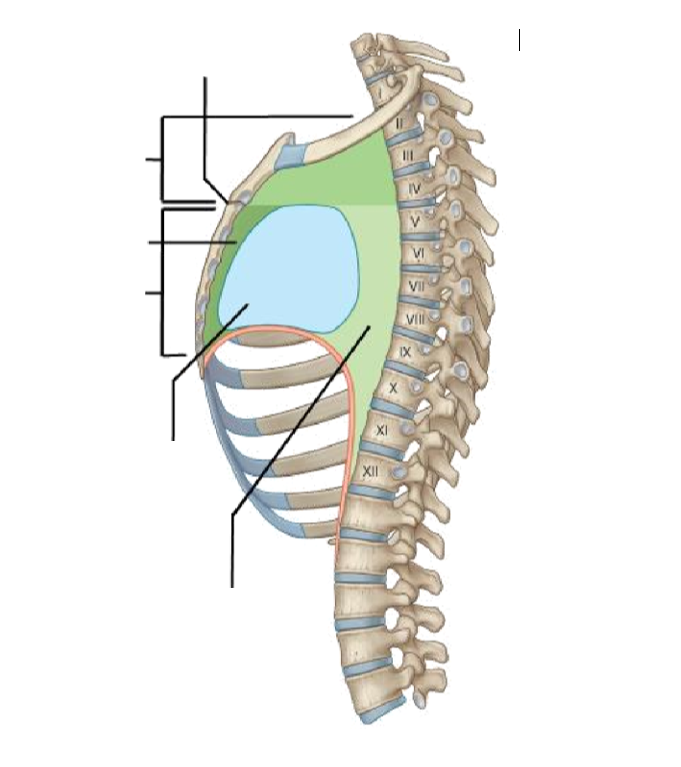
Label this diagram to show the regions of the mediastinum.
Answer
-
Sternal angle
-
Superior mediastinum
-
Posterior mediastinum
-
Middle mediastinum
-
Inferior mediastinum
-
Anterior mediastinum
Question 7
Question
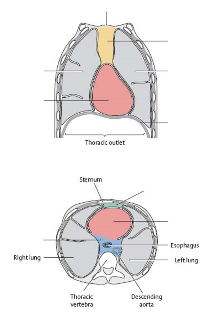
Label these diagrams to show the boundaries of the mediastinum.
Answer
-
Superior thoracic aperture
-
Superior mediastinum
-
Right lung
-
Left lung
-
Middle mediastinum
-
Diaphragm
-
Anterior mediastinum
-
Middle mediastinum
-
Posterior mediastinum
Question 8
Question
What is the level of the sternal angle?
Answer
-
T4/T5
-
T3/T4
-
T2/T3
-
T5/T6
Question 9
Question
Drag and drop the correct answers to give the borders of the superior mediastinum.
Superior - [blank_start]superior thoracic aperture[blank_end]
Inferior - [blank_start]continuous with the inferior mediastinum[blank_end]
Anterior - [blank_start]manubrium[blank_end]
Posterior - [blank_start]vertebral bodies of T1-T4[blank_end]
Lateral - [blank_start]pleurae of lungs[blank_end]
Answer
-
superior thoracic aperture
-
continuous with the inferior mediastinum
-
manubrium
-
vertebral bodies of T1-T4
-
pleurae of lungs
-
inferior thoracic aperture
-
body of sternum
-
xiphisternum
-
vertebral bodies of T2-T4
-
vertebral bodies of T1-T5
Question 10
Question
The aortic arch occurs in the superior mediastinum.
Answer
- True
- False
Question 11
Question
Where is the thymus gland found?
Answer
-
Behind the sternum
-
Behind the costal cartilage on the left side
-
In the jugular notch of the manubrium
-
Below the xiphisternum
Question 12
Question
At what point does your thymus gland stop producing T cells?
Answer
-
Puberty
-
Birth
-
Death
-
Age of 21
Question 13
Question
Fill in the blanks to describe the contents of the superior mediastinum.
- A[blank_start]ortic arch[blank_end] thus including the b[blank_start]rachiocephalic[blank_end] artery, left common [blank_start]carotid[blank_end] artery and left [blank_start]subclavian[blank_end] artery
- Superior [blank_start]vena cava[blank_end] thus including the b[blank_start]rachiocephalic[blank_end] veins and the az[blank_start]ygos[blank_end] vein
- The v[blank_start]agus[blank_end] nerves, p[blank_start]hrenic[blank_end] nerves and s[blank_start]ympathetic[blank_end] trunk
- The t[blank_start]hymus[blank_end] gland
- The t[blank_start]rachea[blank_end]
- The o[blank_start]esophagus[blank_end]
- The [blank_start]thoracic[blank_end] duct
Answer
-
ortic arch
-
rachiocephalic
-
carotid
-
subclavian
-
vena cava
-
rachiocephalic
-
ygos
-
agus
-
hrenic
-
ympathetic
-
hymus
-
rachea
-
esophagus
-
thoracic
Question 14
Question
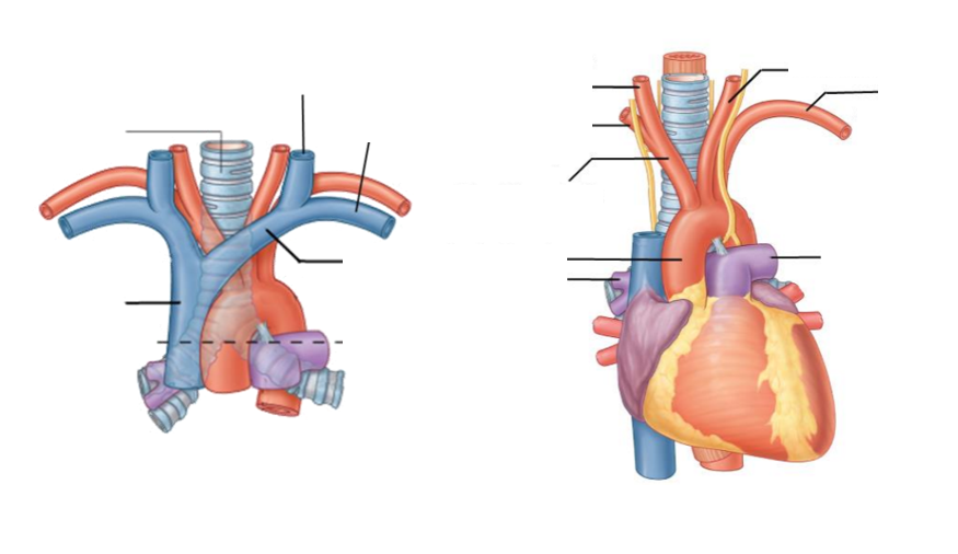
Label this image to show some of the great vessels found in the superior mediastinum.
Answer
-
Trachea
-
Superior vena cava
-
Brachiocephalic vein
-
Left jugular vein
-
Left subclavian vein
-
Aortic arch
-
Brachiocephalic trunk
-
Right common carotid artery
-
Right subclavian artery
-
Left common carotid artery
-
Left subclavian artery
Question 15
Question
Drag and drop the correct answers to give the borders of the anterior mediastinum.
Superior - [blank_start]continuous with superior mediastinum[blank_end]
Inferior - [blank_start]diaphragm[blank_end]
Anterior - [blank_start]body of the sternum[blank_end]
Posterior - [blank_start]pericardium[blank_end]
Lateral - [blank_start]mediastinal pleura[blank_end]
Answer
-
continuous with superior mediastinum
-
diaphragm
-
body of the sternum
-
pericardium
-
mediastinal pleura
-
xiphisternum
-
manubrium of sternum
-
costal cartilages
-
transverse colon
-
superior thoracic aperture
Question 16
Question
The thymus extends inferiorly into the anterior mediastinum in infants and children.
Answer
- True
- False
Question 17
Question
What ligaments are present in the anterior mediastinum?
Answer
-
Sternopericardial ligaments
-
Costopericardial ligaments
-
Costochondral ligaments
-
Pleurapericardial ligaments
Question 18
Question
Drag and drop the correct answers to give the borders of the posterior mediastinum.
Superior - [blank_start]continuous with superior mediastinum[blank_end]
Inferior - [blank_start]diaphragm[blank_end]
Anterior - [blank_start]pericardium[blank_end]
Posterior - [blank_start]T5-T12 vertebrae[blank_end]
Lateral - [blank_start]mediastinal pleura[blank_end]
Answer
-
continuous with superior mediastinum
-
diaphragm
-
pericardium
-
T5-T12 vertebrae
-
mediastinal pleura
-
T6-T12 vertebrae
-
costal cartilage
-
transverse processes of T5-T12
-
superior thoracic aperture
Question 19
Question
Fill in the blanks to give the contents of the posterior mediastinum.
- The [blank_start]thoracic[blank_end] aorta and thus the posterior [blank_start]intercostal[blank_end] arteries, the b[blank_start]ronchial[blank_end] arteries, the o[blank_start]esophageal[blank_end] arteries and the superior [blank_start]phrenic[blank_end] arteries
- The [blank_start]azygos[blank_end] venous system
- The o[blank_start]esophagus[blank_end]
- The [blank_start]thoracic[blank_end] duct
- The [blank_start]sympathetic[blank_end] trunk
Answer
-
thoracic
-
intercostal
-
phrenic
-
ronchial
-
esophageal
-
azygos
-
esophagus
-
thoracic
-
sympathetic
Question 20
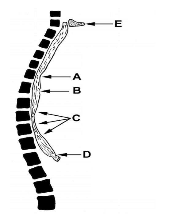
Question
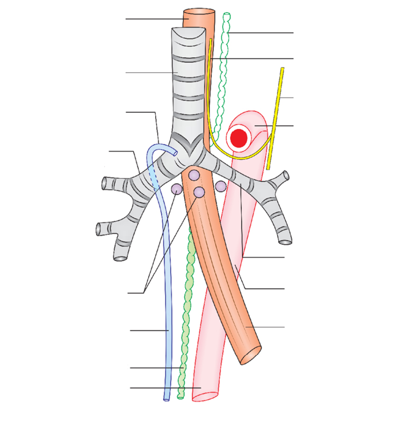
Label this diagram to show the relations of the contents of the posterior mediastinum.
Answer
-
Oesophagus
-
Thoracic duct
-
Thoracic aorta
-
Azygos vein
-
Right main bronchus
-
Left main bronchus
-
Tracheobronchial lymph nodes
-
Aortic arch
-
Vagus nerve
-
Left recurrent laryngeal nerve
Question 21
Question
Where does the left vagus nerve branch to give the left recurrent laryngeal nerve which ascend to the larynx?
Answer
-
Aortic arch
-
Left subclavian artery
-
Pulmonary trunk
-
Left main bronchus
Question 22
Question
The vagus nerves form an anterior and posterior plexus respectively which still have branches that interconnect them.
Answer
- True
- False
Question 23
Question
Between what vessels is the left vagus sandwiched during its descent into the trunk?
Answer
-
Subclavian artery and subclavian vein
-
Left common carotid artery and left jugular vein
-
Brachiocephalic trunk and left subclavian artery
-
Superior vena cava and brachiocephalic trunk
Question 24
Question
Label this image to show how the vagus nerve branches in the thorax.
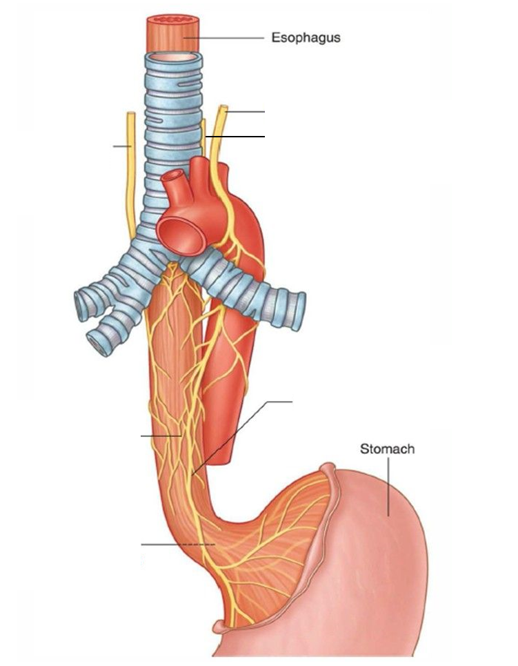
Answer
-
Left vagus nerve
-
Left recurrent laryngeal nerve
-
Right vagus nerve
-
Oesophageal plexus
-
Anterior vagal trunk
-
Posterior vagal trunk
Question 25
Question
Label this image to show the points of potential compression of the oesophagus.
Answer
-
Cricoid cartilage
-
Aortic arch
-
Left main bronchus
-
Left atrium
-
Oesophageal hiatus
Question 26
Question
The upper third of the oesophagus is skeletal muscle then it transitions into smooth muscle more inferiorly.
Answer
- True
- False
Question 27
Question
Label this image to show branches of the thoracic aorta.
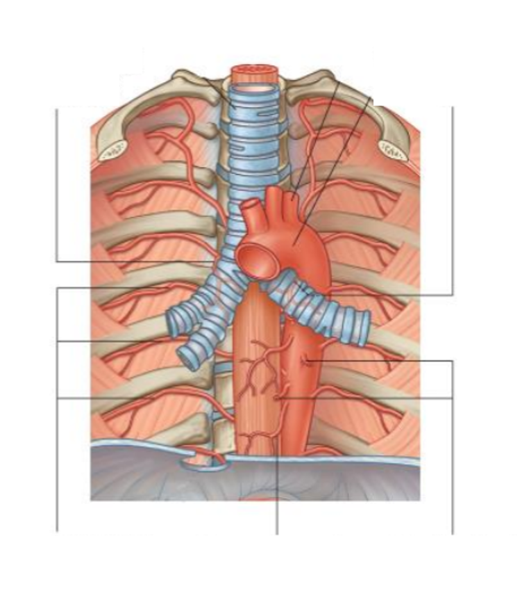
Answer
-
Aortic arch
-
Posterior intercostal arteries
-
Right bronchial artery
-
Left bronchial artery
-
Mediastinal branches
-
Oesophageal branches
-
Left subclavian artery
Question 28
Question
Label this image to show the venous drainage of the thorax.
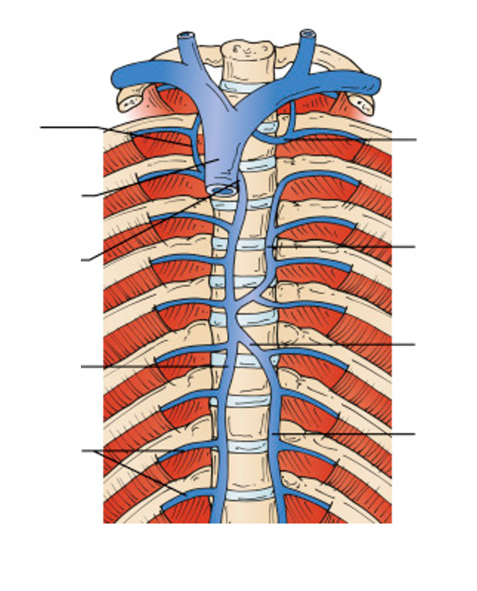
Answer
-
Superior vena cava
-
Left superior intercostal vein
-
Accessory hemiazygous vein
-
Hemiazygos vein
-
Intercostal veins
-
Azygos vein
-
Arch of azygos
-
Right superior intercostal vein
Question 29
Question
What structure does the sympathetic trunk run into the trunk alongside?
Answer
-
Neck of the ribs
-
Shaft of the ribs
-
Intercostal grooves of the ribs
-
Costal cartilages
Question 30
Question
Label this diagram to show how the nerves of the thorax enter via the superior thoracic aperture.
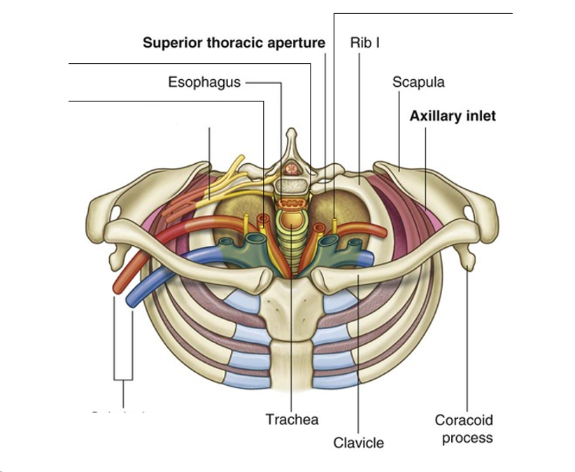
Answer
-
Phrenic nerve
-
Vagus nerve
-
Sympathetic chain
-
Brachial plexus
Question 31
Question
Label this image to show the organisation of the nerves supplying the larynx.
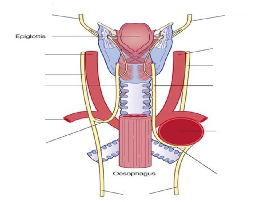
Answer
-
Right superior laryngeal nerve
-
Left superior laryngeal nerve
-
Right common carotid artery
-
Left common carotid artery
-
Right vagus nerve
-
Right recurrent laryngeal nerve
-
Left recurrent laryngeal nerve
-
Left vagus nerve
-
Right subclavian artery
-
Aortic arch
-
Right recurrent laryngeal nerve
Question 32
Question
The internal laryngeal nerves penetrate the thyrohyoid membrane.
Answer
- True
- False
Question 33
Question
Which division of the autonomic nerve system causes vasconstriction in the lungs?
Answer
-
Sympathetics
-
Parasympathetics
Question 34
Question
Which division of the autonomic nervous system has afferent pain fibres that refer lung pain to the sternum?
Answer
-
Parasympathetics
-
Sympathetics
Question 35
Question
Which division of the autonomic nervous system is responsible for bronchodilation and bronchosecretion?
Answer
-
Sympathetics
-
Parasympathetics
Question 36
Question
Which division of the autonomic nervous system is afferent to the visceral pleura for stretch reception?
Answer
-
Sympathetics
-
Parasympathetics
Question 37
Question
Which spinal nerves give sympathetic innervation to the lungs?
Answer
-
T1-T4
-
T1-T5
-
Vagus
-
T2-T6
Question 38
Question
Fill in the blanks to describe the origins of the following plexuses.
The pulmonary plexus has sympathetics from [blank_start]T1-T5[blank_end] and parasympathetics from the [blank_start]vagus nerve[blank_end].
The cardiac plexus has sympathetics from [blank_start]T1-T4[blank_end] and parasympathetics from the [blank_start]vagus nerve[blank_end].
The oesophageal plexus has sympathetics from [blank_start]T2-T6[blank_end] and parasympathetics from the [blank_start]vagus nerve[blank_end].
Answer
-
vagus nerve
-
vagus nerve
-
vagus nerve
-
T1-T5
-
T1-T4
-
T2-T6
Want to create your own Quizzes for free with GoConqr? Learn more.